The quality of dental services 20 years ago left much to be desired. Fillings of dubious quality, loss of teeth, bridges that injure neighboring teeth - the list is endless. The reason for such unprofessional actions of dentists is unknown. Dentistry has changed since then. Specialists acquired the opportunity to gain new knowledge and improve their skills at seminars and conferences. Familiarity with new techniques makes life easier for the dentist and increases the comfort of patients.
These methods include dental photography. The method allows you to track your treatment history. This is a way of regulating relations between clinics and clients, increasing the level of trust in dentistry and raising their prestige.
Article for patients: treatment of wedge-shaped defect and cervical caries using modern methods.
Patients often come to our clinic with problems such as cervical caries and wedge-shaped defect.
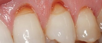
IN THE PHOTO: AN EXAMPLE OF TREATMENT OF A WEDGE-shaped TOOTH DEFECT AT THE BIONIC DENTIS DENTAL CLINIC. BEFORE AND AFTER TREATMENT.
We present methods that were developed in Germany and are of the highest quality!
- eliminate enamel defects for many years.
- do not damage tooth enamel
- BIO compatible
- without the use of acid etching
- do not require excision of healthy tooth tissue.
IN THE PHOTO: AN EXAMPLE OF TREATMENT OF A WEDGE-shaped TOOTH DEFECT AT THE BIONIC DENTIS DENTAL CLINIC. BEFORE AND AFTER TREATMENT.
Key Goals
Dental photography helps achieve the following goals:
- Documenting the stages of treatment provides the patient with information about the procedures performed by the dentist and allows him to evaluate the quality of the work.
- Preventing misunderstandings between patients and dentists.
- The images provide the doctor with the opportunity to consider the upcoming treatment and assess the deficiencies and parameters of the client’s teeth. Thanks to the images, he gets a complete picture of the color, structure and shape of the teeth. Taking into account the data obtained, it is easier for technicians to develop crowns and inlays.
- Correcting the dentist's omissions. The specialist analyzes the work and eliminates errors in a timely manner.
- The use of dental photographs at dental symposiums and master classes demonstrates the level of professionalism of the dentist.
- Compiling a portfolio.
A few words about the wedge-shaped defect and cervical caries.
Problems associated with this disease are usually:
- increased sensitivity of the neck of the tooth to sweets and sours.
- increased sensitivity of the neck of the tooth to cold and hot.
- increased sensitivity of the neck of the tooth to the touch of a toothbrush when brushing your teeth.
- The fillings that were previously installed on the cervical area of the tooth do not hold and fall out in an extremely short time (up to six months).
IN THE PHOTO: AN EXAMPLE OF TREATMENT OF A WEDGE-shaped TOOTH DEFECT AT THE BIONIC DENTIS DENTAL CLINIC. BEFORE AND AFTER TREATMENT.
The reason for these problems associated with increased sensitivity is that there is a very thin layer of enamel on the neck of the tooth and the nerve of the tooth is located nearby. With this anatomical feature, any irritant, even with a small cavity, produces sharp pain. The same feature leads to the fact that with cervical caries and wedge-shaped defects, the nerve of the tooth is very quickly damaged, which leads to pulpitis.
The problem of fixing fillings in this pathology is due to the fact that modern fillings are not fixed in this area due to a deficiency of hard dental tissues.
In our clinic, we have 100% solved these problems based on the most modern European techniques and using high-quality European restoration materials.
IN THE PHOTO: AN EXAMPLE OF TREATMENT OF CERVICAL TOOTH CARIES AT THE BIONIC DENTIS DENTAL CLINIC. BEFORE AND AFTER TREATMENT.
We present to you three methods for treating cervical caries and wedge-shaped defects:
Features of working with a microscope
Dental treatment under a microscope has its own characteristics that must be taken into account when working:
- To work with a dental microscope, specialized training of the doctor and assistant is required;
- Adaptation of the dentist’s eyes to the device lasts about six months; during this period, the dentist may experience pain in the eyes when working with the microscope for a long time.
- The assistant must accurately respond to the doctor’s requests and place the necessary instruments in his hand.
- Conventional dental instruments are not suitable for treatment under a microscope, since their handle or the operator's hand may obscure the field of view; requires the purchase of new tools.
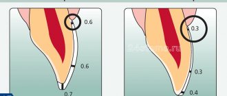
- The disadvantage of a microscope in root canal treatment is its ability to “see” only what is directly in the line of sight; when the canal is bent, the surface becomes inaccessible for viewing. In reality, in 90% of cases, the doctor’s gaze reaches almost to the apical part of the tooth;
- The patient is required to remain as still as possible.
- A dental microscope is an expensive device. This circumstance, as well as the need for special training of the dentist, increases the cost of treatment.
More and more patients understand that dental treatment under a microscope is better and more effective, so they are looking for clinics that have the necessary equipment and specialists. The use of a dental microscope in some cases is the only way to conduct accurate diagnosis and treatment and provide the patient with high-quality dental care.
Treatment with the Sandman kinematic system without drilling.
When using the Sandman system, the tooth is not drilled, and the affected areas of the tooth in the cervical area are cleaned using an air flow with aluminum oxide granules. This system is configured in such a way that it carefully and accurately cleans the surface, but does not injure healthy areas of the enamel at all.
This new European technique allows you to safely treat cervical caries or a wedge-shaped defect without damaging healthy areas of the tooth, which is especially important in conditions of a shortage of tissue to fix the restoration.
IN THE PHOTO: AN EXAMPLE OF TREATMENT OF A WEDGE-shaped TOOTH DEFECT AT THE BIONIC DENTIS DENTAL CLINIC. BEFORE AND AFTER TREATMENT.
Treatment is carried out in the following sequence:
- A topical anesthetic gel is applied to the patient’s gums to reduce the sensitivity of the mucous membrane.
- the treatment area is anesthetized by injection of Ultracaine, which makes all manipulations 100% painless.
- Using the Sandman system, softened, pigmented and infected tooth tissues are cleaned.
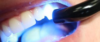
- the bottom and walls of the cavity are processed using a dental laser for sterilization.
- The tooth surface of the tooth is treated with an adhesive system.
- a defect in the tooth enamel in the cervical area is restored using a light restoration system in such a way that it is not possible to notice such a restoration on the tooth.
- The restoration is polished.
On average, treatment of a wedge-shaped defect or cervical dental caries using the Sandman system takes 30-40 minutes.
If there are several adjacent affected teeth, they are treated simultaneously.
The cost of treating one tooth is 14,000 rubles.
IN THE PHOTO: AN EXAMPLE OF TREATMENT OF CERVICAL TOOTH CARIES AT THE BIONIC DENTIS DENTAL CLINIC. BEFORE AND AFTER TREATMENT.
Canal treatment under a microscope
The dental microscope is used in various fields of dentistry, but its use is most effective in endodontic treatment - treatment of tooth cavities and root canals. The work of a dental therapist during endodontic treatment requires increased care, skill and “flair,” therefore the use of a dental microscope increases the success of endodontic treatment several times. Below is a short demonstration of the root canal treatment process under a microscope:
A dental microscope can increase the likelihood of success when:
- Searching for the mouths of canals, including additional and fourth ones;
- Detection of channel branches;
- Detection of pulp residues or carious lesions inside the canals;
- When identifying voids formed during canal filling (the latest techniques, such as injection gutta-percha, when used correctly, eliminate the formation of voids);
- When cracks are detected in the root canals that are not visible on x-rays.
Below is a list of dental operations, the implementation of which has an average and low percentage of successful prognosis with conventional treatment, but when using a microscope, the success rate increases:
- Working with the uncovered root tip;
- Unsealing and re-treatment of poorly treated canals;
- Removal of intracanal pins;
- Removing from the canal fragments of instruments broken during previous treatment;
- Closing perforations. Using the usual method, it is impossible to accurately localize the site of damage to the root canal, and even more so, to bring the instrument to it;
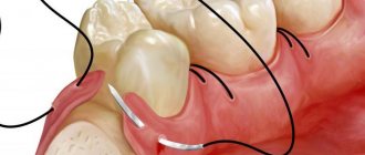
- Treatment of cysts and granulomas. Previously, when cysts and granulomas occurred, teeth had to be removed. Later, methods of therapeutic and surgical treatment appeared, and finally today, thanks to the use of magnifying devices, dentists are able to cure teeth that were previously considered hopeless.
The microscope allows you to reduce the number of medical errors and complications that often occur during root canal treatment in the usual way, “blindly”.
All branches and bends of the main and additional root canals are visible through the microscope, cleaning canals and carious cavities, removing old filling material and filling with new material is greatly facilitated, and objective control of its adherence to the canal walls appears. All this increases the quality of the dentist’s work, the service life of the filled canal and the restored tooth.
Treatment of wedge-shaped defect and treatment of cervical caries with ceramic inlays.
This technique is a premium one. Its distinctive feature is that the cavity in the tooth is closed not with a light-colored restoration material, but with a ceramic inlay. The service life of this ceramic inlay is about 25 years. In terms of aesthetic parameters, it is completely indistinguishable from natural tooth tissue.
Restoring a wedge-shaped defect or cervical caries with a ceramic inlay has several advantages:
- biocompatibility of the material from which the inlay is made. Lithium disilicate, the restoration material in this case, is called synthetic enamel in Germany.
- high aesthetics: the inlay is not noticeable on the tooth due to the chameleon effect that is inherent in lithium disilicate.
- long-lasting shine and no need for periodic polishing.
- Super smooth surface prevents the accumulation of plaque and tartar.
- service life up to 25 years due to the special strength of lithium disilicate.
IN THE PHOTO: AN EXAMPLE OF TREATMENT OF A WEDGE-shaped TOOTH DEFECT AT THE BIONIC DENTIS DENTAL CLINIC. BEFORE AND AFTER TREATMENT.
The process of treating a wedge-shaped defect or cervical caries using a ceramic inlay consists of the following steps:
First visit to the clinic:
- A topical anesthetic gel is applied to the patient’s gums to reduce the sensitivity of the mucous membrane.
- the treatment area is anesthetized by injection of Ultracaine, which makes all manipulations 100% painless.
- Using a turbine and a dental diamond bur, softened, pigmented and infected tooth tissues are cleaned and a place is formed for the placement of the inlay.
- the bottom and walls of the cavity are processed using a dental laser for sterilization.
- The tooth surface of the tooth is treated with a 7th generation adhesive system.
- Impressions are taken from the dentition using silicone impression material or a 3D scanner for transfer to the laboratory.
- a tooth enamel defect in the cervical area is restored with a temporary filling.
IN THE PHOTO: AN EXAMPLE OF TREATMENT OF CERVICAL TOOTH CARIES AT THE BIONIC DENTIS DENTAL CLINIC. BEFORE AND AFTER TREATMENT.
Second visit to the clinic:
- A topical anesthetic gel is applied to the patient’s gums to reduce the sensitivity of the mucous membrane.
- the treatment area is anesthetized by injection of Ultracaine, which makes all manipulations 100% painless.
- Using a turbine and a dental diamond bur, the temporary filling is removed.
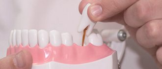
- An inlay made in a dental laboratory is glued into the cavity in the cervical area of the tooth.
- The glue is polymerized using a dental lamp.
- The restoration is polished.
Thus, to make a ceramic inlay for the treatment of a wedge-shaped defect or cervical caries, two visits to the clinic are required. However, the long service life of such a design (several times longer than the service life of the filling) compensates for such a long process.
Each visit takes approximately 40 minutes.
The time between visits is on average 10 days.
The cost of restoration of a wedge-shaped defect or cervical caries of a ceramic inlay made of lithium disilicate is 55,000 rubles.
IN THE PHOTO: AN EXAMPLE OF TREATMENT OF CERVICAL TOOTH CARIES AT THE BIONIC DENTIS DENTAL CLINIC. BEFORE AND AFTER TREATMENT.
These methods for treating wedge-shaped defects and cervical caries within the walls of our clinic will allow you to forget about this problem for many years.
Ozerov Petr Vladimirovich
Chief physician. Dentist, implantologist, orthopedist, surgeon. Laser dentistry specialist
More details
Werner Elena Vladimirovna
Dentist periodontist
More details
Eremina Anna Arturovna
Dentist therapist
More details
Characteristics of the method
The use of devices that allow magnification of images of the operating area provides doctors with significant advantages in medicine.
It becomes possible to assess the condition of each tooth and perform complex manipulations, controlling every movement. Key factors that improve the quality of dental treatment and prosthetics: multiple magnification , good lighting , accurate and convenient positioning of the microscope.
In dental treatment, special dental microscopes are used - electronic optical devices that can magnify images 30 times or more. The microscope is brought to the surgical field using a pantograph, which in turn is attached in various ways: floor (bed), wall (brackets), ceiling; Thanks to the mechanism, the microscope easily takes the exact position above the tooth and is fixed in it. The microscope is equipped with a digital camera connection, which can be used to take photographs and record videos. An image of the treatment process is displayed on the monitor so that the assistant, whose presence is required, can navigate the doctor’s actions. Some devices are equipped with a monocular or binocular tube for an assistant to monitor the progress of treatment.
During treatment using a microscope, the patient is in a chair in the “lying” position, and a microscope lens is installed over the tooth being treated. The doctor sits behind the patient's head or to his right and conducts treatment through eyepieces. The assistant gives the dentist the necessary instruments. For the doctor who conducts treatment under a microscope, a work chair with special armrests and lumbar support is provided - this is necessary so that during long procedures the dentist does not get tired of sitting and there is no hand trembling.
Technical equipment
For fast shooting mode in high quality, the room is properly equipped:
- retractors;
- lamps with UV radiation;
- occlusal and lateral crystalline mirror;
- contrastor;
- cling film to protect the lens during operation.
The assistant is responsible for ensuring the operating condition of the equipment. The dentist is responsible for the quality of the images and settings. Proper distribution of responsibilities helps optimize the shooting process.
It is necessary to prepare the shooting location:
- choose a background (stage table or black matte fabric);
- prepare whatman paper and cardboard.
Lens, camera, macro flash
The clarity of the pictures depends on the quality of the digital camera. It is recommended to use a DSLR camera, which allows you to capture every detail when shooting.
European experts prefer the 70D technique. The camera is suitable for working in rooms with different levels of illumination and is capable of capturing an object from different angles without losing the quality of the images.
Characteristics of the flash for macro photography Canon Macro Ring MR-14EX:
- double lamps for brightness;
- LED bulbs;
- ring flash ensures even illumination;
- reliability and performance.
When choosing a lens, criteria such as quality, ease of use, functionality and affordability are taken into account. The listed characteristics are specific to the Canon EF 100mm f/2.8 USM Macro Lens Review.
Retractors, mirrors, contrastors
Full shooting is not possible without additional equipment.
Contrastors are specialized devices for creating a dark background. With their help, the shooting is focused on the part of the dentition of interest. The black background highlights imperfections. It is recommended to choose anodized aluminum products that are suitable for steam and high pressure sterilization. Treatment is carried out with a soap solution. Chemicals damage the black layer.
Mirrors – used for intraoral photography. Glass, 3 mm thick, is coated with platinoids. Reflects light effectively and is not prone to oxidation.
Mirrors are fragile and require careful handling. The product is processed and sterilized separately from other equipment.
A dental photograph requires at least two mirrors – a lateral and an occlusal one.
Retractors – provide visibility. Devices in the form of hooks, plates, etc. used to retract soft tissues and hold the mouth open. Due to its soft structure, it does not injure mucous membranes.
Shooting technique
Macro photography of the inside of the mouth is a challenging task. To implement it, you must comply with the following rules:
- shooting in manual mode without autofocus;
- installing a contrastor at a distance that prevents glare;
- heating the mirror with hot water before taking photographs to prevent fogging;
- the nose does not fit into the frame with the upper front teeth;
- the dental arch occupies the entire area of the image;
- saliva is removed and chewing surfaces are blown;
- The lighting turns on and the flash is removed.
Dental photography is the solution to dental problems.
How to navigate the names
When choosing records, first of all look at:
- material – ceramics, zirconium, plastic, composite;
- thickness – the method of preparing a dental unit depends on it; the thinner the model, the less turning;
- manufacturer - give preference to a well-known brand whose products are reliable, safe, and time-tested.
In most cases, onlays are placed in the smile area - on the incisors, canines, and premolars. But they can also be fixed to molars. If the tooth is severely damaged, the standard option is not suitable. In this case, the product is installed 180° or 360°, covering the crown on all sides.
What is a classic veneer
This is the name for plates that are fixed only at the front. To attach them, the tooth is ground down, removing the enamel and part of the dentin (hard tissue). This is explained by the fact that:
- The structure of the enamel prevents high-quality adhesion, so it must be removed.
- Dentin needs to be prepared if the thickness of the plate is more than 0.5 mm. Otherwise, the bite will be disrupted, the shape of the face will change, and the patient will feel discomfort.
When you don’t want to sharpen your incisors, canines and molars too much, take lumineers. To fix them, only the enamel is removed.
Working with pictures
Particular attention is paid to standardization and archiving of images. Information is stored from the moment of the client’s first visit to record dental problems until they are eliminated.
Standardization
The photograph is taken on a plain background. The camera is fixed at face level. The patient looks straight ahead.
Standard types of photographs:
- smiling face;
- calm face;
- smile;
- smile on the right side;
- smile on the left side;
- front teeth straight, left, right;
- close-up of front teeth straight, left and right;
- bite planes of the upper and lower jaws;
- upper and lower incisors in clear view.
At the time of the first visit, up to 8 photographs are taken in order to capture important moments for future work. At the discretion of the doctor, an additional series of photographs is taken.
Particular attention is paid to adjusting the thermal mode when shooting. Temperature is measured on the Kelvin scale. To adjust the color temperature, white balance is adjusted in the digital camera software.
A color scheme that is comfortable for the human eye is selected. Natural photographs with optimal color rendition are taken at a color temperature of 5300-5500 K.
Archiving
To work correctly with photos, you need to install optimizing software. Similar software is included with the equipment. The user views the images and consults the patient without additional time.
Archiving of photos is carried out using special programs. FotoStation helps you quickly sort your photos. Special search filters using keywords allow you to quickly find data for each client.
The program is not adapted to Russian-speaking audiences. Active negotiations are underway on the Russification of software.
Since a specialized program is not available to domestic clinics, specialists have to get out of the situation using other methods. A folder is created on the clinic’s shared resource with restricted access to the photo deletion option. In the folder, each doctor places photos of his patients, sorting them by last name. A client base of dental images is gradually being formed, access to which is provided to the entire clinic team. Image storage formats are JPEG and RAW.