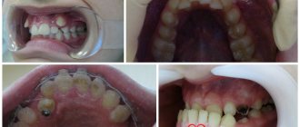
Yellow spots on teeth
Yellow spots occur due to tartar, trauma, caries, as well as problems during pregnancy and living conditions. The problem often occurs in very young children (up to one year old). These include hygienic violations and dry air in which he is forced to constantly be.
In any case, regardless of the cause of this pathology, you need to consult a dentist. Regular brushing, dental checkups, and flossing can help reduce the risk. It is also recommended to have your teeth professionally cleaned at your dentist's office twice a year. Significantly increases the effectiveness of prevention by excluding from the diet foods that contribute to tooth decay. A similar pathology can also occur in adults.
Stages of treatment
Most of the operations, the main task of which is to heal the inflamed pulp, can be divided into a list of conditional stages:
- Excision of dental tissue.
- Extraction of the neurovascular bundle itself.
- Channel cleaning.
- Filling.
The question of how pulpitis is treated in 2 visits with the removal of teeth and nerves can clearly be answered by any more or less qualified dental specialist. However, not everyone, even experienced doctors, had to deal with the most severe forms of this disease. It is not difficult to guess that the key to the success of the entire therapy as a whole is a visit to a specialized specialist who knows how to handle modern equipment and the advanced range of today’s pharmacological drugs. Such professionals, of course, include dental staff.
Disease that causes stains on teeth
A dental disease that manifests itself in the form of multi-colored spots on their surface is called hypoplasia . The disease appears during the gestation period of the future dental patient. It develops in a child if the mother, during pregnancy, had stomach problems, and also had a lack of vitamin D or was exposed to a viral disease.
In such cases, it is necessary to increase the child’s immunity and change his diet. First of all, it is necessary to exclude the possibility of excessive amounts of fluoride entering the body. To do this, you should avoid toothpaste with a high content of it. You need to give your child only purified (bottled or filtered) water, and also give him more vitamins, vegetables and fruits.
With the development of the disease, expressed in the appearance of new spots and an increase in the area of existing ones, remineralization is necessary. The procedure consists of the dentist coating the teeth with a special paste containing substances designed to strengthen the enamel. For hypoplasia detected before the age of eighteen, fluoridation or silvering is also performed.
Black spots
Tooth degradation of this nature can have several causes. One of them is Priestley's plaque , most often caused by disturbances in the microflora of the children's intestines. As a result, both baby and molar teeth are destroyed by bacteria. The disease can only be treated with medication.
Black spots can also be caused by the development of caries. Blackening of teeth is often a consequence of a lack of calcium in the body. For prevention, you need to reduce the amount of sweets you consume.
In addition, you can increase calcium levels by giving your child more milk and using vitamin complexes prescribed by your dentist.
Much less often, but still possible, this pathology can be inherited from parents. Therefore, the child must have personal cutlery (at least a spoon and fork).
University
Teeth have many shades. The individual natural color of teeth is determined by dentin, but is influenced by the color, transparency, thickness and degree of mineralization of the enamel. The blue or pink tint of the enamel is complemented by the color of the dentin, which varies from yellow to brown. This is how different color variants of the norm are formed.
Over time, the color of teeth can change as a result of external or internal staining. External action occurs during the local influence of a number of factors on tooth tissue, while chromogens (compounds that include pigments) are located on its surface. Internal color change is associated with the process of systemic exposure to the body; chromogens are located in the tooth tissues (usually in dentin). Dentists also identify internalized discoloration (as a result of chromogens entering the tooth through cracks and chips).
Features of external color change
External color change is divided into direct and indirect coloration. Direct staining occurs as a result of the penetration of chromogens into the pellicle of the tooth, which leads to a change in the basic color. Indirect staining occurs due to the chemical interaction of various compounds on the surface of the tooth.
Direct coloring. Color-changing chromogens are found in many foods (tea, coffee, spices, vegetables, red wine). Tobacco and many medications also cause staining of teeth. The chromogen penetrates into the pellicle, which absorbs and retains dyes like a sponge. Chromogenic bacteria have been identified that are responsible for green or orange staining of teeth in children (due to poor oral hygiene).
Indirect staining. Associated with cationic antiseptics and metal salts, which are either colorless or give color as a result of chemical interaction with other compounds. For example, blackening of enamel in those taking iron supplements; green discoloration resulting from the use of mouthwash solutions containing copper salts; purple-black coloration that occurs when using mouthwash solutions containing potassium permanganate.
Causes of internal discoloration of teeth
Metabolic. Metabolic disorders (alkaptonuria, congenital erythropoietin porphyria and hyperbilirubinemia) cause discoloration of teeth. Alkaptonuria is an inborn error of metabolism accompanied by discoloration of teeth (brown color). Congenital erythropoietin porphyria is a rare recessive autosomal metabolic disorder that causes a red-brown color to the hard tissues of teeth. Congenital hyperbilirubinemia is characterized by a yellow-green discoloration caused by the deposition of bile pigment in the mineralizing hard tissues of the tooth, especially in the neonatal line, as a result of massive hemolysis due to Rh incompatibility.
Hereditary. Amelogenesis imperfecta is an inherited condition in which enamel formation is impaired. The appearance of the teeth is greatly changed: the enamel is hypoplastic, thin, yellow to brown in color, and may be hypomineralized (depending on the type of amelogenesis imperfecta). It is assumed that the color of the teeth reflects the degree of enamel hypomineralization: the darker it is, the more severe the degree of this condition.
Dentinogenesis imperfecta is a hereditary disorder of dentin. Dentinogenesis imperfecta type 1 is associated with osteogenesis imperfecta (a mixed disorder of type 1 collagen connective tissue). Characterized by the opalescent color of primary teeth, especially if this condition is the result of dominant inheritance.
Type 2 dentinogenesis imperfecta may be more severe in primary teeth than in permanent teeth. The pulp chambers of the teeth become obliterated. When the enamel is chipped, dentin wears out quickly. The clinical appearance (amber, gray, purplish-blue, or opalescent color) is considered to be the result of absorption of chromogens into porous dentin after dentin is exposed.
Dentinogenesis imperfecta type 3 is similar to the first two types of dentinogenesis imperfecta, characterized by multiple pulp exposures in primary teeth.
Researchers note that primary and permanent teeth with type 1 dentin dysplasia have normal outlines and shapes, but are amber-colored. The pulp chambers in primary teeth are often obliterated, while in permanent teeth only crescent-shaped processes of the pulp parallel to the enamel-dentin junction are revealed. With type 2 dentin dysplasia, the teeth are brown and the pulp chambers are affected with the formation of denticles.
Iatrogenic. Tetracycline staining occurs as a result of systemic use of tetracycline. Pregnant and lactating women and children under 12 years of age should avoid taking the drug. The color change depends on the dosage, duration of use, and the age of the patient at the time of prescription. Some authors note a decrease in color intensity over time, explaining this by photo-oxidation under the influence of light. Tetracycline analogues cause various color changes, for example, chlortetracycline is a bluish-gray tint, oxytetracycline is creamy.
Tetracycline discoloration is classified according to the extent, extent, and location of the lesion.
Fluorosis can result from high levels of fluoride in drinking water or from the use of fluoride mouthwashes and toothpastes. This type of discoloration is more common in enamel, ranging from speckles to a diffuse opaque spot appearing on a chalky white or brownish-black background.
Traumatic. The cause of tooth discoloration is the bleeding products of the pulp formed as a result of trauma. Hemoglobin molecules accumulate in an injured tooth; when they break down, iron ions are released, which, when bound with oxygen, form iron oxide. The oxides can then combine with sulfur to form dark gray iron sulfide. Penetration into dentin determines the intensity of the color change.
Root resorption due to dental trauma often presents as a pink, punctate lesion at the enamel-cementum junction, with no other symptoms. Resorption always begins on the root surface and can be internal or external.
Enamel hypoplasia of permanent teeth occurs as a result of damage to the tooth germ of a developing tooth due to trauma or infection of primary teeth, which causes a local enamel defect (postnatal discoloration). The formation of depressions or grooves promotes further internalization of external chromogens.
Dentin hypercalcification can occur following trauma. Temporary disruption of the blood supply to the tooth and damage to the odontoblasts causes excessive, irregular deposition of dentin in the pulp chamber and the walls of the root canal. The tooth gradually loses its transparency and becomes yellow or yellow-brown.
Idiopathic. Molar-incisal hypomineralization (MIH) is a condition of unknown etiology characterized by significantly hypomineralized enamel affecting the incisors and first permanent molars. Enamel hypomineralization occurs asymmetrically, affecting one molar and leaving the opposing molar relatively intact or with minimal subsurface defects. Enamel defects can range from white to yellow or brownish areas. The border between healthy and damaged enamel is always clearly visible.
Currently, possible etiological factors for MGD include infections in early childhood, dioxin in breast milk, and genetic factors.
Age. The enamel undergoes thinning and structural changes, and the deposition of secondary, tertiary dentin and denticles contributes to darkening during the aging process.
Internalized color change
Dyes penetrate into the hard tissues of the tooth through developmental defects or acquired defects as a result of diseases of the hard tissues. The penetration of a dye into the porous structures of a tooth with developmental defects often changes the color of an already stained tooth.
Acquired defects arising due to caries or inadequate quality of restoration materials can lead to discoloration.
• Abrasion, erosion of teeth. As a result of these processes, dyes affect dentin. When enamel is lost and dentin is exposed, teeth appear darker (as the color of the dentin becomes more visible).
• Gum recession. The exposure of dentin makes the teeth more susceptible to the internalization of stains.
• Caries. Accompanied by blackening of the tooth.
• Restoration materials. The gray-black discoloration seen around old amalgam fillings is caused by the migration of tin into the dentinal tubules.
• Endodontic treatment. Preparations containing eugenol give the tooth an orange-yellow tint when the root canal mouth is not properly sealed. Silver posts in root canals give the tooth a gray-black hue due to oxidation.
Solving aesthetic problems associated with tooth discoloration is one of the actively developing areas of dentistry.
The methods are varied and numerous. The modern approach includes the combined use of different techniques, which gives the most effective results. Elena Mirnaya, Associate Professor of the 2nd Department of Therapeutic Dentistry of Belarusian State Medical University, Candidate of Medical Sciences. Sciences Photo: Anna Bergman Medical Bulletin , June 13, 2022
Brown spots
Similar manifestations on tooth enamel are also caused by the development of hypoplasia. The disease is generally very common and, according to some estimates, affects up to 40% of children.
The causes of hypoplasia, in contrast to caries, which destroys still growing teeth, are complications during pregnancy or trauma during birth. It can also develop due to poor nutrition, digestive problems and damage to tooth enamel.
For prevention, it is necessary to monitor the condition of the mother’s teeth. You should stop artificial feeding and monitor the child’s nutrition subsequently, as well as regularly visit the dentist.
Other treatments
In 2022, there are additional ways for doctors to save people from troubles associated with pulp inflammation:
- laser therapy;
- complex of physiotherapeutic processes.
Both mentioned operations, in principle, can also answer the question of how to cure pulpitis without removing the nerve. True, they can only be used in the early stages, when the pathology has not yet had time to develop and acquire serious symptoms. In addition, to use such funds, a person will need to go to a truly modern and advanced clinic with an experienced medical team. For example, a set of works related to the use of a laser is carried out exclusively in the presence of high-quality, proven equipment. Such installations, of course, are present in dental departments.