When a patient comes in with extensive defects in the dental system, the dentist and dental technician are faced with the task of selecting a prosthesis design that would optimally replace missing teeth, restore chewing function, be invisible in the oral cavity and cause a minimum of discomfort. As a rule, in the absence of end supports, there is no talk about fixed prosthetics, so options for removable prosthetics are considered.
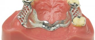
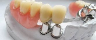
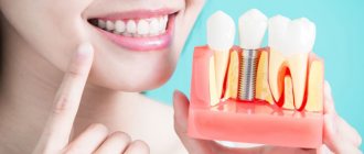
In many cases, such patients can be offered a partially removable clasp (arch prosthesis), which best meets all the stated requirements. It consists of a metal arched frame attached to the lingual or palatal side, so it takes up very little space in the mouth and is invisible to others. An acrylic base with artificial teeth is laid on top of the frame.
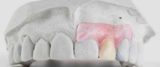
There are dentures with clasp (hooks) and lock (attachments) fastenings. Locks are more advanced and convenient options, due to the fact that they are located on the artificial crowns of the teeth closest to the prosthesis.
Description of the device of locking prostheses
Attachments consist of a patrix and a matrix. The matrix is located on the crown of the abutment tooth, and the patrix is the end part of the clasp and is rigidly connected to it. The locking type of fastening is more preferable, because it does not injure the surrounding periodontium, does not loosen the supporting teeth and is generally not visible even with the mouth wide open. When the prosthesis is inserted into the oral cavity, the micro-lock snaps into place, the structure is firmly fixed, exerting pressure on the alveolar processes, the body of the jaw and gums.
The materials for making attachments are metal and polymers, but in some cases it is more convenient to use a combination of these materials, which gives a more elastic locking fastening. According to the connection principle, locks can be deadbolt, rail, or spherical. The doctor decides together with the dental technician which material and connection principle of the attachment is suitable for a given patient, based on the clinical situation.
Facial epitheses: fundamental approach and basis of therapy
Maxillofacial prosthetics, a discipline that covers the non-surgical restoration of tissue defects in the maxillofacial area (MFA), is today the flagship of orthopedic dentistry. However, maxillofacial prosthetics (MFP) remains a medical discipline little known to both patients and specialists. There is much uncertainty in terminology, clinical practice, and treatment outcomes.
Despite the enormous progress in reconstructive surgery, maxillofacial prosthetics today remains a relevant method of facial reconstruction and is becoming indispensable in many clinical situations. The production of individual face masks, using materials such as silicone elastomers, allows the creation of facial epitheses of a high aesthetic and functional level that meet the requirements of patients and contribute to their satisfactory social interaction. The authors decided to turn to a discussion of advances in the field of facial prosthetics in recent years, especially in the areas of materials science and image processing. The presented methods are widely used in all treatment and diagnostic centers and clinical departments of medical universities.
Introduction
The main purpose of making facial epitheses for congenital malformations or acquired defects is the functional, aesthetic and psychological rehabilitation of patients. Previously, epitheses, quite primitive, were used only for more or less acceptable masking of tissue defects in the maxillofacial area, so as not to make the patient an object of curiosity or rejection in society. Today, they are trying to replace a missing organ using a device whose structure, color and contours almost perfectly imitate the skin, including the superficial venous pattern and various reliefs. Thus, it is possible to create practically invisible structures, among other things, of significant functional value. The psychological acceptance of the prosthesis by the patient is paramount, as this guarantees his personal, family and social integration.
Chapter 1. Fundamental Approach
Terminology
Two categories of terms are used to describe medical-grade face masks: plastic facial prostheses and epitheses. A prosthesis (from the Greek “instead” and tithêmi: “I place”) is “a device used in place of a lost natural organ or part of the body, reproducing the form and, if possible, partially or completely restoring function” [1]. The term “plastic” (from the Greek plastein: to shape, model) defines the ability of a prosthesis to restore the shape of the face. Surgery, on the other hand, is divided into two categories that cannot exist in maxillofacial prosthetics: aesthetic and plastic. The first concerns healthy people, it “decorates” the body and is guided by socio-cultural norms based on generally accepted canons of beauty. The object of the second, as well as prosthetics, are patients with body damage. Plastic prosthesis is also necessary for sick people, both physically and psychologically. Today, the term “epithesis” (short for “epiprosthesis”, “marginal prosthesis”, “epi”: on, on top, at the end) is more often used. This is a medical product for replacing a missing part of the body, repeating the relief and covering an existing defect.
Typology of facial prostheses
The typological classification of prostheses is based on the location of the tissue defect. First of all, there are external prostheses (or ectoprostheses) and internal prostheses (or endoprostheses): the first are external devices, movable, in contact with the skin, mucous membrane or teeth; the second are immobile, surgically implanted into the body. Specialists in maxillofacial prosthetics more often use external prostheses, which are divided into intraoral, when they are in the oral cavity, and extraoral, located outside the oral cavity. The role of the latter is to replace skin defects (external nose, auricle, orbital area). Another type of prosthesis, such as maxillofacial orthoses, which combine static and dynamic designs, is capable of replacing complex defects. The following types of epitheses are most often made: prostheses of the nose, auricle, eyelids and eyes, face masks and complex “multi-story” prostheses, which can combine a facial and dentoalveolar prosthesis (Fig. 1-4) (organigram No. 1).
Rice. 1. Epithesis of the nose (in the process of manufacturing). Rice. 2. Epithesis of the eyelids and eyes (at the mock-up stage). Rice. 3. Epithesis of the auricle. Rice. 4. Epithesis of the naso-orbital region.
Organigram 1
Etiology of facial defects
There are three main reasons for the loss of structures of the maxillofacial area and the occurrence of defects that can be corrected with the help of epitheses: agenesis, surgery of tumor formations and facial trauma. We can add a fourth reason, which is currently not found in Western countries due to the progress of drug treatments, such as inflammatory diseases (noma, for example).
Congenital pathology
Agenesis of facial organs occurs very rarely. They can be observed mainly in the syndrome of multiple developmental defects [2]. According to Dachelott's definition, a defect (or dysgenesis) is “irreversible congenital damage to the morphological structure of a tissue, organ or part of the body as a result of internal developmental disorders” [3]. The congenital nature implies the presence of this pathology from birth. On the other hand, not all congenital anomalies are defects. In addition to true (or primary) defects, there are damages that can imitate a defect (phenocopies), which are called secondary. The term “multiple defects” is used when one individual has at least two anomalies for which, depending on the etiology, a specific syndrome, association or sequence is distinguished. Agenesis mainly affects the auricle. They are observed in Treacher-Collins or Franceschetti-Klein syndromes. We are talking about multiple malformations, including hypoplasia or aplasia of the auricles, atresia of the external auditory canals, anomalies in the chain of auditory ossicles of the middle ear, deafness, hypoplasia of the maxillary and zygomatic bones, thinning of the eyelashes of the lower eyelids and hypoplasia of the lower jaw [4]. Finally, congenital syphilis, as a result of vertical transmission of infection from mother to fetus, can cause dysmorphosis in the face and neck, in particular atrophy of the lower jaw, accompanied by ptosis of the tongue (it does not currently occur in Western countries) [5].
Consequences of surgical excision of oncological formations
This is the most common “reason” for consultations regarding maxillofacial prosthetics. Melanomas, epidermoid carcinomas, basal cell carcinomas, or squamous cell carcinomas, are the malignant tumors most commonly treated with surgical excision. Benign neoplasms (myxomas, cysts) in rare cases can also be locally aggressive. Their surgical treatment can lead to significant losses of the structures of the maxillofacial area. Despite the development of therapeutic technologies in oncology, surgical excision is very often an additional treatment method for patients with tumors. The possibilities of reconstructive surgery, unfortunately, are very limited: its success is difficult to predict due to the consequences of postoperative radiotherapy, which also makes it difficult to monitor the tissues surrounding the primary lesion [6]. Surgical intervention often affects not only a certain organ of the face, but also the integumentary tissues, mainly the buccal and frontal areas. Due to the presence of zones of natural resistance of the maxillofacial tissue, the boundaries of surgical excision are identical in many patients. For example, amputation of the external nose leaves a triangular-shaped defect and an open nasal cavity. In the area of the eyelids and eyes, there are two types of surgical excision: enucleation and exenteration. The first is limited to the removal of the eyeball, while the second involves the removal of the entire contents of the orbit. Some types of resection of the jaw bones involve the removal of cheek skin, which leads to the production of combined or multi-level prostheses (a combination of epitheses, jaw obturators and dentures) (Fig. 5-8).
Rice. 5. Loss of eyelid and eye structures. Rice. 6. Loss of structures in the orbital and nasal areas. Rice. 7. Loss of ear structures. Rice. 8. Loss of structures in the left half of the craniofacial region.
Face and neck trauma
Facial injuries are the cause of polymorphic tissue defects. Reconstructive surgery is the main method of treating this pathology, however, organ amputations or extensive losses of structures of the maxillofacial area often require subsequent prosthetic rehabilitation. In traumatology, injuries can be the result of professional, street, home or sports accidents: this is the so-called civil traumatology [7]. Military and/or ballistic injuries more often result in complex defects directly related to the small caliber of projectiles and the high speed of their movement, which leads to massive tissue destruction [8].
We must also not forget about the consequences of chemical and thermal burns.
Infectious diseases
Some diseases: tuberculosis and lupus erythematosus, noma, leprosy or tertiary syphilis have previously contributed to the appearance of defects in the maxillofacial area. At the moment they are not observed in Western countries (organigram no. 2).
Organigram 2
Basics of therapeutic treatment. Indications
The progress achieved in reconstructive surgery (musculocutaneous or musculocutaneous pedicle flaps, free grafting) allows for aesthetic and functional restoration of extensive defects of the maxillofacial area at a high quality level, regardless of their origin: tumors, trauma or surgical interventions [9 , 10]. Maxillofacial prosthetics alternates with surgery, in particular, with large losses of tissue structures. Prostheses are indicated for elderly patients if their general condition is not satisfactory, as well as in the presence of contraindications to surgery or general anesthesia, or while awaiting reconstructive surgery.
Radiotherapy is an unfavorable factor for reconstructive surgery, and in this case, preference is given to epitheses, as in the presence of some malignant neoplasms, when the movable design of the prosthesis allows visual monitoring of the condition of the tissues surrounding the operated area. Thus, prosthetics are especially indicated for large complex defects [11]. To achieve optimal aesthetic results and stabilize the prosthesis, surgeons sometimes thin the flap to avoid excessive contouring or install retention systems, formerly tunnels and now implants (Fig. 9).
Rice. 9. Support rod on an extraoral implant (collection of J. Dishan).
Advantages of a prosthesis
Prosthetic rehabilitation allows you to:
- accurately restore a missing or defective organ using the moulage technique;
- work in an outpatient setting;
- give an “almost immediate” good aesthetic result, effectively solving the psychological problems of patients;
- make a removable prosthesis and monitor the condition of the tissues along the periphery of the defect;
- create an “immediate prosthesis” in the preoperative preparation phase, in anticipation of or in addition to treatment with reconstructive surgery;
- find a satisfactory aesthetic solution in the presence of extensive, complex in structure and volume defects;
- quickly give a positive answer in almost all clinical situations.
Limitations for prosthetics:
- functional problems: inertia of the prosthesis, epithesis/skin transition zone, tightness of fit;
- mechanical problems: functional integration, fixation and coupling of the epithesis, difficulty of its installation if the retention systems are not adapted to the patient;
- physiological problems: condensation phenomenon, lack of peripheral vascular pressure;
- care problems: the complexity of hygiene due to the porosity of medical silicones and the general condition of the patient (old age, asthenia, problems of understanding, etc.), his remote location from the prosthetics center;
- rapid aging of the prosthesis design;
- problems of a psychological nature: the removable structure provides for the preservation of the original defect, the result is a negative psychological reaction to one’s reflection in the mirror and discomfort during visual contact (acceptance of a new picture of one’s body comes with time) [15].
Materials Science
Silicone is the main material for making epitheses, since its elastic properties are close to the natural structure of the skin.
The birth date of silicones is considered to be 1930, when researchers from DowChemical and CorningGlass, who were trying to develop new electrical insulators, were able to combine the properties of glass and organic plastics [16].
In 1942, DowCorning launched commercial production of silicone elastomers in the United States.
In 1948, the same company officially patented the Silastic brand.
In 1959, the DowCorning Center was created for medical research, searching and developing indications for the use of silicones in medicine and surgery.
The medical silicone production branch of DowCorning was created a little later.
In 1962, Silastic Médical silicone elastomers were made available to medical professionals.
Silicone is a general term to define a subset of a large family of polymers. All silicones, regardless of their physical state, have common chemical, physical and mechanical properties that do not change over time [14] (Table No. 1).
Table No. 1. Physico-chemical and mechanical properties of medical silicones
Chemical properties | Physical properties | Mechanical properties |
| Chemical inertness Resistant to pH changes Oxidation stability Resistance to other chemical substances | Thermal stability (-50 °C, +260 °C) Electrical insulation No modifications in vacuum, pressure changes or radiation exposure Gas permeability Hydrophobicity No foaming | Elasticity Tensile strength Friction resistance |
Characteristics of medical silicones:
- chemical inertness;
- non-wettability;
- permeability to gases;
- radiocontrast;
- possibility of sterilization;
- variety of shapes and consistencies;
- resistance to deformation;
- insolubility and lack of metabolization;
- do not harden, do not tear, withstand loads;
- pyrogen-free;
- do not provoke inflammatory reactions, lack of tightness;
- are not an allergen;
- are not carcinogenic.
The main characteristics of materials for maxillofacial prosthetics are biocompatibility, ease of use, structure similar to natural leather, possibility of coloring and precision of execution. All these criteria lead to an almost exclusive use of silicones in relation to acrylate plastics, which still find specific applications. Most often, silicones are used in maxillofacial prosthetics, which consist of dimethylsiloxane polymers reinforced with amorphous silicon oxide and platinum as a catalyst. Physicochemical properties depend on their molecular structure: weight, type and concentration of polymer chains (long, short) and the number of cross-links between siloxane groups. The strong Si-O bond and flexibility of siloxane chains give silicones low viscosity, high chemical inertness, good UV resistance, thermal stability and a significant service life [20]. In terms of mechanical properties, these are soft, elastic and tear-resistant materials. They provide precision in reproducing structures, ease of operation and quickly polymerize without special tools. Silicones can be completely painted over due to their initial transparent structure. All of the listed characteristics make them materials of choice, but not ideal for making epitheses [21].
The main disadvantage of elastomers is their porosity, which promotes microbial colonization (in particular fungal). Such microbial invasions are responsible for changes in the color and shape of the surface structure. The porosity and heterogeneous nature of silicones necessitate frequent replacement of epitheses, on average once a year. Prevention of colonization, for example by Candida albicans, is carried out by acting on the material itself and carries the risk of destroying its spatial structure. Most often, fungal colonies are concentrated in areas of contact of the epithesis with the warm and moist environment of the upper respiratory tract [22].
The list of references is in the editorial office.
Advantages
In addition to the already mentioned advantages of the clasp prosthesis, one can also name its other advantages:
- Possibility of simultaneous replacement of several dental defects located in different parts of the jaw;
- The chewing load is distributed over all teeth, gums and bone structures, and this brings the functionality of the prosthesis closer to physiological and does not entail rapid loss of bone tissue, which means that replacing the prosthesis with a new one is required much less frequently;
- The presence of an arc, rather than a plastic base, significantly reduces the size of the structure, makes it more compact and gives more freedom to the tongue;
- Due to the fact that the arch only partially covers the palate or gum of the lower jaw, the degree of disturbance of taste, tactile sensations and speech therapy disorders is reduced;
- The metal frame ensures structural strength and long service life of the product.
Manufacturing process
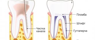
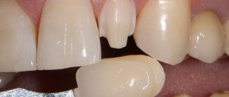
The production of the prosthesis begins after examining the patient, making a diagnosis and making a diagnostic model of both jaws. It is necessary so that the technician and dentist can calculate the number and placement of attachments, and then design a model of the future prosthesis. After this, they begin to sanitize the oral cavity and prepare the supporting teeth for covering them with crowns (treatment of caries, depulpation, installation of pins, grinding). The next stage is taking a silicone impression, which is sent to the laboratory. Temporary crowns are attached to treated and prepared teeth.
Based on the dental impressions obtained, the dental technician makes a frame for the supporting teeth with a matrix and gives it to the doctor for fitting and fitting in the patient’s oral cavity. At the same visit, the color of the future ceramic veneer of ceramic crowns is determined. At the next visit, the finished crowns are fixed with temporary material, the impression is taken again, this time to make the clasp. The crowns, along with silicone impressions, are sent to the laboratory, and temporary crowns are placed on the ground teeth.
In the laboratory, a wax model of the clasp with teeth is first cast, then the final version of the prosthesis is made and carefully verified on working plaster models of the jaws with the patient’s teeth. The finished product is transferred to the clinic.
The dentist fixes metal-ceramic crowns with permanent cement, checks the quality of the locks, makes minor adjustments and places the prosthesis on the patient. At the end of the appointment, the doctor teaches the patient the rules for putting on and removing the denture, caring for it and the oral cavity, and also sets a date for a follow-up examination.
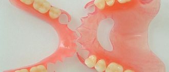
Flexible orthopedic structures
An innovation in dental removable prosthetics has been the use of thermoplastics. Such materials include acry-free, nylon, polyamide. They make it possible to produce structures that are as close as possible in color and texture to the natural tissues of the oral cavity. Unlike standard acrylic prostheses, their counterparts made of thermoplastic materials have a flexible structure, that is, a certain degree of elasticity. All this led to such obvious advantages of these designs as:
- high precision fit to the gum;
- increased strength and resistance to breakage;
- relatively light weight;
- no allergic reactions when used;
- high aesthetics due to the natural appearance and the use of non-metallic clasps that are not noticeable to others.