647
The oral cavity is not the most convenient area of the human body for diagnosis and treatment. Poor visual visibility of some areas of the RP, inconvenience experienced by the patient when the doctor manipulates his mouth - all this increases the difficulties associated with the treatment itself.
The use of traditional dental instruments (mirrors, etc.), in general, helps to successfully diagnose and treat dental diseases.
However, modern dental equipment such as electron microscopes and intraoral cameras offer dentists unprecedented opportunities.
General information
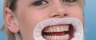
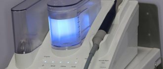
An intraoral camera is a small dental device designed for a detailed examination of the patient’s periodontium and teeth in the most convenient mode for him and the doctor.
The device is a miniature video camera built into an oblong, oval-shaped holder (endoscope) with approximate dimensions of 20x20x200 mm.
The device allows you to display any area of the oral cavity on a real and enlarged scale on the monitor screen, take a picture from it in the form of an image or video file, and save them in the memory of the device or computer.
This class of devices appeared at the end of the last century, and today they are in great demand, significantly increasing the accuracy of diagnosis and the success of treatment.
The assembled device (as a system) consists of 3 main components:
- The device itself (holder, endoscope) , at the very tip of which there is a built-in miniature but quite powerful camera.
- Computer or separate monitor.
- Interface block connecting the endoscope to the computer/monitor with cables.
In some designs, there is no interface block and the holder is connected directly to the computer or monitor.
The endoscope consists of the following elements:
- miniature camera;
- the tip into which it is inserted;
- tip holder;
- a nozzle placed on the handpiece when performing a certain procedure;
- hygienic cover.
A cable is connected to the back of the endoscope, connected at the other end to the interface block or directly to the port of the computer or monitor. Wireless systems are also available that transmit information from the device to a computer using a radio signal or Wi-Fi.
Depending on the design, the signal can be transmitted via various interfaces - USB, RCA (“tulip”), Y/C, S-video, VGA.
When working with a specific patient, a clean, disposable cover is placed on the endoscope to eliminate the risk of infection.
The device does not place high demands on the computer's characteristics - processor frequency, amount of permanent and RAM memory, the presence of advanced interfaces, high screen resolutions, etc.
The lowest characteristics that budget computers have today are enough.
Etiology of acid necrosis of teeth and tactics of treatment of pathology.
Head here to take a closer look at the SAF system.
At this address https://www.vash-dentist.ru/lechenie/zubyi/nesovershennyiy-dentinogenez.html we will talk about the disease “dentinogenesis imperfecta”.
Indications for intraoral dental scanning
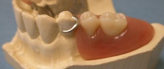
To create a high-quality prosthesis or propose an effective treatment regimen, it is important for the dentist to see the situation in the oral cavity. An intraoral scan will show how teeth, soft tissues, bone structures, and the temporomandibular joint are located in space relative to each other. The use of a 3D scanner replaces several procedures for examining the patient’s oral cavity, modeling with maximum accuracy all the missing elements of the dentition.
Indications for dental scanning are the following pathologies:
- abnormal arrangement of teeth, their unusual shape or size;
- bite defects;
- absence of one or more dental units;
- TMJ diseases;
- damage to hard dental tissues.
Making digital impressions using a 3D scanner on 2 jaws is in demand for creating individual whitening trays, plates, splints, and designing aligners. Dentists at the President clinic in Maryino also recommend signing up for the procedure at the preparatory stage before implantation or surgery.
A special advantage of the technique is the absence of contraindications. Dentists prescribe dental scanning for both children and adults. The occurrence of allergies is also excluded, since the doctor does not use auxiliary materials to which the patient’s body may react negatively.
Main characteristics
The main requirement is acceptable quality of pictures and videos, which is determined by the following parameters:
- Resolution is the number of image elements (pixels) per unit area of the monitor. The higher the resolution, the finer details are displayed in the image, the higher quality the camera is considered. The resolution of the most advanced models reaches 12-15 megapixels.
- Photosensitivity is the ability to create high-quality images in low light conditions. The higher the light sensitivity, the better. If this parameter is indicated in lux, then the lower its value, the more photosensitivity the device has.
- Color rendition. Should be as close to real as possible.
The most important parameters include functionality - the number of operations that can be performed with their help.
Various functions are provided by special attachments - devices that are placed on the tip of the endoscope. The more attachments, the wider the functionality of the device.
Other important characteristics include the following:
- Scale of magnification of details. Usually it ranges from 10 to 40, in some types it reaches 120.
- Possibility and ease of connection to several workstations.
- Type of connection with a computer or interface unit. Communication can be wired or wireless. The latter is more convenient, but also less reliable and requires regular battery recharging.
- The number of images stored in the device’s memory. You can store an almost unlimited amount on your computer's hard drive.
- Method of switching and controlling operating modes. It is carried out using buttons on the holder and/or a foot pedal. In some models, the foot pedal is provided as an option and is not included in the basic package.
- Price.
General parameters that you should pay attention to also include dimensions, ease of use, waterproofing (the device operates in a humid environment), and reliability.
When you have an intraoral camera,...
The status of the clinic is growing, the number of patients is increasing, and therefore financial flows are increasing. Your investment works for you - modern dental equipment quickly pays for itself and brings a stable income even in a highly competitive environment. After all, what could be more attractive to a person interested in fast and high-quality dental treatment than a real picture seen with his own eyes.
High technology rules the world, and dentistry is no exception.
Functional
The use of devices significantly expands the dentist’s capabilities in diagnosis and treatment. It allows:
- in detail, from different angles, with maximum convenience for the doctor and minimal inconvenience for the patient, examine and assess the condition of the teeth and periodontium.
- enlarge the resulting image to view small details;
- clearly demonstrate to the patient the condition of his teeth, thereby increasing motivation for treatment;
- correctly and timely diagnose the disease;
- choose the color of the prosthesis (crown, veneer) that ideally matches the color of the patient’s natural teeth;
- draw up a treatment plan;
- implement it, in some cases using a camera as a technological tool (for example, a photopolymerizer for a composite);
- monitor the effectiveness of treatment by comparing the achieved result with the initial state;
- use saved video files and pictures for research work;
- send them to any interested party (another doctor, patient).
Some models have additional capabilities, such as photopolymerization of the composite material, detection of soft and plaque on teeth and, thanks to this, diagnosing caries in the early stages.
Additional built-in functions of some devices also include the ability to:
- obtaining a mirror image and a “freeze frame”;
- reproduction of X-ray images in digital form in order to improve their visual perception;
- obtaining the “picture-in-picture” effect.
Intraoral cameras are useful for most dental procedures. Specifically, in addition to diagnosing and treating standard dental diseases, they can be used for:
- osteotomies;
- gingivoplasty;
- removal of granulations and cysts;
- treatment of periodontal abscesses;
- implantation and prosthetics.
In general, it is easier to list those points where the device cannot be used than those where its use gives a high effect.
How dangerous is hypercementosis of the tooth root and is it necessary to remove it?
In this publication you will find objective reviews of plaque indicators.
Here https://www.vash-dentist.ru/lechenie/zubyi/tipyi-rezorbtsii-kornya.html we will consider the causes of pathological resorption of the roots of temporary teeth.
The use of intraoral cameras in dentistry Grandmed (St. Petersburg)
Treatment with miniature dental cameras in our clinic is carried out by specialists of all specialties. The device helps therapists control caries treatment, including avoiding the removal of healthy dental tissue. The intraoral camera is most in demand by orthopedists and orthodontists, who allow them to draw up an accurate plan for multi-stage and complex treatment, and in addition, promptly identify emerging problems (for example, overhanging crown edges).
During the appointment, the specialist inserts a camera into the patient’s oral cavity and receives a tenfold magnified image of the teeth on the screen. The intraoral camera is controlled by buttons on the tip. The resulting images are displayed on an LCD monitor, and the camera allows you to record 4 pages, each of which has 4 images (they can also be viewed in full screen mode). Images can be compared with each other, saved on removable media and printed.
Selection rules
The most important selection criteria include:
- image quality (resolution, photosensitivity, color rendition, zoom scale);
- functionality;
- brand;
- optics supplier (the best include Carl Zeiss, Olympus, Schneider-Kreuznach Variogon, Nikon, Canon);
- price.
The most important parameter is functionality; the number of operations performed, and, therefore, the quality of diagnosis and the effectiveness of treatment depends on it.
How much does treatment using an intraoral video camera cost in St. Petersburg? Price in Grandmed
The decision on the need to use an intraoral camera in the diagnosis and treatment of various dental pathologies is made by the doctor during a preliminary consultation or directly during the procedure. The images obtained with the camera complement other types of x-ray examinations.
| Service | Price |
| Diagnostics using an intraoral video camera (consultation with a doctor + treatment plan) | as part of the consultation - free of charge |
Popular models
The Russian dental market contains devices from many international manufacturers of video equipment for dental offices. Among them:
- the German company Durr Dental, known worldwide for the VistaCam brand;
- American TPC Advanced Technology (model TPC 899B Advance CAM, etc.);
- French Sopro and Satelec Acteon Group (Sopro 717, Sopro 617, etc.);
- Chinese Pragmatic (MD camera);
- South Korean Good Doctors (Whicam I, Whicam II, Whicam III, Whicam IV);
- and others.
The abundance of offers allows you to purchase devices of different functionality and cost , and select the optimal solution for any services provided by a particular clinic. Below are the main characteristics of some models produced by the above manufacturers.
TPC 899B Advance CAM
A simple budget model from TPC Advanced Technology that can be installed in any office. Despite the low price, it generates high-contrast, clear images.
Main technical characteristics:
- Number of megapixels – 2;
- the presence of a “freeze frame” function;
- 4-image mode;
- connection to a computer via USB port.
Equipment:
- the device itself;
- connection cable;
- 50 disposable covers.
The warranty is 2 years, which is a good indicator for devices of this class.
Camera MD-2000
An inexpensive model from a Chinese manufacturer, which nevertheless has fairly good functionality.
Main technical characteristics:
- resolution – 5.0 MP;
- connection to a computer – via Wi-Fi or VGA connector;
- there is a video recording function;
- The 350 mA battery lasts for 35 minutes of battery life.
Equipment:
- MD-2000 camera;
- VGA cable;
- wired IR receiver;
- Remote Control;
- 50 disposable covers.
Positive features include the presence of Wi-Fi (connection up to 10 m), large battery capacity, and a convenient shape of the endoscope.
VistaCam
The German model VistaCam IX provides high quality images, wide functionality, and easy integration into dental offices even without the use of a desktop computer.
Wide range of possibilities is provided thanks to 5 attachments.
- Cam . Main interchangeable head used for live inspection and imaging.
- Proof . Visualizes dental and soft plaque, allows you to detect caries in the initial stage.
- Proxy . Designed for diagnosing approximal caries.
- Macro. Allows you to create images at a scale of 120:1.
- Poly . Used for photopolymerization of composite compositions.
Direct removal of the camera from the tip and Plug and Play connection ensures it works on multiple desktops. Connection to a computer is carried out using a USB 2.0 port. In addition to the control buttons on the device itself, the device can be controlled by a foot pedal.
Whicam I
The Korean wired model of the mid-price range is one of the latest generation devices. High resolution (1280x720 pixels) and 6 powerful LEDs provide high quality images.
A big plus is the ability to connect to various ports (USB, VGA, S-video, RCA) of a TV, computer or laptop). There is a “freeze frame” function.
The device memory can store 8 (in normal mode) or 32 (in 4-frame mode) images. When saving pictures in the computer's memory, their number is limited only by the capacity of the hard drive.
SOPRO cameras
French Sopro models are relatively expensive devices, prices vary in the range of 160-300 thousand rubles. Satelec and Sopro companies produce devices with different characteristics and capabilities.
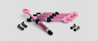
In particular, the Sopro 717 model is equipped with Sopro Shade attachments, which allow you to select the color of the prosthesis (crown or veneer) that ideally matches the shade of natural tissues. An image is taken of the patient's teeth, which is recorded in half the window of the Sopro Shade attachment.
After this, the device is brought to the color template, which fills the second half of the window. By comparing the color of both halves (ideally, the line between them should disappear), you can accurately determine which value on the color scale corresponds to the natural color of the teeth.
With the Sopro 717 you can take both intraoral (at a distance of 5-30 mm) and extraoral (at a distance of 30 mm-infinity) images. Connection is made via USB, Video and S-Video ports.
The delivery set includes an adapter for different power sources (220-6VDC) and cables for connecting to various computer/monitor ports.
The video provides additional information on the topic of the article.
Price
The cost depends mainly on their functionality and brand. Products from Western European and North American companies are more expensive than South Korean or Chinese ones.
Approximate prices of some popular models are shown in the table below.
| Manufacturer | Brand | Price, rub |
| TPC Advanced Technology, USA | TPC 899B Advance CAM | 16 000 |
| Pragmatic, China | MD-2000 | 18 800 |
| Durr Dental, Germany | VistaCam iX | 240 000 |
| Good Doctors, South Korea | Whicam I | 61 000 |
| Satelec and Sopro Acteon Group, France | Sopro 717 | 350 000 |
| Sopro 617 | 180 000 |