The most common form of gingivitis is catarrhal gingivitis, which affects the gums, causing superficial inflammation. Most often, this disease affects children and young people under 35 years of age. In older patients, as a rule, chronic catarrhal gingivitis is diagnosed.
Teeth polishing with pastes - 1,200 rubles.
Polishing one tooth with paste - 50 rubles.
Coating with a fluoride-containing product (1 row of teeth) - RUB 1,500.
Coating with fluorine-containing varnish (1 tooth) - 250 rubles.
Retraction of hypertrophied gums in the area of 1 tooth - 600 rubles.
At CELT you can get advice from a dental specialist.
- The cost of a dental consultation is 1,000
- The cost of an orthodontist consultation is 2,000
Make an appointment
Causes of gingivitis
Gingivitis does not always progress as a separate disease. It often appears in the background:
- Imbalance in the hormonal system.
- Disturbances in the gastrointestinal tract.
- Diseases of the cardiovascular system.
- Infections and inflammatory processes.
Internal factors in the development of pathology are as follows:
- Fall of immunity.
- Growth of wisdom teeth.
- Lack of vitamins P and C.
- Allergy.
- Diabetes.
- Metabolic disorders.
- Chronic gastrointestinal diseases.
- Severe mental disorders.
Among the external causes of gingivitis:
- Smoking or improper breathing.
- Burns and mechanical damage to the mucous membrane.
- Incorrect installation of the seal.
- Irregular brushing of teeth.
- Infections.
In more than 50% of cases, the development of pathology occurs due to pathogens that penetrate the oral cavity with food and liquid. If you brush your teeth incorrectly and irregularly and do not use floss, a thick plaque will appear. Often inflammation begins due to a burn or mechanical damage to the gums. A lack of nutrients in the diet and serious stress can reduce immunity, which will also lead to the development of pathology.
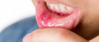
Ulcerative-necrotizing gingivitis
Otherwise, the disease is called Vincent ulcerative-necrotizing gingivitis. It occurs acutely or occurs chronically.
The source of the development of the disease is said to be a sharply increasing amount of bacterial plaque at the border of the gums and tooth enamel, with a predominance of spirochetes and fusobacteria in the biomass. In this case, there is an initial ulceration of the mucous membrane, and then the tissues die. The provocateurs of ulcerative gingivitis are often a decline in immunity and serious illnesses with a chronic clinical picture.
The acute form is accompanied by high fever and bleeding from under the gums. Loss of appetite, the smell of rotting flesh emanates from the mouth. The gums look affected, with ulcers, the interdental papillae acquire a gray tint and become necrotic. In a chronic course, the same thing happens, but the picture is blurred and not expressed so intensely. Changes are developing gradually.
It is impossible to treat ulcerative gingivitis at home; a visit to the dentist is required to establish a diagnosis and prescribe antibiotic drugs. Dental plaque is removed using professional means. It will not do without applications and rinses. To regenerate the mucous membrane, active gels are prescribed, for example Solcoseryl.
Symptoms of catarrhal gingivitis
The sooner treatment for acute catarrhal gingivitis begins, the lower the risk of subsequent complications. Symptoms that should promptly contact your dentist include the following:
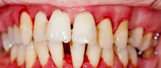
- bleeding, redness and swelling of the gums;
- itching in the gums;
- increased body temperature;
- bad breath, unpleasant taste;
- pain in the gums when eating or pressing on them;
- general malaise, fatigue.
Indications
You need to immediately begin treatment for catarrhal gingivitis if you find:
- Swelling of the gums.
- Bleeding after eating or brushing teeth.
- Acute pain and burning in the gum area.
- Bad breath.
- Putrid or bloody taste in the mouth.
- Changes in gum color and density.
Contraindications
It is not recommended to start treatment if you have:
- Psychological and nervous diseases.
- Cardiovascular pathologies.
- Problems with blood clotting.
- Chronic diseases in the acute stage.
- Allergic reactions to anesthesia.
A specialist will give you a full list of contraindications during a face-to-face consultation.
Case history: Chronic localized catarrhal gingivitis
Passport part
1. Full name — Vasiliev Ivan Alexandrovich
2. Date of birth - 04/13/61
3. Profession - honey. entrepreneur
4. Home address - Skobelevskaya str., no.3. bldg. 1 kv. 39
Complaints upon admission
Reception date: November 11, 2004. The patient complains of bleeding gums while eating and brushing teeth with pink discoloration of the oral fluid, and an aesthetic defect: pigmented dental plaque.
Life history (Anamnesis vitae)
1. Previous diseases - chicken pox.
2. Concomitant diseases - no
3. Denies the presence of tuberculosis, hepatitis and AIDS. There are no chronic diseases.
4. Drug intolerance - no history of allergies
5. Occupational history - no occupational hazards or diseases
6. Bad habits - denies the use of alcohol, tobacco, and drugs.
7. Living conditions: lives in a 2-room apartment with his wife and 2 children. The family environment is good. I did not work in hazardous industries.
History of the development of this disease (Anamnesis morbi)
When collecting anamnesis, it was established that 2 years ago he noticed bleeding gums when brushing his teeth.
The present condition of the patient (Status praesens)
1.- General condition of the patient The general condition is satisfactory. The skin is pale pink, normally moisturized, elastic. No dryness, rash, scratching, hemorrhages, peeling or ulcers were detected on the skin.
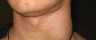
2. External examination of the maxillofacial area. The configuration of the face is not changed, the skin is pale pink and normally moisturized. There are no skin rashes or swelling. The red border of the lips is without pathological changes; lips are normally moisturized; There is no dryness, crusts, cracks, erosions or ulcerations. Regional lymph nodes (submandibular, mental, cervical) are not enlarged, not fused with surrounding tissues, and painless on palpation.
3. Examination of the oral cavity. The mucous membrane of the lips, cheeks, hard and soft palate is pale pink, moderately moisturized, without pathological changes; no swelling is observed. .The tongue is of normal size, the mucous membrane is pale pink, moderately moist. The dorsum of the tongue is clean, there are no desquamations, cracks or ulcers. There is a slight overlap. Soreness, burning, and tongue are not observed. The palatine tonsils are not enlarged, there are no purulent plugs in the lacunae, and there is no plaque.
4. Examination of the gums. The marginal part of the gums and interdental papillae are swollen and slightly hyperemic. Bleeding is observed from the top of the papillae when pressing at their base and probing. The dentogingival junction is not broken. There are no pathological periodontal pockets.
5. Dental examination Dental formula: Orthognathic bite. All teeth up to the equator of the crowns are covered with a dark brown coating from strong tea. Soft plaque of a light brown color is localized in the cervical area of all teeth. There is supragingival tartar of brown color, dense consistency and subgingival tartar on all teeth. The depth of the vestibule is normal. No anomalies in the position and shape of the teeth were identified. There are no non-carious lesions on the teeth.
Additional research methods
Forms of catarrhal gingivitis
Depending on the nature of the lesion, there are:
- acute catarrhal gingivitis - characterized by rapid development and one-time inflammation, limited in time;
- chronic catarrhal gingivitis is a continuation of acute; in the absence of treatment, it is practically asymptomatic, but with regular exacerbations.
The scale of inflammatory processes determines two forms of gingivitis:
- localized - part of the gum is inflamed, no more than 1 - 3 teeth;
- generalized - the entire gum on one or both jaws is inflamed.
The severity of the disease is as follows:
- mild - the periodontal papillae are affected;
- medium - the free area of the gums is affected;
- severe - the alveolar region of the gum is affected.
Classification
From the point of view of the nature of the course of the disease, dentists distinguish between acute and chronic gingivitis. The localization of the pathology is classified into point and diffuse forms - in the first case we are talking about one to three teeth, in the second - about damage to the entire jaw. There are also three stages of development - mild, moderate and severe, each of which affects the choice of treatment method.
Diagnosis of the disease
Diagnosis of catarrhal gingivitis involves examination by a doctor and laboratory tests.
The latter allow you to determine indicators such as:
- bleeding gums;
- degree of inflammation;
- amount of microbial plaque.
When conducting an examination, the dentist pays special attention to the following factors:
- how full the blood vessels of the gums are;
- red gums;
- integrity of the periodontal junction;
- the presence and amount of plaque and tartar;
- presence of signs of destruction of the interalveolar septa.
Treatment of the disease
Treatment of chronic catarrhal gingivitis involves a set of measures aimed at eliminating the already emerging consequences of the disease, as well as the causes that caused it. Typically, it begins with a professional teeth cleaning at the dentist's office.
The course of treatment is selected individually and depends on the form, severity and nature of the disease. Often the patient is prescribed mouth rinsing with antiseptic solutions and the oral cavity is sanitized. It involves treating caries, filling teeth, and replacing incorrectly installed fillings. This approach eliminates the possibility of relapse of the disease in the future.
Drug therapy consists of treating the oral cavity with antiseptic solutions and applications of special ointments. When treating the chronic form, it is possible to massage the gums.
It is worth noting that treatment is often carried out in combination with a diet that excludes foods that irritate the gums and is rich in vitamins A, B, E.
Treatment of chronic catarrhal gingivitis of any severity has been successfully carried out by dentists at the CELT clinic for several years now.
Recommendations after treatment
To avoid the occurrence of catarrhal gingivitis after treatment, you need to follow 7 rules:
- Lead a healthy lifestyle and support your immune system. Consume a vitamin-mineral complex and a sufficient amount of proteins, carbohydrates and fats.
- Prevent the transition of chronic diseases to the acute stage. Treat chronic diseases of the cardiovascular and hormonal systems, gastrointestinal tract.
- At least 2 times a year, carry out professional cleaning of the oral cavity to remove soft and hard plaque and prevent the formation of tartar.
- Use a toothbrush with medium hardness. Too soft does not sufficiently clean teeth from plaque, and hard ones can injure the enamel and cause pain with high sensitivity.
- Properly clean the oral cavity and the space between the teeth using floss and irrigator.
- Visit the dentist regularly in order to promptly detect inflammatory processes and eliminate them at an early stage.
- Choose the right toothpaste with sodium, fluoride, potassium nitrogen and other components. Entrust this task to your doctor.
If you follow these recommendations after treatment, you will confidently avoid a recurrence of catarrhal gingivitis.
Make an appointment through the application or by calling +7 +7 We work every day:
- Monday—Friday: 8.00—20.00
- Saturday: 8.00–18.00
- Sunday is a day off
The nearest metro and MCC stations to the clinic:
- Highway of Enthusiasts or Perovo
- Partisan
- Enthusiast Highway
Driving directions
Hypertrophic gingivitis
The essence of the disease is reflected in the name - the gums and gingival papillae grow and become oversized. This occurs either through tissue swelling or due to internal proliferation of connective tissue with the formation of scars - fibrosis.
Accordingly, hypertrophic gingivitis can be edematous and fibrous.
The incentives are:
- Hormonal changes in the body during pregnancy;
- Injury to the gums by the edges of dentures and crowns;
- Progression of catarrhal gingivitis;
- Changes in hormonal levels during adolescence.
Edema form of gingivitis
This type of disease is often associated with internal changes in metabolism in women and growing children. The interdental papillae look loose and engorged. At the beginning of treatment, the oral cavity is sanitized, plaque and stones are removed, then anti-inflammatory procedures begin.
Sclerotization is most often used. After applying anesthesia, a special solution containing calcium chloride, magnesium sulfate and glucose is injected into the patient’s gums. The course takes 6-8 days with one or two day breaks.
Advanced swelling of the gums can change; the patient’s mucosa becomes scarred with the formation of fibrous cords.
Fibrous form of gingivitis
The source of troubles are internal changes in living tissues. Surgery is indicated here; unfortunately, fibrosis is not amenable to conservative therapy. First, external irritants are corrected - tartar is removed, the shape of the crown is corrected. Then the hypertrophied tissues are excised. Subsequently, healing bandages are applied.