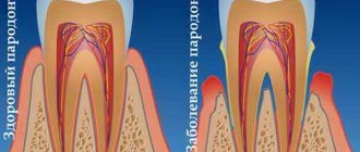
Periocoronaritis is a dental disease characterized by inflammatory processes in the soft gum tissues that surround the erupting units (in the vast majority of cases, wisdom teeth). It manifests itself as intense pain, the inability to open the mouth normally, swelling of the gums, as well as an unpleasant odor from the oral cavity. Inflammation develops from the accumulation of plaque in the opening of the gum through which the unit erupts, or due to the difficulty of the process itself. The latter causes pressure on the adjacent unit along with damage to the bone and gum tissue.
The CELT Surgical Department of Dentistry invites you to undergo treatment for pericoronitis in Moscow. Our multidisciplinary clinic has been operating in the paid medical services market for more than two decades and enjoys a good reputation among patients. We have all the necessary certificates and licenses and provide treatment with the conclusion of an official contract and provision of a guarantee. You can find out the price of excision of the hood for periocoronitis or removal of a wisdom tooth by going to the “Services and Prices” section.
Consultation with a dentist-therapist - 1,000 rubles.
Treatment of pericoronitis with medication - 1,000 rubles.
Surgical treatment of pericoronitis (excision) - 2,500 rubles.
At CELT you can get advice from a dental specialist.
- The cost of a dental consultation is 1,000
Make an appointment
Etiology of pericoronitis
Most often (in more than 90% of cases), this disease is provoked by wisdom teeth, which begin to develop on the bulky area of the gums, which is located behind the third molars. The reasons for pericoronitis in this case lie in the fact that the width of the dental arch in modern humans has decreased by ten to twelve millimeters, but the teeth have retained their previous sizes. As a result, there is a lack of space for the eruption of units, which makes it difficult and manifests itself as inflammation.
Another reason lies in the too thick walls of the dental sac around the dental crown and gum mucosa, which also seriously complicate the teething of units. The reduction in growth-forming factors should not be discounted. During tooth growth, its position does not change, but part of its surface is hidden under the mucosa - and it is there that food fragments, pathogens and dental plaque begin to accumulate. Together, they create an ideal environment for the development of specific inflammatory processes, which are maintained due to regular trauma to other teeth during chewing. The result is scarring and an increase in the periodontal gap.
It is worth highlighting separately the factors that contribute to the development of dental periocoronaritis:
- early loss of primary teeth;
- malocclusion;
- abnormal increase in units – macrodentia;
- their complete or partial absence is adentia.
Causes of pericoronitis. Etiology and pathogenesis
There are many opinions regarding the causes of pericoronitis. With the growth of the lower jaw in the prenatal period, the distal surface of the alveolar process decreases. For this reason, the eighth tooth does not have enough space to erupt.
Another reason may be the initial incorrect location of the tooth germ in the jaw bone. Therefore, during the eruption of a molar, the gums become inflamed.
Etiologically, inflammation of the gums around an erupting tooth develops against the background of active activity of the normal microflora of the oral cavity. Anaerobic and facultative anaerobic bacteria occupy the majority of human saliva.
Pathogenesis: during the process of tooth eruption, the gums above the masticatory cusps atrophy, and the rest of the crown remains under the soft tissue. Food debris, saliva and bacteria get into the space between the tooth and gum. Humidity, warmth and an abundance of nutrients are favorable factors for the active life of bacteria.
Chronic trauma from chewing food leads to the formation of wounds and erosions on the gum surface. As a result, an inflammatory process develops surrounding the crown of the tooth.
Symptoms of periocoronaritis
The disease is characterized by intense pain symptoms localized in the area of the unit. Often it radiates to the temporal or ear region. The patient suffers from the inability to chew and swallow food normally, and even to open his mouth normally. As the inflammatory processes develop, they cover the surrounding tissues: pain is most often caused by the involvement of the gum hood, less often by the incorrect position of the growing units. If they are located at an angle, damage to the gum and bone tissue occurs, and there may also be pressure on the adjacent tooth, which often leads to its destruction.
Pericoronitis of the wisdom tooth also manifests itself:
- increased temperature in the affected area, sometimes in the whole body;
- enlargement of regional lymph nodes;
- the appearance of halitosis;
- unpleasant aftertaste when consuming food.
The lack of adequate treatment leads to the development of a purulent focus in the maxillofacial area of the face. The patient constantly secretes pus, the pain intensifies even at rest, and complications of pericoronitis develop, such as purulent melting of the subcutaneous tissue (phlegmon) or osteomyelitis.
Symptoms of pericoronitis
All of the above factors negatively affect the development and eruption of wisdom teeth, but at the same time, pericoronitis itself is caused by the activity of bacteria that accumulate in the resulting gingival fold. As for the symptoms of the disease, until the wisdom tooth begins to actively erupt, the patient does not feel any concern, however, when performing an orthopantonogram, the risk of pathology can often be predicted. At more advanced stages, wisdom tooth pericoronitis has quite obvious symptoms:
- itching in the area of the wisdom tooth (in the early stages);
- swelling, redness, pain;
- formation of a gingival hood;
- elevated temperature;
- enlarged lymph nodes;
- abscesses, purulent discharge, unpleasant odor.
Classification of pericoronitis
Based on the shape of the flow, they are distinguished:
- Acute pericoronitis is typical for the primary manifestation of inflammation, and in the future, in the absence of treatment, for exacerbations of the chronic form;
- Chronic pericoronitis - characterized by repeated inflammatory changes in the mucosa around the erupting unit.
Depending on the clinical form of soft tissue inflammation, the following are distinguished:
- • Purulent pericoronitis in acute form – manifests itself with intense pain symptoms, deterioration of the general condition and an increase in temperature up to 30°C. Upon palpation, the doctor detects an increase in the submandibular lymph nodes. There is swelling of the mucous membrane in the area of the hood, cheeks and soft palate, pus flows from under the hood;
- Acute catarrhal pericoronitis - manifested by pain during chewing, body temperature remains within normal limits (as well as the tissues around the jaw do not swell), the patient can open his mouth normally. At the same time, the hood looks swollen, the entire crown of the tooth is under it, and exudate flows out from under it.
Types of pericoronitis
| Type of pericoronitis | Description |
| Catarrhal | The initial form of the disease, in which the inflammatory process has just begun, and the gingival hood has not yet formed. |
| Ulcerative | Noticeable swelling, formation of ulcerative rims, bleeding of the gums in the affected area. |
| Acute pericoronitis | An acute form of the disease, rapidly developing and progressing to more severe stages. |
| Retromolar | Deep inflammation affecting the periosteum and going deep. Visually expressed by severe swelling. |
| Chronic purulent | It affects soft and hard tissues in the area of the wisdom tooth, as well as neighboring parts of the jaw. At this stage, the risk of developing phlegmon and osteomelitis is highest. |
Diagnosis of pericoronitis
The diagnosis is made by the dentist after an examination, taking into account the patient’s symptoms and complaints. In order to determine the direction of growth of the unit, an x-ray examination is often required. The main task of the doctor is to determine the form of the disease, its prevalence, nature and severity. In addition, he must identify contraindications to treatment:
- individual intolerance to medications or materials used in it;
- the presence of concomitant pathologies that will complicate treatment;
- inflammation of the tissues and organs of the oral cavity.
Diagnostics
Thanks to the results of a diagnostic examination, it is possible to distinguish a simple difficulty in the eruption of the third molar from carious complications and trigeminal neuralgia.
Stages of diagnosing gum inflammation near an erupting tooth:
- Collection of patient complaints.
- Visual inspection of the gum mucosa.
- X-ray examination in lateral projection.
How to treat pericoronitis?
Treatment tactics for pericoronitis are selected by CELT dentists on an individual basis, depending on the diagnostic results and the patient’s indications. It is aimed at:
- pain relief;
- relief of inflammatory processes;
- restoration of the functionality of the dental system;
- excluding the development of complications.
Therapeutic manipulations are carried out on an outpatient basis, taking into account the severity of inflammatory processes, the local and general picture of the pathology and radiographic data. Treatment of the catarrhal form of acute periocoronaritis involves:
- antiseptic treatment of the area under the hood;
- application of antiseptic and anesthetic dressings;
- lifting the hood using a gauze strip soaked in iodine solution.
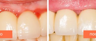
Treatment of pericoronitis
Treatment of pericoronitis is carried out in outpatient settings. The range of dental measures depends on the severity of gum inflammation, symptoms of the disease and the results of a diagnostic examination.
The first step in eliminating the inflammatory process is washing the wound and antiseptic treatment of the subgingival hood. For mild pericoronitis, treating the space between the tooth and gum with an aqueous solution of chlorhexidine from a syringe with a blunt needle will be sufficient.
In case of purulent inflammation, an operation is performed during which the swollen gum above the tooth is dissected, and a special therapeutic bandage with iodoform is applied under the gum. After the inflammation has subsided, a decision is made to remove the subgingival hood. The tissue is cut using a laser under local anesthesia.
Laser treatment is considered less traumatic than instrumental surgery and prevents blood loss during surgery.
In cases where the wisdom tooth is in an incorrect position under the gum, extraction is performed. Removal of third molars is also indicated in the following cases:
- an impacted tooth that was completely embedded in the gum;
- partial eruption of a tooth with an incorrect position;
- horizontal position of the third molar and its emphasis on the second molar;
- the presence of a cyst at the root apex;
- inability to clean plaque from teeth;
- caries and its complications.
Extraction of the eighth tooth in most cases is complicated by alveolitis (inflammation of the socket). To prevent postoperative consequences, the patient is given recommendations for oral care at home:
- Intraoral baths with a 0.05% solution of chlorhexine, hydrogen peroxide or Rotokan infusion.
- Treating the gum surface with Cholisal gel.
- Local applications of vitamins A and E to accelerate tissue regeneration.
- The use of broad-spectrum antibiotics: Amoxiclav, Metronidazole.
- The use of vitamin and mineral complexes.
It is important to remember that many medications have their own indications and contraindications, so prevention of secondary infection should be carried out under the strict supervision of the attending physician. Self-medication and ignoring gum inflammation can worsen the severity of the disease.
Treatment of pericoronitis during pregnancy is more gentle than the standard principle of therapy.
As a rule, in such cases, laser excision of the gums is performed under a low dosage of anesthetic, and during the rehabilitation period, antibiotics are replaced with less toxic drugs. To avoid harm to the fetus, surgery is performed in the second trimester.
Traditional methods of treatment at home
Traditional medicine for inflammatory dental diseases involves the use of medicinal herbs in the form of intraoral baths and rinses.
Chamomile, oak bark and sage soothe the inflamed area of the gums and disinfect the subgingival space. All herbal ingredients are mixed together and infused in a water bath for 15 minutes. After the substance has cooled to room temperature, thoroughly rinse the side of the diseased tooth. The course of treatment is 7–10 days.
When purulent exudate accumulates, rinsing with soda-saline solution is performed to clean the wound. To prepare the liquid you need:
- 1 tsp. table salt;
- 1 tbsp. l. soda;
- 250 ml warm water.
The procedure is carried out every 4 hours for 3–4 days. After the course, you must consult a doctor for preventive purposes.
Classification, ICD-10 code, reasons
According to the International Classification of Diseases, 10th Revision (ICD-10), pericoronitis is classified as an impacted or impacted unit. In the first case, the tooth changes its position in the row as it erupts, but there is no obstacle from the neighboring unit. In the second, the change in the position of the unit being cut through is directly related to the collision from the neighboring one.
The most common cause of the condition in question is the incorrect location of the “wisdom tooth” - parallel to the growth line of the row units, and this is what prevents it from taking the correct place. The second possible cause of pericoronitis is incomplete eruption (partial), when the rudiment of the unit is in the bone.
There are several types of condition:
- catarrhal - it all begins with swelling of the soft tissues and their redness, itching and slight pain, burning sensation, tingling;
- ulcerative - the surface of the inflamed gums becomes covered with small ulcers with a whitish coating, tissue necrosis is possible;
- purulent - the pathological process is accompanied by the appearance of serous and then purulent fluid, the pain syndrome is pronounced, characterized by “pulsation”, and a foul odor emanates from the mouth.
The listed types refer to acute pericoronitis. If there is no treatment, the acute form quickly transforms into chronic. The clinical picture becomes mild, but the purulent contents continue to increase, and a passage may form through which the pus flows into the oral cavity. Chronic pericoronitis is periodic relapses, constant intoxication of the body, increased body temperature and fever.
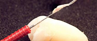
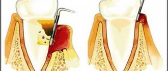
Excision of the hood over the wisdom tooth
If there is an acute period of the disease or it develops again, the doctor usually recommends a simple surgical operation - excision of the hood. This is the name given to the muco-subperiosteal flap, which partially covers the tooth. A pocket is formed under it, in which food debris accumulates. By rotting, they turn into a nutrient substrate for pathogens. The situation is aggravated by constant mechanical impact on the gums.
Careful excision of the hood over the wisdom tooth allows you to open the surface of the molar and eliminate the source of infection.
Procedure to complete:
- Primary elimination of inflammation with medications.
- Anesthesia (usually local) and excision of the hood with a surgical scalpel.
- Antiseptic treatment.
After excision of the mucous hood is completed, patients are advised to refrain from food and hot drinks for 1-2 hours, as they can cause pain and infection of the wound. To completely eliminate the consequences of the disease, the doctor prescribes anti-inflammatory drugs, rinses, and physiotherapeutic procedures.
Symptoms
The disease is quite pronounced, so it is difficult not to notice it.
- The earliest sign is swelling , redness and a significant increase in the size of the gum tissue. These symptoms may be so severe that they make it difficult to open your mouth. The area of edema can involve not only the affected gum, but also neighboring areas, including the cheeks.
- The next clear sign is pain - sharp, radiating to the ear and temple area, as well as to neighboring areas of the jaw. The pain intensifies when pressing on the affected area, when chewing, brushing teeth, or talking. Often the pain can become so intense that the patient cannot open his mouth.
- Over time, another symptom appears - bad breath , sour taste. Even regular brushing of teeth and the use of rinses and chewing gums does not help get rid of these sensations, since the source of the odor is located under the gum hood, and it is impossible to remove it from there with ordinary means. Smell and taste may occur even at the initial stage of the disease.
- Upon palpation, enlarged submandibular lymph nodes on the affected side are noticeable. As the process spreads, other groups may become involved. The patient's general condition gradually worsens, the temperature may rise, and general intoxication syndrome may develop.
Causes
The only cause of the disease is pathogenic microorganisms that provoke the occurrence of the disease. However, there are certain factors that determine the development of pericoronitis .
The main ones among them are:
- long and complex tooth eruption , more often third molars, also called eights and wisdom teeth;
- periodic injury to the gingival hood covering the tooth due to its swelling when the jaws close;
- the occurrence of ulcerations or erosions at the site of injury.
Development mechanism
With prolonged teething, plaque and food debris accumulate in the pocket of the fabric hood (between the crown of the adjacent tooth and the swollen gum itself).
Cleaning this area is problematic even in normal situations. In case of difficult teething, plaque accumulates quite quickly. In addition, it is a breeding ground for aggressive microorganisms.
When too many of them accumulate, their waste products provoke inflammation in the adjacent tissues. Moreover, the spread of the pathological process in periodontal tissues occurs more quickly than its occurrence .
Photo: wisdom tooth pericoronitis
Also among the reasons for the development of pericoronitis, one can also name the fact that it is a factor that provokes difficulty in the eruption of wisdom teeth . This occurs in more than half of people.
What are the contraindications for plasma lifting in dentistry? What determines the choice of solution?
In a separate publication we will talk about the symptoms of various forms of periodontitis.
Follow this link https://www.vash-dentist.ru/lechenie/desnyi/gingivit/opisanie-kataralnogo.html you will find photos of the symptoms and forms of catarrhal gingivitis.
In the process of human evolution and improvement of living conditions, the dentofacial apparatus has undergone some changes. In particular, the size of the jaw decreased.
Considering that the size of the teeth themselves has remained virtually unchanged, the last teeth - the eighth - very often do not have enough space on the jaw for normal growth.
Therefore, in many cases it turns out (this can be seen on the x-ray) that one of the following violations occurs:
- The tooth “lies” on its side and grows, accordingly, in the same way.
- The figure eight may partially remain in the jaw branch.
- Eruption occurs in the right direction, that is, parallel to the others, but the crown does not come out completely and remains partially hidden by the gum hood.
What are the possible complications?
Improper teething, accompanied by pericoronitis without treatment, can lead to the following serious complications:
- Formation of necrotic ulcers along the gingival margin. Subsequently, necrosis can spread to other tissues of the oral cavity;
- Damage to healthy neighboring teeth;
- Phlegmon of the perimaxillary spaces;
- Periostitis, which easily spreads to nearby areas;
- Osteomyelitis;
- Abscess formation.
Any of these pathologies can become life-threatening, so treatment of inflammation of the mucous membrane must be timely.
Treatment of pericoronitis is not difficult. A timely visit to the dentist and diagnostics will allow you to immediately carry out the correct treatment. For example, if a tooth initially erupts incorrectly, then removing it at the very beginning of the process will avoid further discomfort.
If for one reason or another it was not possible to visit a doctor on time, then you should come for a consultation when the first signs of inflammation appear. The purulent form is much more difficult to treat and causes more inconvenience. In addition, pus is the main cause of complications.
Classification
Pericoronitis is divided into acute, which occurs most often, and a rarer form - chronic pericoronitis. Despite the fact that the latter type is not reflected in ICD-10, it occurs with improper treatment of pericoronitis.
According to the flow, the following types of pericoronitis are distinguished:
- Catarrhal – characterized by slight swelling and pain when touched;
- Purulent - constant pain and the presence of purulent discharge;
- Ulcerative – leads to the formation of ulcerative defects on the gums;
Retromolar is distinguished as a separate form - it is distinguished by its location, which does not allow it to be easily seen and treated, which makes it difficult to combat this disease and increases the likelihood of complications.