1740
Medicine is constantly moving forward, and dentistry is no exception. Its arsenal is constantly updated with new technologies, drugs and high-tech equipment.
One such advancement is the microscope. Thanks to this device, most problems that were previously beyond the power of dentists are solved, and 80% of previously removed teeth can be saved.
Features and Applications
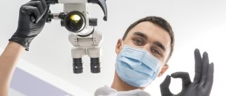
A dental microscope is a system of lenses with multiple magnification (from 3 to 25 times), mounted on a special tripod, and making it possible to conduct a detailed examination and perform complete and high-quality treatment.
If previously the doctor worked almost blindly through a mirror, now with optics he can see the treated area better. Thanks to the microscope, it became possible to more carefully and thoroughly treat the affected cavity without affecting living tissue.
Many models of the device are equipped with additional functions , for example, all stages of treatment are recorded, a well-lit and clear image of the surgical field is displayed on the monitor, and light filters are activated for accurate diagnosis.
You can also study in detail the color, relief, structure of teeth, the condition of tissues and canal walls, determine the quality of standing fillings and the degree of fit of orthopedic structures.
The endodontist can independently adjust the desired lighting and sharpness , obtaining a clear image of even the deepest canal with all its branches.
The scope of application of the device is extensive, i.e. it is indispensable in work requiring high precision performance, namely:
- diagnostics;
- endodontics
- periodontology;
- dental operations;
- restoration;
- surgery;
- prosthetics.
All manipulations performed under a microscope do not differ in technique from those usually performed by a dentist, but they are more comfortable for the patient.
During treatment, he is in a supine position, which is more psychologically comfortable and convenient. The lens of the device is placed at a distance of 20-25 cm from the face, and about another 35 cm is the device itself with eyepieces. Now the doctor’s face is located at a considerable distance (up to half a meter) and does not hang over the patient as before.
An assistant monitors the entire process together with the endodontist. He also provides the doctor with all the necessary instruments and medications.
Purpose and rules for applying rubber dam in dentistry, types of material.
Come here if you are interested in why a tooth might break.
At this address https://www.vash-dentist.ru/lechenie/zubyi/karies/klassifikatsii-po-mkb-10.html we suggest that you familiarize yourself with the features of the classification of dental caries according to ICD 10.
Our teeth are like stones
Tooth enamel is the outer protective shell of the tooth, which is 97% composed of hydroxyapatite crystals (Fig. 4) - Ca10(PO4)6(OH)2 - and foundation proteins: amelogenins and emellines, on which during the development of enamel (amelogenesis ) and hydroxyapatite crystals are strung [2], [3], [6], [7].
Figure 4. This is what the “closest relative” of our enamel looks like - hydroxyapatite. It’s good for him, because he doesn’t know anything about caries!
"Wikipedia"
Hydroxyapatite, although the hardest part of man, is still very sensitive to acids. Acids “stuck” in dental plaque, especially in the area of fissures (Fig. 5) - folds of enamel - begin to destroy the mineral, leading to demineralization of dental tissue, in other words, to softening. The mineral decomposes, forming calcium salts with other acids [3].
Ca10(PO4)6(OH)2 + acid → 10Ca2++ 6HPO42– + 2H2O
Notably, hydroxyapatite can remain dissolved for up to two hours after ingestion. In addition to carbohydrates, the main role here is played by fruit acids. That is why after eating fruit you feel a feeling of cleanliness of the enamel. Over time, saliva neutralizes the effects of acids due to its buffering properties [2]. The more often high-carbohydrate foods are consumed, the more constant the acidification process in the oral cavity becomes. Thus, tooth decay does not depend on the amount of sugar you take, but on the frequency of its consumption: do not eat a bag of sweets during the day, but eat a large dessert once a day.
Figure 5. Enamel folds - fissures - are the favorite place for the “seeds” of caries. It is here that demineralization processes occur especially actively.
drawing by Anastasia Prokhorova
Advantages
Treatment under a microscope has the following advantages:
- The efficiency of diagnosis increases , i.e. all pathological conditions can be detected in the early stages. The device allows you to examine even shallow and smallest lesions, eliminating the need for radiography.
- During treatment, healthy tissue is preserved as much as possible. Working only with a mirror, the doctor removes healthy tissue along with the affected tissue. Now, having a detailed picture, he excises only the infected areas, leaving healthy ones.
- The degree of fit of the orthopedic system or filling is accurately assessed . Checking this indicator and adjusting it under the control of the device allows you to improve the quality of the installation.
- It became possible to treat canals with complex structures. Their diameter is a thousandth of a millimeter.
It is very difficult to examine entrances without optical equipment, walk through them, process them, and then seal them.Undetected canals remain uncovered and untreated, and this leads to further development of infection and early tooth loss. Optics helps to detect all channels and seal them.
- The device is necessary during retreatment , especially if unsealing of canals is required. The doctor cleans out the failed composite, removes long-standing pins and identified remains of dental instruments broken during the last treatment.
- Ergonomics during long-term treatment . In order to examine remote, hard-to-reach areas, a specialist no longer needs to adjust to the lighting, take an uncomfortable position and strain his eyes. Better ergonomics – more productive and better treatment.
- Preventing medical errors and minimizing the risk of complications. With a microscope, the likelihood of side effects, complications and injuries is reduced to zero.
- The specialist receives continuous and complete control over the manipulation being performed. Seeing every part of the surgical field, he accurately detects hidden lesions, eliminates only problem areas, and performs all actions correctly and as accurately as possible.
- Convenience and psychological comfort for the patient during procedures - he lies in the dental chair, the level of fear and anxiety is reduced.
- Some types of manipulations can be performed through an artificial crown without having to remove it.
- Documentation of treatment is possible. The images and video recordings taken help the patient understand the condition of the oral cavity and the need for a certain manipulation.
All of the above advantages guarantee the effectiveness and high quality of therapy.
Indications for using the microscope
There are no specific diseases in dentistry that cannot be treated without high-precision optics, but the quality of medical care greatly benefits from its use. This is especially true for complex cases, for example, dental treatment with any anomaly in the development of the jaw or with an atypical structure of the root canals (when they are highly branched or tortuous). The use of electronic optics when re-sealing canals is justified, because this requires first unsealing1 and cleaning the canals without damaging their walls. The doctor must make sure that no particles of the previous filling, medication, pin, etc. remain in their branches.
The use of a microscope in the treatment of cysts, granulomas and other neoplasms at the apex of the roots allows for high-precision resection or cystectomy without damaging the surrounding tissues, and most importantly, preserving the tooth. This is a very important point when removing a cyst or root tip, because improper operation or injury to adjacent tissues can lead to re-infection and growth of the cyst.
And of course, such a device is used in all organ-saving and minimally invasive (low-traumatic) endodontic operations.
Indications and restrictions
Indications for using a microscope are the following conditions:
- caries at any stage;
- inflammation of the periodontium or pulp chamber;
- poorly performed or incomplete treatment (overtreatment);
- leaving a fragment of an instrument in the root canal;
- dental restoration;
- perforation (punching) of the pulp;
- a large number of channels and their complex structure;
- elimination of granulomas or cysts without extraction of the tooth itself;
- identification of hidden caries under an old filling, between adjacent elements;
- detection and sealing of perforated roots;
- unsealing canals that were previously closed with cement or resocin-formalin;
- removing the stump inlays on the pin or the pins themselves;
- identifying hidden cracks;
- lightening of units darkened from the inside due to inflammation in the canals.
The device is also necessary to reduce the number of diagnostic images, determine the exact quantity, quality and parameters of root canals, as well as to control each stage of their cleaning and processing.
There are no restrictions on the use of a microscope for diagnosing and treating identified pathologies. All existing contraindications relate only to the disease that is diagnosed and which needs to be eliminated.
Complications of caries -
If the deep form of caries is not treated in time, then the infection penetrates into the pulp of the tooth. In this case, inflammation of the nerve in the tooth develops, which is called pulpitis. If, in turn, pulpitis is not treated in a timely manner, then the inflammation extends beyond the boundaries of the tooth - into the tissue surrounding the tips of the roots of the diseased tooth. In this case, granulomas or cysts (purulent sacs) appear at the tops of the roots. This disease is called periodontitis.
Important aspects
The use of optics is most relevant and justified in endodontics , i.e. in the treatment of dental canals. Without a microscope, doctors relied only on their knowledge of anatomy (they acted by touch), and radiography came to their aid.
But here you need to understand that x-rays are one of the diagnostic methods, and all responsibility for the quality of treatment lies with the dentist. The advent of optics has greatly facilitated his work, and now a specialist can treat canals with great accuracy.
A microscope is also indispensable for diagnostics. It helps to detect diseases at their earliest stages, see hidden caries, and find the smallest cracks invisible to the eye. There is no need for additional procedures, which means it protects the patient from unnecessary material costs.
No less often, optics are used to re-treat canals in cases where the previous treatment was of poor quality, and in order to eliminate the problem, the canals need to be unsealed.
Not so long ago, after a poorly performed manipulation, a tooth had to be removed. The microscope helps to examine the structure and direction of the canal , see the old composite in it, determines the exact direction of treatment, and reduces the likelihood of complications and injury to a minimum.
When working with the device, the doctor is always sure that the canals are completely traversed, well processed and sealed. It can also be used to find and remove fragments of a broken instrument left in the tooth cavity.
The use of a microscope in surgical dentistry
The microscope is also effective during surgical operations.
The use of a dental microscope makes it possible to make surgery minimally invasive, since it becomes possible to use microinstruments specially designed for microscopic dentistry, which are significantly smaller in size compared to traditional ones, and therefore cause less damage to oral tissue. Patients tolerate both the operation itself and the postoperative period more easily.
Progress of the procedure
Despite the fact that the use of microscopes in dentistry has only recently begun, the technique has already received high marks among experts and patients. The device has completely changed the idea of dental treatment and helps save them even in the most severe cases.
But no matter what procedure is performed by optics, the doctor will definitely carry out the following before starting it:
- visual examination of the oral cavity;
- advise the patient about the course of the upcoming treatment and its results;
- will prepare the oral cavity by carrying out professional cleaning and sanitation.
Treatment of caries
Before starting the procedure, the tooth is isolated with a rubber dam (a special latex plate). It protects the surgical field as much as possible from the penetration of saliva and ensures the cleanliness of this area.
To completely cope with the disease, enamel and necrotic dentin tissue are first mechanically removed. Tissues affected by caries, located next to healthy elements, are cleaned out under a microscope.
When filling canals, the doctor also uses optics to control the quality and completeness of adhesion of the filling material to the walls of the formed cavity.
Therapy of pulpitis and periodontitis
Infection entering the pulp from a carious cavity leads to the development of an acute form of inflammation of the dental nerve. Its defeat is accompanied by severe pain and swelling.
The treatment technique for pulpitis or periodontitis consists of removing this nerve, carefully treating the canals, and then closing them with filling material.
Hidden canals, without treatment of which the pathology cannot be dealt with, are diagnosed by dentists only in 50% of all cases. Microscopic examination reveals them 100%, which means the cure is complete.
Does Analgin help with toothache, and how to take it correctly.
In this publication we will talk about Icon treatment of caries.
Here https://www.vash-dentist.ru/lechenie/zubyi/plombyi/iz-chego-skladyivayutsya-tsenyi.html you will find prices for dental fillings.
Root canal treatment
Root canal therapy involves sealing the cavity with a filling. Receiving an enlarged and high-quality image of the canals, the endodontist processes their apices.
Due to the fact that everything can be seen under a microscope, the doctor has the opportunity to carry out manipulations in a gentle manner , without affecting healthy tissue.
Removing Infected Material
During complex dental procedures and operations, situations occur when a small part of the instrument or material remains in the gum or tooth cavity.
The size of these fragments is so small that they cannot be seen visually. Left in the cavity or gum, the remaining material over time leads to the development of infection and other consequences.
It is impossible to extract particles without unsealing and without a microscope.
Identification and elimination of defects
In their practice, dentists often encounter various defects of teeth and gums that were previously treated, such as improper filling, microcracks in the enamel, hidden caries.
All these defects are small, not visually detectable, which means it is impossible to identify them at an early stage without special equipment.
Using a microscope, you can identify even the smallest sources of future pathologies and eliminate them in a timely manner.
How to determine caries at home -
- The presence of pain syndrome, which we wrote about above.
- The cavity may have sharp edges and irregularities, which can be perfectly felt (in some cases) by the tongue.
- Bad breath may indicate the presence of caries. Food debris is retained in the carious cavity, which is difficult to clean; under the influence of the microflora of the oral cavity, they begin to rot, causing an unpleasant odor.
- Inspection of visually accessible surfaces of teeth (for example, in a mirror). At the same time, even if the surface layer of enamel is preserved, darkening may be visible underneath it. Such darkening may indicate caries.
Prices
Of course, the cost of treating dental pathologies under a microscope exceeds the payment for the same disease, the treatment of which was carried out according to the standard protocol. Thus, the cost of treating some diseases is:
- pulpitis will cost 10 thousand rubles;
- periodontitis - 13-15 thousand rubles;
- granulomas and cysts – from 10 thousand rubles;
- caries - about 8 thousand rubles.
Typically, this price already includes payment for processing and filling of root canals. The approximate payment for retreatment is 4-6 thousand rubles, diagnostic measures - 1 thousand rubles. Prices at different clinics will vary slightly, as they largely depend on the following factors:
- clinic status;
- location;
- cost of drugs.
The high cost of the service is explained by the high cost of the device, as well as the need to improve the dentist’s qualifications. But when it comes to preserving the tooth and minimizing complications, such material expenses are justified.
The video provides additional information on the topic of the article.
Cost of dental treatment under a microscope
The concept of the cost of dental treatment under a microscope, in most cases, does not make sense, because... There is a dental service that has a set price, it can be performed either with a microscope or without a microscope. In most cases, the cost of the service itself increases, this is due to two factors - depreciation of the microscope itself (the cost starts from 30,000 USD) and the large labor costs of the doctor.
However, the increase in cost will be minimal, and potentially, when using a microscope, the patient receives treatment at a qualitatively higher level, the result of which can be used longer; if you want, the return on investment when treating teeth using a microscope will be higher .
Reviews
A microscope is a universal dental equipment that gives a real chance to save a seemingly hopeless tooth and diagnose pathologies at the initial stage of their development.
It allows you to make the treatment process predictable, improve quality and accurately predict its outcome.
If you have similar experience and would like to express your opinion on the effectiveness and advisability of optical treatment, please share them in the comments to this article.
If you find an error, please select a piece of text and press Ctrl+Enter.
Tags toothache treatment dental treatment under a microscope
Did you like the article? stay tuned
Previous article
On the effectiveness of tooth restoration in dentistry using an anchor pin
Next article
Intraligamentary anesthesia is a step towards painless treatment
Aesthetic restorations using a microscope
What criteria must the restoration meet:
The restoration, like a filling, must restore occlusion, not interfere with the natural closure of teeth, repeat the anatomical shape of the tooth in detail, and be reliable enough for the tooth to perform the function of biting or grinding food. Reliability is achieved through the complete implementation of all steps when placing a filling. Defects can occur when cleaning the surfaces of a prepared tooth. Using a microscope, before and after etching, the dentist is able to visually verify the absence of tissue particles or foreign materials. This undoubtedly improves the prognosis of treatment;
The restoration must match the natural color of the teeth and be invisible. Not only the color, but also other optical properties of the enamel (opalescence, transparency) must be reproduced, which is obviously easier to do if the work area is increased several times;
The junction of the restoration with the biological tissue of the tooth. A large gap at the junction will concentrate stress, become a place for the accumulation of bacteria and plaque and the development of caries, i.e. The more accurately the dentist “attaches” the edge of the filling to the tooth, the longer it will last. In this context, a microscope is an ideal instrument for monitoring the size of the joint. Similar logic is suitable for installing ceramic inlays or crowns.
When restoring the form and function of teeth through restoration, it is important to consider as many details as possible. This is possible by increasing the restoration area. With traditional treatment, the dentist's view is limited to what is reflected in the dental mirror or what he sees with his eyes. The use of a microscope allows you to expand the capabilities of the dentist and improve the quality of the filling being installed.


