Excessive tooth mobility - This is a common symptom indicating the development of severe dental diseases.
Unfortunately, many patients underestimate the danger of this pathology and postpone visiting the doctor until the last minute, depriving themselves of the opportunity to receive timely dental care. Meanwhile, in the absence of treatment, tooth mobility progresses and becomes irreversible. It is for this reason that any delay in starting restorative therapy significantly reduces the chances of maintaining the integrity of the dentition.
Symptoms accompanying mobility
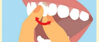
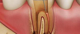
In fact, there is only one symptom - when you touch a tooth with your finger, tongue or any object, as well as during eating, its mobility is clearly felt. As a rule, horizontally, that is, a shift to the right-left or forward-backward. Other symptoms are mainly associated with the underlying disease and, as a rule, we are talking about inflammation of the gums, exposure of the roots, the formation of copious amounts of deposits under the soft mucous membranes, even rotting of the contents of periodontal pockets.
Classification of tooth mobility
In modern dental practice, to determine the degree of unnatural mobility of teeth, the classification developed by Honored Scientist of Russia David Abramovich Entin is most often used.
According to this typology, the following levels of mobility are distinguished:
- I degree (instability of position in relation to the coronal parts of adjacent teeth in the oral-vestibular or bucco-lingual directions, amplitude of movement less than 1 mm);
- II degree (mobility in similar directions with an amplitude of more than 1 mm, the appearance of movements in the distal-palatal direction);
- III degree (ability to move in all directions, including vertical);
- IV degree (adding rotational movements of the diseased tooth around its own axis).
Causes of mobility
There are quite a few reasons that cause abnormal mobility. The main ones are the following:
- age-related changes, hormonal imbalances,
- lack of vitamins and minerals,
- inflammatory processes of the gums, i.e. diseases of periodontal tissues – periodontitis and periodontal disease. These are the main reasons why teeth begin to become loose. In turn, their development is caused by poor oral hygiene,
- the presence of gaps in the row, as the teeth begin to shift towards the empty space,
- injury to the jaw system or the tooth itself, its root,
- complications of chronic diseases associated with disruption of the endocrine or immune system,
- purposeful loosening of the tooth,
- incorrect bite, as well as uneven pressure on one or a group of elements of the dentition.
Timely identification of the cause of the problem will allow you to save your teeth by restoring the functionality of the ligamentous apparatus and jaw bone cells.
What to do if loosening occurs?
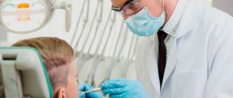
If you notice your teeth are moving, immediately make an appointment with a dentist. The specialists at Dr. Granov’s clinic are ready to help you at any time. We urge you not to ignore your symptom, since subsequently the treatment of the provoking pathology can become very complicated.
While you are at home, try to follow three basic rules:
- Never loosen the tooth with your fingers or tongue. Try not to touch it at all, so as not to aggravate the current situation;
- Rinse your mouth with warm water (or a mild saline solution), but do not brush your teeth with a brush or toothpaste;
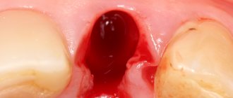
- If a tooth falls out, see a doctor immediately. If this has just happened, you still have the option of emergency implantation. The more you delay the process, the less opportunities there will be for high-quality restoration.
When a tooth falls out, we must not forget that parts of it may still remain inside the gums. And this can also have very dangerous consequences for you if you continue to ignore the warning signs.
Causes and diagnosis
Healthy people have physiological tooth mobility, which is necessary so that during eating, chewing loads are distributed evenly throughout the entire dentition. We are talking about pathological mobility when the displacement of tooth roots in the sockets becomes noticeable visually or according to the patient’s subjective sensations. There are three degrees of abnormal mobility: in the first, the teeth can only move back and forth, in the second - also to the sides, in the third they can move around their axis and vertically (as if raised in the socket). The doctor identifies the pathology through examination and x-ray diagnostics.
The causes of this condition may be:
- Periodontal diseases (periodontitis, periodontal disease). Insufficient or improper oral hygiene plays an important role in the development of such diseases, since poor care contributes to the accumulation of plaque and the deposition of tartar, and these are predisposing factors for the development of oral diseases.
- Resorption (sparseness) of jaw bone tissue . Often occurs as a result of tooth extraction and inflammatory processes.
- Bite defects . Abnormal positions of teeth in rows can cause their pathological mobility.
- Injuries and mechanical damage.
In order for the treatment of tooth mobility to be high-quality and successful, it is important for the dentist to first establish the exact cause of this condition.
Signs of pathological mobility
Violation of the ligamentous apparatus, in which the tooth becomes loose, is accompanied by accompanying symptoms:
|
What to do if an adult’s tooth is loose and hurts when pressed?
Normally, our teeth have little mobility. Dentists call it physiological; it is necessary to absorb the loads during chewing, as well as for their more even distribution over the entire dentition. But such mobility is so insignificant that it is impossible to notice it. Therefore, if an adult suddenly discovers that a tooth in his gum has begun to loosen, this is already a symptom or an indicator of pathology that requires the participation of a dentist. In the absence of timely medical care, such violations very often lead to irreversible loss of a tooth.
Selective grinding of teeth
- one of the most common methods in the system of complex therapy of periodontal diseases. According to some data, 95.8% of patients with periodontal pathology need it.
Objectives of selective teeth grinding:
1. Elimination of premature occlusal contacts
2. Elimination of moments that block and interfere with the movements of the lower jaw
3. Elimination of deformation of the occlusal surface of the dentition.
There are different methods of grinding teeth, the most popular are the Jenkelson and Schuller methods.
The technique proposed by Jankelson (1979) is considered a functional method, however, premature contacts are removed in central occlusion.
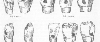
According to Jenkelson's classification, premature contacts are divided into 3 classes:
1st contacts on the vestibular slopes of the buccal cusps of molars and premolars and the vestibular surface of the lower incisors
2nd contacts on the oral slopes of the palatine cusps of the upper molars and premolars;
3rd contacts on the vestibular slopes of the palatal cusps of the upper molars and premolars.
Indications for selective grinding:
— Presence of premature contacts of antagonist teeth in central, anterior and lateral occlusions;
— Absence or uneven wear of hard dental tissues;
— Deformations of occlusal surfaces;
— Bite abnormalities
Method of selective grinding of teeth:
-carried out before therapeutic and surgical procedures or in parallel with them.
- Preliminary grinding of teeth involves shortening protruding teeth and is intended to eliminate significant deformations of the occlusal surfaces that occur due to defects in the dentition. If significant shortening is necessary, depulpation of the latter is indicated.
- The final sanding is carried out in a certain sequence. First, premature contacts in various types of occlusion are eliminated, then when moving the lower jaw from central to anterior and lateral occlusions.
- Before grinding, an occludogram is obtained, which is stored to monitor the results of grinding. After carrying out this manipulation, it is necessary to polish the ground surfaces and coat them with fluorine varnish.
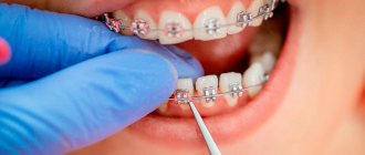
Orthodontic preparation for orthopedic treatment for periodontal diseases
In patients with periodontal pathology, the clinical picture is often complicated by deformations of the dentition; in addition, malocclusions in some cases can themselves lead to periodontal pathology, so orthodontic preparation before orthopedic treatment is of great importance.
Structurally, orthodontic appliances have some differences from classical ones, these include:
— Application of minimal forces to move teeth.
- Longer active treatment and retention period.
Temporary splints can be used as retention devices.
Features of non-surgical treatment of loose teeth at the Healthy People clinic
The Healthy People clinic employs professionals, dentists with extensive experience, who perform all necessary procedures with high quality. The center's doctors perform diagnostics, identify why the swaying occurs, and prescribe effective treatment. If necessary, the patient is referred to see specialists.
A timely visit to the clinic makes it possible to eliminate mobility and carry out dental treatment, treatment of pulpitis without expensive operations, restoring their health and beauty.
The cost of treatment can be found out by calling the Healthy People clinic or looking at the website in the prices section.
Functional traumatic overload of the periodontium
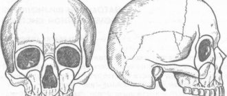
-occupies a special place among local causes in the etiology and pathogenesis of periodontal diseases. When chewing and swallowing, at the moment of closure of the dentition, the periodontium of each tooth perceives a force load, which under normal conditions is absorbed by special periodontal devices (cemento-alveolar, interdental fibers, etc.). Then it is transformed and transmitted to the bone structures of the jaws, the temporomandibular joint and the skull. This physiological load helps normalize trophism and metabolism, stimulates the processes of growth and development.
With pathological processes in the periodontium caused by common causes (vitaminosis, diabetes and other disorders of endocrine regulation, diseases of the gastrointestinal tract, cardiovascular system and nervous systems, etc.), the resistance of periodontal tissues decreases.
As a result of weakening of the periodontium, the usual occlusal load begins to exceed the tolerance of its structures and turns from a factor stimulating development into a traumatic one, disrupting the trophism of the periodontium and destroying its tissues. Traumatic occlusion occurs, which subsequently plays a leading role in the course of this disease.
The term “traumatic occlusion” was proposed by PR Stillman in 1919. Other terms have been proposed to characterize and define periodontal overload:
- “traumatic articulation”
- “functional traumatism”
- “pathological occlusion”
- “functional traumatic overload of teeth”, etc.
According to the mechanism of development, three types of traumatic occlusion are distinguished:
— Primary
— Secondary
— Combined
Primary traumatic occlusion develops against the background of unaffected periodontium as a result of the action of an occlusal load that is excessive in magnitude and/or direction.
Thus, primary traumatic overload of healthy periodontium can occur due to excessive magnitude, abnormal direction and duration of action of the occlusal functional load and steam function of the masticatory, facial muscles and tongue. More often than not, overload is caused by the simultaneous action of several causes.
Secondary traumatic occlusion:
Its pathogenesis is based on pathological changes in periodontal tissue. At the same time, degenerative and inflammatory processes develop in the supporting tissues of the teeth throughout the entire dentition, which are accompanied by:
1). resorption of bone tissue of the alveolar process.
2). Gingivitis.
3). destruction of the periodontium with the formation of a pocket.
4). suppuration from it.
Resorption of the bone tissue of the sockets leads to a disruption of the normal biological patterns of the structure and function of the periodontium. From this moment on, a radical change occurs in the biomechanical relationship between teeth and surrounding tissues.
Thus, a change in the ratio of the extra- and inside the alveolar part of the tooth is one of the pathogenetic mechanisms in the development of traumatic occlusion.
The clinical picture of secondary traumatic occlusion is diverse and depends on the patient’s age, the form of the underlying disease (periodontal disease, periodontitis), its severity and stage of development, the presence of dentition defects, malocclusion or tooth position, pathological abrasion and other traumatic factors.
If for primary traumatic occlusion orthopedic intervention is sufficient, then for secondary occlusion complex therapeutic (local and general), surgical and orthopedic treatment is required. The prognosis also varies. In case of primary traumatic occlusion, after eliminating the overload of the teeth in all periodontal tissues, reparative processes occur.