Features of dental x-rays for children
Previously (about 10-15 years ago), x-rays of baby teeth were not a common procedure. Some parents even believed in the myth that baby teeth have neither a root nor a nerve, so the causes of toothache must lie somewhere on the surface, that is, on the crown. This is absolutely not true, and childhood toothache can be caused by the same reasons as in an adult (more on this below).
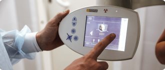
When asked whether children undergo dental x-rays, we can confidently answer that yes, they do. Moreover, to accurately diagnose the problem and build the correct treatment plan, this is necessary. Another thing is that preference should be given to relatively safe types of x-ray examination, for example, digital.
There are several recommendations for parents to listen to when going for an x-ray with their child:
- You need to come to the clinic in advance so that you have time to psychologically prepare your child for the procedure. You need to tell him that the x-ray will not cause any pain, so you shouldn’t be afraid of it.
- The child's clothing should be loose, easily removable and without complex metal decorations.
- As for girls, you need to give them a simple hairstyle, without using metal pins, bobby pins, etc.
What do dentists tell patients?
- What we do know is that radiation from dental exams has not yet been scientifically proven to increase the risk of cancer.
- We know that the human body is always bombarded with X-rays from cosmic rays from space and radon gas in the air constantly from the time we are born until the time we die, so our bodies are accustomed to repairing the damage caused by low amounts of radiation.
- We know that dental radiation has always been considered safer than other types of medical radiation because it targets areas of the body that are simply not sensitive to radiation. And if we protect areas of concern, such as the thyroid gland, and make sure the lenses of the eyes are not exposed to X-rays, we help the patient feel safe and protected.
Types of X-rays for children
Like adults, children may be prescribed different types of x-rays.
Sight radiograph
A targeted radiograph is performed using a special digital visiograph. A specific problematic tooth or several adjacent ones (maximum 4) are removed.
Panoramic radiograph
The panoramic image shows the entire oral cavity: the upper and lower dentition, teeth that have not yet erupted, and the jaws. It also affects the sinuses. An orthopantomogram (this is what a panoramic image is called) is often prescribed to young patients when it becomes noticeable that their teeth are erupting incorrectly: with an inclination, rotation, and so on. In this case, X-rays help to understand whether there is an anomaly in the development of the jaw bone. There are cases when, after the loss of baby teeth, permanent teeth do not appear for a long time. An x-ray will help identify the cause of this deviation.
Answers to frequently asked questions about x-rays of baby teeth in children
We offer expert answers from specialists from the Smile Factor network of clinics to frequently asked questions by parents.
Can an x-ray be prescribed to a child “just in case”?
Unfounded medical (and dental) research is prohibited by SanPiN.
But, most often, diagnosis according to indications is at the discretion of the attending physician. Any parent can ask clarifying questions and learn more about the assigned test and what it is for. What is better - 2D or 3D scanning result?
It all depends on the goals of the diagnosis.
In some cases, a 2D image is sufficient, while in others a CBCT scan is required to obtain a 3D image. Taking into account how CT scans of the jaw and teeth are done, the procedure is not much different from a simple x-ray in terms of safety and is indicated for obtaining more extensive data about the child’s dental system in the images. Are x-rays harmful to a child?
Modern X-ray machines (and most often these are ultra-modern tomographs) produce the minimum possible radiation dose during operation, which does not cause any harm to the child’s body. To achieve the maximum recommended radiation exposure of 1000 µSv per year, a child needs to undergo about 400 x-ray examinations. It becomes clear that so much research is simply not needed. And the radiation that a baby receives during a CT scan is equal to what he receives after a week of watching TV.
Indications for testing
To determine the extent of caries damage
Children's teeth are susceptible to caries. We cannot assume that caries of baby teeth is not dangerous, since they will fall out anyway. Pathology can spread to permanent teeth even before they erupt. Therefore, it is very important to identify caries and carry out effective treatment.
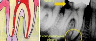
To identify periodontitis and pulpitis
Periodontitis (tooth root pathology, pulpitis) is an inflammation of the pulp where the nerve is located. It is clear that such diseases cannot be detected during the initial examination, because they are hidden in the tooth itself or inside the gums. The child’s main complaint is pain in his baby teeth, and the ability to identify its cause is through an X-ray photo.
When planning endodontic treatment
Endodontic treatment is a set of procedures aimed at preserving a tooth. When emerging caries causes complications, such as pulpitis or periodontitis, the doctor needs to perform therapeutic manipulations inside the tooth. To understand how much pathology has affected the tooth, what percentage of it is destroyed, you need to take an x-ray.
To assess the condition of the permanent tooth buds
An assessment of the condition of the permanent tooth buds is also required before endodontic treatment. It is necessary in order to understand whether the permanent teeth have been affected by pathology and how close they have already descended to the milk teeth.
To diagnose and determine treatment regimens for pathologies of occlusion or teething
At an early age, it is still relatively easy to correct an incorrectly formed bite or correct the position of erupted teeth. This can be done using braces or other special systems. However, to begin orthodontic intervention, you need to understand how serious the pathology is. X-rays help with this.
To determine the reasons for the delay in the appearance of permanent teeth
If a child does not have baby teeth before one year of age, the doctor may order an X-ray to predict their appearance. Of course, such a developmental pathology as adentia (when there are simply no rudiments of baby teeth) is extremely rare, but it is still worth excluding it completely and understanding how soon the first tooth can appear.
What can a dental x-ray show?
Carrying out X-rays of a child’s teeth or jaw is aimed at identifying pathologies and determining the anatomical features of the development of the dental system. Dental x-rays are performed before treatment, during treatment or upon completion for control.
X-rays may show:
- decrease in tooth weight due to inflammation of soft tissues;
- foci of carious lesions in any area;
- changes occurring in dental roots;
- purulent processes flowing into an abscess;
- rudiments of permanent teeth;
- anomalies in the structure and development of teeth and jaw;
- impacted teeth;
- supernumerary teeth;
- damage to the jaw bones;
- and other.
The result of the image is assessed exclusively by a specialist. Based on the information received, the doctor can determine the diagnosis, prescribe or adjust treatment, or evaluate the effectiveness of the measures taken.
How is research conducted?
Panoramic dental x-rays are done for children, just like for adults:
- The child stands inside the orthopantomograph.
- He clamps the plastic tube between his teeth and keeps his lips closed.
- The device blade is moved as close to the patient as possible.
- A picture is taken (while the device rotates around the child’s head). This takes no more than 20-30 seconds, during which you cannot move or breathe.
A targeted photograph is taken as follows: the child sits on a chair and approaches the device.
Next, he wraps his mouth around the digital sensor and clenches his teeth. A picture is taken (takes a few seconds); during this process you cannot move or breathe. To protect the body from exposure to x-rays, a special apron is worn.
Are there any risks?
Modern X-ray machines are characterized by a low dose of radiation, but since exposure to X-rays cannot be completely ruled out, it is recommended that X-rays be taken no more than 2 times a year for children with baby teeth and no more than once every 1.5 years for adolescents. The need for diagnosis is assessed by the attending physician. If the risk from x-rays is lower than the risk of complications from dental pathology, then the specialist may prescribe a study with a more frequent frequency.
There are no risks of side effects after radiography if all the rules of the procedure are followed. There are no contraindications to the study. The exception is the patient's serious condition or previous irradiation.
For children under the age of one year, an X-ray of the teeth/jaw is taken if other diagnostic methods have turned out to be uninformative or there are contraindications to their use. Thus, children are examined mainly only in case of mechanical injuries.
Contraindications
If there is bleeding
Bleeding in the mouth, caused, for example, by problem gums, can affect the quality of the X-ray image. First, the doctor must prescribe hygiene procedures that can stop the bleeding, and only then send for x-rays.
If you feel unwell
If your child is sick, has a fever, or simply feels unwell, you should not take him for an x-ray. It is better to postpone this procedure until complete recovery.
Operating principle
The technique is based on X-ray radiation, which is combined with special programs and equipment. X-rays are a special type of radiation that can pass through the tissues of our body. These rays pass through some tissues (for example, abdominal organs) very easily, but through others (for example, muscles, ligaments or bone tissue) - worse, since they are partially or completely absorbed.
When taking a panoramic photo, the body receives a certain amount of radiation. Some of the rays are retained by tissues, while the rest passes through them. All information is sent to a special computer, which analyzes the received data and forms a contour image of the organs located in the path of radiation. As a result, the most accurate image of the oral cavity is obtained, on which the roots of all teeth, periodontal tissue, etc. are clearly visible.
Dental Imaging
Extraoral imaging allows the dentist to see a complete picture of the inside of the patient's mouth. Panoramic and 3D imaging assist in diagnosis, treatment planning and evaluation of certain dental conditions.
Dental systems using flat panel detectors have the advantage of providing highly detailed images for more accurate diagnosis. Modern high-speed, high-sensitivity flat panel detectors are an attractive option for dentists and are used for imaging in:
- Periodontal bone loss or disease detection
- Extraoral exams
- Planning a dental implant
- Visualization of abnormal teeth
- Jaw and face assessment
- Cleft palate assessment, dental cavity diagnosis
- Endodontic diagnostics, including root canals
- Diagnosis of dental trauma
And also offers beam computed tomography, also known as CBCT. Dental CBCT systems allow manufacturers to optimize their system to provide fast images while reducing radiation dose to patients. CBCT provides three-dimensional information and images compared to the two-dimensional image obtained with conventional x-ray images. 3D imaging can be especially useful in diagnosis, treatment planning, and evaluation of certain dental conditions.
Our discounts and promotions