An inflammatory formation affecting the upper part of the tooth root is called granuloma. It is formed during the development of periodontitis. Tooth granuloma is tightly attached to the root, being the initial stage of a granular cyst (cystogranuloma), from which it differs, in fact, only in its slightly smaller size. If the formation is ignored and not treated, it will begin to increase.
Tooth granuloma: symptoms of the disease
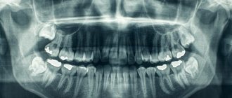
Only a doctor can make a diagnosis based on an x-ray, where a darkening will be visible in the root area. Any darkening in the image is a sign of the presence of a cavity, but if a granuloma occurs, it means that periodontitis, which is chronic in nature, begins to develop.
For a long time, the tooth may not bother a person at all. Sometimes pain may occur when biting or when eating hot food: these are the first signs of the development of chronic periodontitis. An exacerbation of the disease is observed during periods of weakening of the body's immunity: then the pain intensifies significantly, which is especially noticeable when biting. During such periods, as a rule, the gums become swollen, and in the area of inflammation it becomes painful to touch.
Why does granuloma appear on the root of a tooth?
At the tops of the roots, such formations can appear for several reasons:
- Poorly performed treatment of pulpitis. If caries is started, the affected cavity gradually becomes quite deep. When microorganisms enter the pulp, its inflammation begins, accompanied by acute pain, which may stop over time, indicating the death of the nerve.
But the development of the disease does not end: bacteria through the root canals extend beyond the boundaries of the affected tooth, as a result of which a focus of inflammation appears near the upper parts of the roots, called periodontitis. The further course of the disease can occur according to several scenarios, one of which is the formation of granuloma.
It is necessary to understand that a tooth with a granuloma does not necessarily have to be affected by caries, since the root can become inflamed near a tooth that has already been treated. If the doctor has not completely removed the tissues affected by caries by placing a filling on top of them, then there is a high risk of developing pulpitis with the subsequent formation of granuloma.
- Filling of root canals performed with violations. A granuloma that appears on the root of a tooth whose canals were filled some time ago indicates the unsatisfactory quality of the procedure performed. Due to inattention, haste or inexperience, the doctor could not fill the canals to the very top. Dentists often refuse to admit their own mistake, because in this case they will have to treat the tooth for free. Such unfortunate specialists can evade until the last minute, pretending that they do not understand the causes of pain, recommending taking a course of antibiotics.
Symptoms of a dental cyst
The formation of a dental cyst occurs slowly. The first stage is the formation of a granuloma - it is difficult to diagnose and does not cause unpleasant symptoms. There may be only slight pain in the gums and teeth during chewing; as a rule, it does not cause inconvenience.
Only X-ray diagnostics can reliably determine the presence of a developing cyst. Since there is no cause for concern, the scan is most often performed for other reasons - for example, before installing braces or a prosthetic for another tooth.
As the cyst grows, the patient begins to feel an increase in symptoms and notes the appearance of new signs:
- pain in the gums and teeth can radiate to the opposite dentition;
- body temperature rises;
- swelling of the soft tissues forms around the tooth, swelling of the cheek on the causal side.
Other symptoms may indicate complications that have already begun.
Diagnosis of granuloma on the tooth root
At the early stage of development of the disease, it will not be possible to notice any visual changes. The first signs of pathology become noticeable as the size of the infected area and the amount of pus increase.
To treat the disease with therapeutic methods, it is necessary to examine the affected area in detail: a complete picture of the granuloma must be obtained, which will allow one to detect key signs of the disease that differ from other diseases.
When suppuration occurs, the gums become very red and swollen, and pain appears, which can radiate to the head area in general and the ear in particular. This is the first symptom that should make you wary.
The following methods can be used to make an accurate diagnosis:
- use of classical x-rays;
- the use of radiovisiography or, in simpler terms, computer x-ray.
In the picture, the affected area looks like a dark spot with a clear border in the upper part of the tooth. The size of the spot indicates the following:
- 5-8 mm – the probability of having a dental granuloma is high;
- more than 8 mm – formation in the form of a cyst.
In rare cases, a large granuloma, up to 1.2 cm in size, may occur. Therefore, X-rays may not be enough for diagnosis: it is recommended to perform a biopsy of tissue cells from the area affected by the disease.
In most cases, granuloma is discovered during treatment of other dental diseases: the doctor may pay attention to increased swelling and swelling of the gums. In addition, the bone tissue near the top of the tooth may also bulge.
An increased risk of developing granuloma is observed in patients with crowns and pulpless teeth. Such people are advised to undergo regular examinations in order to promptly identify any changes affecting the gums and teeth.
Types of dental cysts
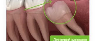
- Retromolar. A common cause of its appearance is chronic tissue inflammation due to complicated teething, most often molars. Wisdom or “eight” teeth are more susceptible to disease than others - they often grow in the wrong direction due to a lack of space in the jaw, which has already formed by adulthood.
- Eruption cyst. These are forms of the retromolar variety, but comparatively softer. It is a small formation that forms during teething. It is believed that the appearance of the disease is “due” to a local decrease in protective forces when baby teeth are replaced by permanent ones.
- Follicular. It is a consequence of the pathology of the development of the molar tooth and appears from the follicles during the formation of dental tissues.
- Radicular. This is the most common type of cyst; in this case, it is the result of a chronic inflammatory process or injury.
- Residual. Occurs after tooth extraction if a root fragment remains in the socket. Its presence provokes inflammation and the formation of a purulent vesicle.
- Keratocyst. The result of pathological formation of periodontium. The cyst in this case appears from the epithelium, which is required for the formation of periodontal tissues. As a rule, it causes difficulty in teething.
- Cyst of “eye” teeth. Appears during inflammatory processes occurring in the maxillary sinuses.
Tooth granuloma: treatment
Usually treatment is only therapeutic in nature, but its strategy may vary depending on whether the canals have been filled before.
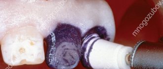
- If the canals have not been filled, the tissues affected by caries are drilled out, and the old filling is removed. This is required to carry out high-quality mechanical treatment of the canals, which are expanded and treated with antiseptic agents. If the granuloma is small (formations up to 3 mm are considered such), then the canal is sealed immediately. If the size of the granuloma exceeds 3 mm, the treatment period increases, since it is necessary to put a medicine into the canal, which includes potassium hydroxide - this substance leads to a decrease in the granuloma or even to its complete disappearance. A temporary filling is placed for the period the medication is placed (approximately 2-3 weeks). Then a repeat x-ray is taken, which should clearly show a significant reduction in the inflammatory formation. If the dynamics are obvious, the canals are sealed and a permanent filling is placed.
- If the canals have been filled, then the first stage of treatment is their unsealing. The further sequence of actions is similar to that described in the previous paragraph. The only thing is that if there is a crown on the tooth, it will have to be removed, and after the treatment is completed, it will have to be made and placed again. Some patients do not want to spend money on re-installing a crown: the solution is to perform a root resection operation, when the upper part with the granuloma attached to it is cut off through a small incision in the gum.
Granuloma between tooth roots: treatment with antibiotics
The number of people putting off visiting a doctor until the last minute is large. There are many reasons for this: fear of pain (although modern means of anesthesia make it possible for the patient to feel nothing at all during treatment procedures), lack of free time, and reluctance to part with money. But all these reasons and excuses come to naught when the pain becomes unbearable, becoming chronic. As practice shows, as soon as a person does not sleep for a couple of nights, the issue of the need to visit the dentist is resolved by itself.
However, many people try to cure granuloma on their own using strong antibiotics, which is basically impossible. Antibiotics may be prescribed to relieve inflammation and stop the formation of pus, but nothing more. But the action of antibiotics may not affect pathogenic microorganisms located in the root canals, and it is the infection in their unsealed areas that is the only reason for the formation of granulomas.
If a patient comes to the doctor with a suspicion of granuloma, and the doctor, driven by some of his own considerations, does nothing, recommending taking antibiotics, it is better not to delay time, but to immediately contact another dentist. Some doctors simply do not like to re-treat someone’s poorly treated teeth, since repeating the same operations takes much more time, while others, if the patient comes back again, do not want to admit that they made a mistake, which for them is tantamount to realizing their own lack of competence.
Tooth granuloma: surgical treatment
If therapeutic treatment does not help, then surgery comes into play: the operation is performed in the presence of a destructive process affecting the gums, and anesthesia (both general and local) is used to carry it out.
There are several surgical treatment options.
Root resection
Elimination of granuloma occurs in several stages:
- a passage to the top of the tooth opens by peeling off the gum shell;
- the root canals are cleaned and subsequently filled with medicinal substances;
- excision of the granuloma is carried out;
- the area is filled with synthetic fabrics;
- the tooth is filled.
On average, the operation takes about an hour.
Hemisection
If a tooth has many roots, then this procedure is prescribed (if there are complications that prevent the root from being saved).
The treatment procedure includes:
- removal of roots under the crown;
- filling the empty space between the roots with a special dental product;
- installation of a crown;
- monitoring the condition of the tooth using x-rays.
The method is simple, while the functionality of the tooth is preserved. If necessary, some time after the procedure, the patient can resort to prosthetics, provided that the root system remains healthy.
Cystotomy
The method is used when removal of large granulomas is required.
To implement it:
- a channel is created between the affected area and the oral cavity, through which pus absorbed by tampons is removed;
- after cleaning, the cavity is treated with antibacterial agents;
- sutures are placed;
- the cavity, free from suppuration, is filled with bone cells.
Types of X-ray
After the examination, the doctor can prescribe the patient one of four possible types of x-rays
.
Prikusnoy
This method allows you to reflect the crown part of the tooth in the image. It is used to detect periodontitis and interdental caries. Bitewing can be used to obtain images of the upper and lower teeth.
Sometimes such a picture can be taken after prosthetics and crown installation to see how correctly the procedure was performed.
Sighting
With the help of a targeted image, it is possible to discern a specific affected tooth or several. Moreover, such a picture cannot include more than 4 teeth.
Panoramic
Using panoramic images, you can monitor the quality and effectiveness of the treatment already performed. They allow you to see a complete picture of the state of the entire dental system, and this is not only teeth with obvious problems (for example, caries, chips, etc.), but also roots, periodontal tissue, paranasal sinuses and the lower jaw joint.
On a panoramic image, the doctor will be able to see the presence/absence of filling material, hidden carious cavities, inflammation of the peri-root tissues, cysts, tumors, as well as teeth that have not yet erupted.
Digital or 3D X-ray
This type of x-ray is considered the most modern and safe. Using 3D X-rays, you can get a clear image of the entire row of teeth and a specific tooth. The result is a three-dimensional image that is displayed on the monitor.
Measures to prevent granuloma after an extracted tooth
Preventive measures are aimed at preventing the development of the disease. They include:
- high-quality cleaning of teeth and gums on a daily basis;
- timely treatment of bleeding gums;
- regular visits to the dentist at least once a year (it is better to do this twice);
- replacing toothbrushes (old brushes accumulate bacteria that can provoke the development of the disease);
- contacting the dentist at the slightest pain associated with the gums or teeth;
- treatment of caries, periodontitis and pulpitis - quite often these diseases are the catalyst for the development of granuloma;
- the use of medicated toothpastes and herbal decoctions for rinsing;
- eating foods rich in calcium and other trace elements.
Complications after granuloma at the site of an extracted tooth
Lack of timely treatment and hope that the disease will go away on its own is fraught with serious consequences. Among the most common complications it is worth highlighting:
- development of periodontitis and further formation of a fistula;
- the occurrence of alveolitis is a consequence of the presence of an inflammatory process;
- formation of purulent flux;
- suppuration of the perimaxillary tissue;
- entry of pathogenic bacteria into the lymph nodes, from where they penetrate the cardiac system and internal organs (kidneys, liver and brain);
- development of facial asymmetry;
- the emergence of new foci of infection;
- infection of the body due to the penetration of pathogenic microorganisms into the blood vessels.
Timely removal of granuloma is a guarantee to avoid unpleasant health consequences.
Reasons for the formation of a dental cyst
Conditionally, the reasons can be divided into two groups:
- Poor oral hygiene - the development of caries, pulpitis, periodontitis, inflammation of the soft tissues of the oral cavity, which results in a cyst.
- Jaw injuries - facial injuries, complicated teething, violation of the technology for installing dental prostheses, filling complex, narrow, curved root canals during endodontic treatment, excessive loads on the teeth as a result of spasms of the masticatory muscles, malocclusion, etc.
These phenomena and diseases can cause inflammation, the focus of which is located at the root of the tooth. Still, the most common cause of a cyst is considered to be a carious lesion - ignoring caries leads to the formation of a cavity in the crown, damage to dentin and the inner soft part - the pulp, and subsequently inflammation of the periodontal tissues. The body strives to “isolate” healthy tissue around the tooth root from the inflammatory process, as a result of which a sac with purulent contents is formed. Why is a dental cyst dangerous? The answer to this question depends in part on the specific type of education.