Professional dental care includes the removal of caries, cystic areas and many other procedures. In accordance with the protocol for providing medical care, the dentist is also required to monitor the condition of the salivary glands
. In clinical practice, their damage is quite rare. Most often, poor performance of the excretory ducts occurs due to injury or incorrect oral therapy. Quite often this happens due to insufficient hygiene.
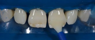
Bougienage or treatment of the salivary gland involves intubation of a dental instrument or probe in order to examine the organ and administer appropriate substances. Saliva contains a large number of enzymes that take part in the processing of food and the removal of metabolites from the body. If appropriate manipulations are not performed, this can lead to an increased likelihood of developing caries, pain when eating and a malfunction in the digestive system.
Indications for use
Bougienage of the salivary gland ducts is performed with the following symptoms:
- Unpleasant painful sensations.
- Sharp pain when eating certain foods.
- An involuntary feeling of fullness in the mouth after eating.
In case of malfunction of the excretory duct, you must immediately contact a specialist. Otherwise, the clinical picture may worsen, and the pathological process will become chronic.
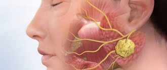
Salivary gland blockage
The salivary glands are ducts that secrete a special secretion into the oral cavity - saliva.
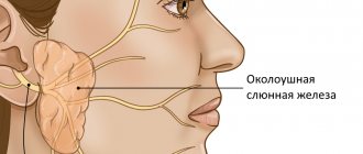
There are paired large salivary glands: submandibular, parotid, sublingual (they are closely related to the digestive system); as well as minor salivary glands: buccal, labial, molar, lingual, glands of the soft and hard palate. They are located in the submucosal layer of the cheeks, lips, palate, and tongue.
Kinds
According to the specificity of the secretion secreted by the glands, they are divided into protein, mucous and mixed. The largest paired salivary glands are the parotid glands, located in the back of the angle of the jaw, in front of the ears. The minor salivary glands are distributed throughout the oral cavity. If there is not enough saliva produced, the oral cavity becomes dry. In this regard, there is a high risk of developing caries, since with a deficiency of saliva, natural protection against such dental disease is no longer created.
Causes
It happens that the excretory duct of the salivary gland is blocked by various mineral deposits, which are also called stone. Blockage of the salivary gland provokes the normal outflow of saliva, as well as an enlargement of the gland. The cause of inflammatory phenomena can be infection with various bacteria and microbes.
When the duct is blocked, the swelling usually intensifies before eating, especially when eating pickled foods that have high acidity. It provokes salivation. When the ducts are blocked, saliva cannot be released freely. This often results in painful sensations.
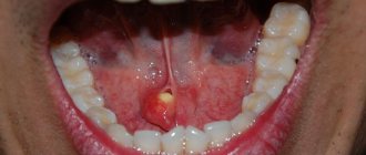
A complication of blockage of the salivary gland is mucocele - the formation of a cyst, which is caused by the retention of secretions resulting from blockage of the outlet of the so-called Blandin-Nun gland.
Symptoms
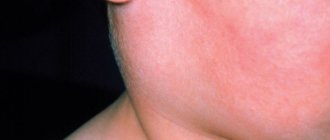
Blockage of the salivary glands is manifested by swelling, a change in the color of the mucous membrane of the lips, which acquire a bluish tint. As a rule, this is how inflammation of the minor salivary glands manifests itself. If the major salivary glands are blocked, swelling and pain occur in the neck and cheeks, especially after eating.
Diagnosis and treatment
The Dento Style clinic network provides comprehensive diagnostics and treatment of various pathologies of the salivary glands. Our specialists have an individual approach to each patient. If the salivary ducts are blocked, the patient is examined and the affected area is palpated. Methods such as probing are used. It allows you to identify a salivary stone or a narrowing of the duct.
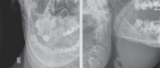
Hypo- and hypersalvation can be detected using sialometry (a method that allows you to measure the amount of secretion released per unit of time). X-ray and computed tomography are also highly effective.
As a treatment, the doctor prescribes the use of drugs that enhance salivation. In the case of an inflammatory process, antibacterial therapy is prescribed, as well as physiotherapeutic procedures.
The dentist first tries to mechanically squeeze out the stone by pressing on the duct. However, if such manipulation is not possible, surgical intervention may be required.
Diagnostic methods for bougienage of the salivary glands
- Sialometry. Allows you to conduct a quantitative and qualitative study of secreted unstimulated and stimulated saliva. With this technique, a special tube is inserted into the flow part.
- Radiosialography. The study is performed by filling the ducts with special substances. The saliva is then assessed for the presence of inflammation or an autoimmune disease. For contrast diagnostics, iodine-containing substances are often used.
- Pantomosialography. An innovative procedure that involves simultaneous contrasting of four salivary glands. With its help, it is possible to identify hidden pathological processes in the organ.
Additionally, X-ray diagnostics, thermosialography and other methods can be performed. It all depends on the symptoms of the disease and the characteristics of the inflammatory process in the oral cavity.
Causes
- The presence of inflammatory processes in the oral cavity
- Mechanical factors, for example, trauma to the ducts from sharp edges of teeth or crowns
- Stagnation of saliva secretion
Currently, there are four methods aimed at eliminating stones from the salivary glands.
1. Interventional sialendoscopy.
The doctor uses a special instrument with a camera at the end - an endoscope - to remove stones from the salivary ducts. This procedure is performed under local anesthesia.
2. Extracorporeal lithotripsy.
Under the influence of ultrasonic influence on stones, they are crushed.
Thanks to this method, it is possible to extract and wash the ducts with a special solution, which will prevent the development of the inflammatory process.
3. Dissection of the duct is done if the stone is large and cannot be removed using a simple method.
4. Extirpation of the salivary gland.
This operation is used only when irreversible changes are observed in the parenchyma of the gland. The doctor performs the procedure in a hospital setting under general anesthesia.
Common diseases of the salivary glands
- Narrowing of the flow channel. An inflammatory process or traumatic injury can lead to scarring of the tissue. As a therapy, a conical probe is inserted into the flow path. The number of sessions can range from 12 to 25 depending on the clinical picture.
- Chronic sialadenitis. In this case, the outlet flow channel does not work fully. This happens most often due to infection, the penetration of a pathogenic bacterial environment. Treatment involves drug therapy using antibiotics of the appropriate spectrum of action.
- Violation of the integrity of the duct. As a therapy, a probe is inserted into the cavity to prevent clogging of the duct and ensure normal patency.
These are the most well-known pathologies. There are other cases in clinical practice. Most often, a narrowing of the flow path with severe swelling is diagnosed. A full course of dilatation allows you to achieve the required therapeutic effect.
Treatment using bougienage of the salivary gland duct
The salivary glands can be susceptible to the following diseases:
- Stricture (narrowing) of the salivary gland duct - occurs due to tissue scarring due to frequently recurring inflammatory processes or traumatic damage to the gland. Bougienage is performed by inserting a thin conical probe (bougie) into the duct for 15 minutes. The next day, the procedure is repeated, slightly increasing the diameter of the inserted probe. The number of sessions can vary from 15 to 30.
- Sialadenitis is an inflammation of the salivary gland resulting from its infection. Causes functional disruption of the glands. It is treated with medication; instillation of antibiotics into the duct of the salivary gland is also prescribed, which is carried out after bougienage.
- Also, with the help of bougienage, the integrity of the parotid gland duct is restored. The procedure protects the end of the damaged duct from becoming overgrown. Bougienage should be carried out daily with the requirement that a larger diameter probe must enter the created cavity.
Prevention of diseases of the salivary glands includes, first of all, strengthening the immune system, timely detection and treatment of foci of infection, and proper level of oral hygiene.
Features of the procedure
If the flow channels are narrowed, appropriate therapy is prescribed using a probe to bougienage the salivary gland
. The procedure itself is as follows. First, an instrument is selected, a probe of the appropriate diameter, then it is inserted into the flow channel and left there for 12–16 minutes. In this case, the diameter of the probe increases each time. Thanks to this, it is possible to expand the duct as painlessly as possible. Before the procedure, the patient is advised to refrain from eating for 2.5 hours.
In order to consolidate the result, it is recommended to adhere to a special salivary diet; in addition, electrophoresis with potassium iodide and the required physiotherapeutic manipulations may be prescribed. The recovery period takes from several weeks to several months.
The specialists of the AlfaDent clinic will help solve any dental problem. Doctors use high-quality pharmaceuticals and advanced techniques. Timely detection of pathology and its treatment will help maintain the elements of the dental system and salivary glands in a healthy condition. We will help you get rid of any disease painlessly and with minimal time.
Sialolithiasis (salivary gland stones) - symptoms and treatment
Sialolithiasis, or salivary stone disease (Greek: sialon saliva + lithos stone) is a disease characterized by the formation of stones in the salivary gland and its chronic inflammation.
Normally, a person has three pairs of salivary glands: sublingual, submandibular and parotid. There are also small glands in the oral cavity, such as the palatine, buccal and lingual glands. Their task is to produce and secrete saliva. The submandibular gland most often undergoes the process of stone formation in the gland ducts and subsequent inflammation (89 - 95% of all cases), then in descending order is the parotid gland and extremely rarely the sublingual gland; sometimes stones can form in the minor salivary glands [3].
The disease is often detected at the age of 20-45 years - calculi (stones), which began to grow in childhood, by this age reach sizes that impede the flow of saliva and cause complaints. Sialolithiasis is more common in men than in women [11]. Salivary stone disease is rarely observed in children [1].
The formation of saliva is the initial stage of food digestion. Saliva contains enzymes, antibacterial substances and dissolved minerals. The exact reasons for the formation of stones in the salivary glands are unknown. Only factors that favor the development of pathology are identified:
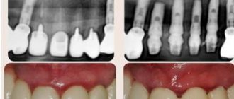
- Mechanical effects on the salivary glands in the area of the excretory duct , for example, due to injuries from teeth and crowns. A change in the lumen of the gland duct due to injury disrupts the outflow of saliva, which contributes to the formation of stones.
- Inflammation , in which microflora accumulates in the gland and pus appears. The disorder appears due to the presence of an inflammatory process in nearby tissues or the entry of pathogenic microorganisms into the gland duct. If the cause persists, then over time the stones increase due to deterioration in the outflow of saliva, metabolic disorders and exacerbation of the process. The sublingual and submandibular glands are susceptible to inflammation.
- Narrowings and kinks of the salivary glands and ducts.
- Impaired calcium metabolism due to bad habits, taking medications, poor quality drinking water, systemic diseases (for example, problems with the thyroid gland).
- Lack or complete absence of vitamins - most often a deficiency of vitamin A leads to the disease.
- Increased blood clotting.
- Entry of a foreign body into the gland duct. Bacteria actively multiply around it, forming a stone. This could be a solid fragment of food, a bone, a stone remaining in the grain, a fish scale, etc.
Mostly one stone is formed, but sometimes multiple stones are also found. It is extremely rare for several glands to be affected at the same time. The weight of stones can vary from fractions of a gram to several tens of grams. Stones come in various shapes: oblong, round or irregular; in their center there are often foreign bodies. Salivary stone mainly consists of inorganic salts - phosphates and calcium carbonates. With sialolithiasis, chronic inflammation and disruption of tissue nutrition are observed inside the gland tissues, which leads to the formation of connective tissue surrounding the lobules of the gland and its dilated ducts.
The development of sialolithiasis is also promoted by the use of anticholinergic drugs. Taking these medications inhibits the secretion of saliva, which leads to the accumulation of various food debris in the oral cavity. At the same time, the risk of them entering the gland duct increases and the number of bacteria in the oral cavity increases, which contributes to the formation of stones. These reasons increase with a decrease in water consumption, but studies have not been conducted to identify the relationship between these factors.
How to recognize?
“The formation of salivary gland stones can be asymptomatic for a long time, and the formations themselves can be detected during a standard routine examination. There is also the option that the patient himself will find them - he just needs to run his tongue or feel such stones,” says Ilya Antonov.
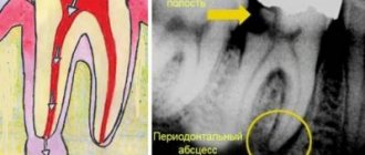
Next, swelling appears, notes dentist Antonov, sharp pain appears, dry mouth, a specific taste in the mouth develops, pain can spread to the neck area, and a headache may also occur. Facial asymmetry appears in the area of the salivary gland blocked by the stone. “If you skip this stage of the disease, then an abscess with pus will form, which can leak into the oral cavity. A cyst, malignant or benign tumor may also form,” warns Ilya Antonov.