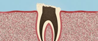
Tooth removal – represents the removal of the pulp (popularly the removal of a nerve). Pulp is a bundle of blood vessels, lymphatic vessels, nerve endings and loose connective tissue that fill the tooth cavity and nourish it from the inside. Thanks to the pulp, the process of blood supply and mineralization of the tooth occurs. When the pulp is removed, these processes will not occur.
On average, a tooth with a nerve “lives” longer than a pulpless one. The dentist’s task is to save the pulp, if possible, because removing the nerve is a necessary measure of dental treatment. As an option, the patient is offered a more loyal solution - to preserve the nerve in the tooth and seal it, and in case of pain, depulpate it. Depulping a tooth allows you to save the tooth, but has some negative consequences:
- removal of the pulp deprives the tooth of mineralization and proper blood supply, which shortens its “life”;
- Tooth enamel becomes more fragile and fades, and the strength of the tooth decreases.
Depending on the damage to the pulp, the dentist decides on its complete or partial amputation. There are cases when you have to deal with advanced inflammatory processes that lead to severe destruction, when the nerve cannot be saved - the nerve must be removed.
Some patients try to avoid the depulpation procedure, preferring to endure the pain and/or take painkillers. This should not be done! In such cases, pulpitis can become chronic without any special manifestations - asymptomatic. And due to provoking factors, a purulent abscess may occur, which threatens the development of periodontitis.
Why is pulp needed?
Pulp is a soft connective tissue that fills the internal cavity of the tooth. It ensures the supply of essential nutrients. The pulp contains many nerve endings, as well as blood and lymphatic vessels. This is where the perception of various sensations occurs. And pain too.
The pulp performs a plastic function, participating in the formation of dentin. And also protective, which consists in the disposal of dead cells, the formation of tertiary dentin and the synthesis of immunoglobulins. We are interested in its sensory function, which can be carried out thanks to a fairly large number of nerve fibers.
How is the nerve removed?
There are several ways to remove a nerve. Which one is needed in each specific case is chosen by the doctor in accordance with the clinical picture. The choice of treatment method depends on how damaged the tooth is and how deep the inflammatory process has penetrated.
Based on the results of the examination and examination, the dentist decides what kind of removal is needed: complete or partial. If the removal is partial, then the nerve is removed only in the crown part of the tooth, and the root is not affected. Partial removal of the nerve is used quite rarely; most often the dentist is forced to perform an operation to completely remove the nerve.
Pulpitis and causes of pain
Pulpitis is an inflammation of the internal soft tissues of the tooth, namely the pulp. The cause of inflammation is usually infection. It is expressed, in most cases, by acute throbbing pain and increased sensitivity.
The tooth cavity is initially closed and sterile. If its integrity is violated, penetration and spread of infection occurs.
Causes of pulpitis:
- Caries;
- Mechanical damage to the tooth, trauma. Violation of integrity may lead to infection;
- Low level dental care. Insufficient experience of the attending physician or poor-quality materials can cause inflammation in the soft tissues of the tooth;
- Unprofessional prosthetics, in particular incorrect grinding of the tooth;
- Retrograde infection in the case of osteomyelitis, sinusitis and other diseases;
- Inflammation of the gums;
- Blood poisoning.
In what cases are the nerve and pulp removed?
- Pulpitis and spread of carious destruction;
- Periodontitis;
- Several carious cavities at once or one, but large carious cavity;
- Mechanical trauma to the tooth (impact, chipped tooth, bleeding from the roots).
Of course, if there is an opportunity to keep the tooth “alive” and leave the nerves and pulp, the doctor will certainly take advantage of it. However, with pulpitis or severe damage, there is one difficulty: there are no 100% methods that would cure pulp inflammation. That is, if once it becomes inflamed, then with a high probability it will be an irreversible process. Of course, if you looked into a tooth with a microscope, you could accurately determine whether the pulp can be cured, or whether it is easier to remove it - but the problem is that you cannot look at a living patient under a microscope, and dentists are not pathologists after all. Therefore, to avoid risk, the pulp is almost always removed.
Also, in some cases, the pulp is removed before prosthetics.
- especially if the original, natural tooth has been severely damaged. In addition, when using metal-ceramic prosthetics, most of the tooth is still filed down, and during this process, the nerve can be damaged - so it is also easier and more reliable to remove.
How does pulpitis manifest?
As a rule, this is a sharp, throbbing pain, quite intense at night. Intensifies when exposed to high or low temperatures. As well as sour, salty or sweet foods. Pressure on the tooth also causes pain. There may be bad breath caused by actively reproducing bacteria. The pain usually spreads to neighboring teeth and even to the other jaw.
Sometimes soft tissue swelling occurs and the cheek swells. This is how flux manifests itself, which signals the occurrence of purulent processes.
Do not neglect visiting a doctor. After all, pulpitis is not just toothache. It can lead to many serious complications, ranging from tooth loss to blood poisoning.
How to independently determine pulp inflammation?
Sometimes, with pulpitis, it is difficult to determine the direct source of pain - that is, the most painful tooth - since the pain sometimes radiates throughout the entire jaw. Most often, a diseased tooth reacts to cold, so the easiest way to determine it yourself is to touch all teeth in turn with something cold. For example, a piece of ice from the refrigerator. Of course, this is the most artisanal method, and it is not suitable for normal diagnosis, but at a doctor’s appointment you can at least point out the tooth that definitely hurts more than the others.
This diagnosis includes an x-ray examination of “suspected” teeth. A high-quality orthopantomogram allows you to 100% accurately identify a pulpitic tooth: in such an X-ray image, the border of the tooth surface and the border of the inflamed pulp will be located very close to each other. There are other obvious signs indicating pulpitis that can be seen on such an image - a hidden carious cavity at the root of the tooth, enlargement or dilation of blood vessels, etc.
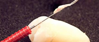
Surgical
Sometimes inflammation of the dental nerve requires depulpation. In this case, the inflamed pulp is removed, cleaned and then filled with special preparations. The result of the filling is checked using an x-ray. A temporary filling is installed. And after a few days, if no complications arise, a permanent filling is installed.
Modern medicine has various options for anesthesia. Therefore, this procedure will be comfortable and painless.
In some cases, when significant tooth decay has occurred, it will be necessary to remove it.
Contraindications for depulpation
When is it impossible to remove a dental nerve?
- stomatitis, inflammatory, purulent processes in the oral cavity;
- cardiovascular diseases;
- ARVI, sore throat, influenza;
- infectious hepatitis;
- hemorrhagic diathesis;
- acute leukemia;
- psychoemotional disorders;
- pregnancy.
Depulpation methods. How is the dental nerve removed?
Vital method
The vital depulpation method is used in adults and children - for all forms of pulpitis. This is a one-session procedure performed under local anesthesia. The dentist prepares the tooth, removes the pulp, cleans and fills the root canals, and then places a filling.
Devital method
The devital depulpation method is used to completely remove the coronal and root parts of the pulp. It differs from the vital method in the duration of treatment. An arsenic-free paste is applied to the pulp, after which a temporary filling is installed. The permanent filling is installed at your next visit to the dentist. In modern clinics, this method of treatment is used less and less.
Regardless of the method, the depulpation procedure is always carried out under anesthesia and the use of x-ray control of the tooth before and after depulpation. In 99% of cases, the vital method is used, in which complete, painless dental treatment can be completed in just one visit to the doctor.
From the history of depulpation or how were dental nerves removed before?
Depulping with arsenic is an outdated, quite painful and time-consuming method. The essence of depulpation with arsenic is that the dentist, using a dental drill, expands the root canal of the diseased tooth, opening direct access to the pulp. Then arsenic is placed in the canal and the tooth is covered with a temporary filling. Within a few days, arsenic affects the pulp, causing its death. A few days later, the patient visits the dentist again, who removes the temporary filling and removes the dead nerve from the canal. Then the dentist cleans the root canal and places a permanent filling. If the tooth continues to hurt after removing the nerve, the nerve may not have been completely removed. If pain after depulpation is caused by improper treatment and/or poor-quality canal treatment, the patient needs to visit the dentist again.
One of the main disadvantages of depulping with arsenic is its unsafety. With prolonged contact with a tooth, arsenic can lead to its complete destruction, because Arsenic is a poison, so arsenic paste was applied for a short period of time.
In modern dentistry, preference is given to safer methods of pulp removal.
Features of depulpation
Features of depulpation of baby teeth
It happens that pulpitis develops on baby teeth in children. In children, the depulpation procedure has its own characteristics:
- The minimum dosage of anesthesia is safe for the child;
- Careful opening of the crown;
- The most careful removal of the nerve ending, so as not to damage the primary processes of the molars;
- Thorough disinfection after depulpation to avoid re-inflammation;
- A filling for a baby tooth (based on mummifying compounds) – gives a disinfecting effect.
Depulpation of wisdom teeth
The difficulty of removing nerves from wisdom teeth lies in the inaccessibility of the teeth and the complexity of the structure of their canals. Therefore, if inflammatory processes occur in the pulp of wisdom teeth, it is better to remove the wisdom teeth.
Possible complications of depulpation
Complications that may arise after depulpation appear for 2 reasons:
- Anatomical features of dental canals and pulp.
- Incorrectly performed depulpation procedure.
Anatomical features of dental canals and pulp
Often the dental canals are very curved, in such cases access to the pulp can be difficult and it is difficult for the dentist to reach the nerve. In this regard, the pulp may not be completely removed, or cavities may remain in the canals.
The use of X-rays allows us to study the shape of the canals before removing the nerve and select the most appropriate instruments, which minimizes the likelihood of the above-mentioned treatment complications.
Incorrectly performed depulpation procedure
- Careless cleaning of the canals will preserve and intensify the inflammatory process due to incomplete removal of damaged tissue.
- Using an inappropriate pulp extractor channel size or improper handling of the instrument.
- Broken dental instrument, retention of its remains in the dental canal (this error may lead to the need to remove the tooth).
- Residual pulpitis occurs against the background of incompletely removed pulp.
- Perforation of root walls;
- Allergic reaction to filling material.
Poor disinfection of the canal when removing the nerve can trigger the process of suppuration. Which, in the absence of adequate treatment, can develop into a periodontal abscess. Such a complication leads to the need for tooth extraction.
Other possible complications:
Painful sensations for several days after depulpation - the duration is individual for everyone. If pain persists for a long time, it is necessary to consult a doctor to re-open the canals and carry out disinfection.
Specific problems can arise if the material is applied incorrectly: if the filling extends beyond the boundaries of the root apex, the jaw nerve may be pinched.
Recommendations
Recommendations after depulpation:
- refuse, for at least 2 hours, increased physical activity;
- rinse your mouth with antiseptics prescribed by your doctor during the day;
- hold off on eating for the first 3 hours (drink boiled, warm water through a straw);
- give up smoking and alcohol for five days, also do not take (solid, hot/cold, spicy) foods that cause irritation of the mucous membrane;
- It is forbidden to drink cold drinks, ice cream, or apply ice to the affected area.
Help at home
Acute toothache requires immediate contact with a dental clinic. And home treatments are in no way a substitute for visiting a doctor. They should be used only if the patient is outside the city and there are no medical facilities nearby. Or at night, when you need to wait several hours before seeing a doctor.
First of all, the patient should be offered anti-inflammatory drugs. They also have analgesic and antipyretic effects. Among them are Solpadeine, Nimesulide, Paracetamol, Ibuprofen and their analogues.
Traditional methods of treatment include rinsing with soda solution or chamomile infusion. They have an anti-inflammatory and antiseptic effect and will help slightly relieve pain.
It is worth remembering that these methods have a temporary effect. They are not curative and do not eliminate the cause of the disease. Therefore, you should not delay contacting a specialist. This will help avoid serious complications and tooth loss.
Removing the nerves of the front teeth: what to pay attention to
A beautiful snow-white smile always attracts attention and encourages communication. That is why during preventive examinations you should pay special attention to the health of the front teeth, which are located on the so-called. "smile lines"
Removing the nerves of the front teeth is not a technically difficult procedure; it is comparable in complexity to similar operations on other teeth. In this case, it should be taken into account that a pulpless tooth becomes a “dead” tooth - without receiving the necessary elements, it darkens and becomes fragile over time. Naturally, in this case it will be necessary to restore the beauty of the dentition over time.
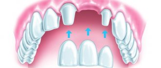
Aesthetic dentistry at the Eurodent clinic offers several options for restoring the appearance of teeth:
- Teeth whitening (including pulpless teeth)
- Installation of veneers (lumineers, ultra-veneers);
- Artistic restoration of teeth;
- Installation of crowns (metal-ceramics, zirconium oxide).
In order to avoid the unpleasant consequences of removing the nerves of the front teeth, you should undergo regular preventive examinations by the dentist. The specialist will be able to notice “problems” before the appearance of alarming symptoms and eliminate them without any problems.
In order to find out how much it costs to remove a nerve in a tooth , you can come for a free consultation at the Eurodent clinic.
With us you can undergo high-quality diagnostics and receive qualified dental care. Based on: 19 votes
What diseases cause inflammation of dental canals?
Root canals can become inflamed due to the development of the following diseases:
- caries;
- pulpitis;
- periodontitis.
An accurate diagnosis of inflammation of the pulp and canals of the tooth can only be determined by a dentist after X-ray diagnostics and a visual examination of the oral cavity.
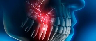
Traumatic neuropathy. Etiology.
Traumatic neuropathy most often occurs in the case of surgical interventions on teeth (traumatic extraction of teeth, removal of filling material beyond the apex of the tooth root, anesthesia with injury to the nerve trunks, removal of bone or tumor of the jaws), as well as in cases of surgical interventions on the paranasal sinuses and infraorbital canal.
Damage to the first branch of the trigeminal nerve, as a rule, is practically not observed. Most often, the third branch of the trigeminal nerve is affected, which is apparently due to the anatomical location of the inferior alveolar nerve, which makes it easily accessible during a variety of traumatic dental procedures. This is especially true for dental interventions on third molars. The cause of traumatic neuropathy of the inferior alveolar nerve can also be filling the mental canal during the treatment of pulpitis of the 4th and 5th teeth of the lower jaw.
Combined damage to the I and II branches of the trigeminal nerve can occur after inflammatory diseases of the brain with the development of adhesions or in the case of sinusitis, when the maxillary and frontal sinuses are simultaneously involved in the inflammatory process.
Clinic.
Patients complain of constant aching, sometimes pulsating pain in the area of innervation of the injured nerve, a feeling of numbness and “crawling goosebumps”. In case of injury to the mandibular nerve, tooth alignment occurs, which is associated with damage to the motor part of the nerve; patients cannot eat or talk. Trigger zones on the face and in the oral cavity are not identified.
During an objective examination, hypoesthesia or anesthesia (hyperpathy is also possible) of the skin and mucous membrane in the area of nerve innervation is detected. On palpation, pain is noted at the exit points of the II and III branches of the trigeminal nerve, as well as in the case of vertical percussion of the teeth and deep palpation of the lower jaw.
Diagnostics.
The main diagnostic criterion is the occurrence of pain after interventions on the dental system. The disease is characterized by clinical polymorphism and significant duration. During weather changes, stressful situations and in the presence of somatic diseases, exacerbation of the pain syndrome may occur.
In the event of scar changes in the nerves or retraction of the nerve into the scar of soft tissues (after gunshot wounds, in the case of defects of soft and bone tissues after resection of the jaws), constant aching pain of unexpressed intensity with persistent sensory disturbances is observed.
Signs of pathology
For many, the concept of an exposed nerve in a tooth is inextricably linked with severe pain. Sometimes the pain is simply unbearable, and it also radiates to the jaw and temples, ears, cheekbones and eyes. Very often, severe discomfort occurs at night and is long-lasting. Pain occurs not only from all sorts of irritating factors (for example, eating cold and hard foods), but also arbitrarily.
However, if the disease, most often pulpitis, becomes chronic, then the unpleasant sensations seem quite tolerable, the pain becomes muted, and at times it does not bother you at all. That is, all symptoms disappear.
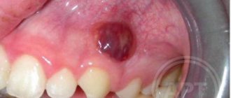
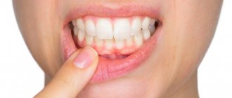
If the nerve in the tooth is exposed, what other symptoms, besides acute pain, can be noticed. As a rule, in the causative unit of the row there is a large carious cavity where pieces of food can get stuck. Near the affected element, the gums often swell, turn red and become painful. A small swollen pimple or ulcer may appear on the mucous membrane, from which pus is released.
Neuropathy of the buccal nerve.
The causes of the disease can be periostitis, inflammatory diseases of the teeth and gums, traumatic removal of teeth in the lower jaw.
Clinic.
The pain occurs subacutely, is constant, and its intensity gradually increases. First it occurs on the front surface of the gum, transitional fold, and then spreads to the entire front surface of the teeth of the lower jaw and covers the entire area of innervation of the buccal nerve. Numbness is uncharacteristic; an objective examination reveals a decrease in all types of sensitivity in the area of innervation of the mucous membrane of the cheek and vestibular surface of the gums, as well as the skin of the corner of the mouth.
Causes of pathology
Could the most sensitive part of the tooth be exposed? Yes, access to it opens when hard tissues are destroyed, which most often happens due to untreated caries. First, bacteria and their metabolic products corrode the enamel-dentin layer, forming a large cavity. And then they penetrate into the pulp through the dentinal tubules[1], causing inflammation and provoking pulpitis, periodontitis and other more severe complications, for example, periostitis, abscess. Sometimes the pulp is exposed under the influence of directed mechanical force, injuries and blows received in the area of the maxillofacial apparatus.
On a note! Some complain about exposed dental nerves and are interested in how to relieve pain that occurs from cold, sour, sweet and hot foods, or mechanical stress. This pain is of a sharp, short-term nature. In fact, in this case, we are most often not talking about caries and pulpitis, but about such a phenomenon as enamel hyperesthesia.
Root canal treatment
The treatment plan for dental canals consists of several stages:
- First, access to the problem area is freed: using a special dental instrument, the filling or the area of the crown damaged by caries is removed.
- Then the contents of the pulp are removed, and the canals are cleaned mechanically using antiseptic drugs.
- After this, the root is prepared for filling. At this stage, the dentist can form the correct conical shape of the canal passage.
- Then the canals are carefully sealed. If baby teeth are treated, the dentist uses a special filling paste, which gradually dissolves as the root dissolves.
- After this, a filling is placed on the crown.
This treatment regimen is standard and does not depend on exactly how many canals there are in the diseased tooth. The main thing is that all dental canals are cleaned, treated with an antiseptic and carefully closed. If treated incorrectly, it may be necessary to remove the tooth and visit an oral surgeon.
Teeth can be single-channel, two-channel, three-channel and even eight-channel. If one of the ducts becomes inflamed, it is necessary to clean and seal not only it, but also all other canals, since the infection could penetrate into them.
How are permanent teeth different from molars?
The most obvious difference is the number of teeth. A person has 28-30 molars, but only 20 primary teeth. There is a difference in the size of the crowns and even the roots of the teeth. According to dentists, baby teeth are smaller than molars. This is explained by the fact that children have a much smaller jaw than any adult. Consequently, it is not able to accommodate teeth the size of molars.
The thickness of dentin and enamel also differs significantly. In baby teeth they are much thinner. But if the dentin and enamel of primary teeth are inferior in thickness to molars, then in the case of pulp volume, it is the non-permanent teeth that take precedence. An interesting fact is that it is not necessary to visit a dentist to remove a baby tooth. But if we are talking about removing a molar incisor, you cannot do without the help of a qualified dentist. After all, this process takes much longer and is characterized by pronounced pain, increased body temperature, and slight bleeding from the gums.