- home
- Dental news
- Teeth filing
|
|
A unique teeth filing procedure has been used to create a perfect smile since 200 years ago. At that time, such a unique treatment was used to prevent caries by completely removing damaged hard tissues. Now the technology is actively used in dentistry to eliminate aesthetic defects and straighten teeth.
Filing is recommended for patients whose one or more teeth differ in size or stand out from the general row. A popular indication for the procedure is the presence of small chips on the fangs or incisors.
What is an impacted tooth?
Retention is an abnormality in tooth eruption, in which the tooth erupts partially or remains completely in the tissues of the jaw.
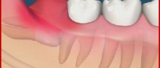
An impacted tooth is a tooth that has failed to erupt and “sleeps” in the gum or even in the jaw bone. There are cases when only part of the tooth crown erupts and is visible, and the rest of the tooth is covered by the gum - such teeth are called semi-impacted.
Most often, impacted canines and wisdom teeth are found - the notorious eights, but there are cases of impaction of the central and lateral incisors, and the first - a little more often.
What are the types of tooth impaction?
There are two types of retention - partial, in which part of the tooth is visible from the gums, and complete - in which the tooth is completely located under the gum tissue and/or jaw bone.
Such teeth can also be positioned differently, both being in “their” place in the dentition and occupying an incorrect position outside the dentition.
- Vertical
- Horizontally.
- At an angle to the jaw
If a tooth does not erupt in the right place beyond the border of the dentition, they speak of tooth dystopia.
There are even reverse retentions, when the tooth lies “upside down,” that is, upside down with its roots.
There may be only one impacted tooth in the dentition, or two impacted teeth may be located symmetrically. This problem concerns both milk and permanent teeth.
Veneers without grinding – is this possible?
The need to prepare teeth before veneering depends to a greater extent on the indications and to a lesser extent on the type of restoration. Lumineers and their Russian analogue ultraneers have a minimum thickness. Therefore, there is no need for grinding before fixing them to the tooth. However, if the teeth are large and require correction of shape and size, preparation is indispensable. Thus, veneers without grinding can be placed for the following indications:
- Need for minor tooth discoloration.
- Elimination of gaps (diastemas) between the front teeth.
- Masking cracks in tooth enamel.
- The need to restore a chipped cutting edge of a front tooth.
The need to correct the shape of the teeth, add volume to them, as well as visually change the angle of their location or align the cutting edge will in any case require preliminary preparation.
What complications do impacted teeth cause and why do they need to be “extracted”?
It is difficult for the patient to immediately understand what danger “hidden” teeth can pose, but it is really serious.
Semi-retinated teeth cause inflammation of the “hood” with which they are covered and adjacent tissues, and this provokes purulent inflammation.
Fully impacted teeth that lie in the jaw can behave like a foreign body; they put pressure on neighboring teeth, causing their displacement and a number of other problems.
In this case, the patient experiences pain and fever in case of inflammation, a feeling of pressure and discomfort in the jaw, neuralgia or numbness of part of the face.
If we list the diseases that can result from impacted teeth, it will become clear that this is a real “time bomb” that can detonate at any moment.
What diseases can be caused by an impacted tooth:
- periodontal cyst,
- caries of neighboring teeth and their roots,
- pulpitis,
- pericoronitis,
- periostitis,
- periodontitis,
- purulent lymphadenitis,
- inflammation of the trigeminal nerve,
- odontogenic sinusitis,
- abscess,
- phlegmon,
- resorption (resorption) of the roots of adjacent teeth,
- bad habits: mouth breathing, infantile swallowing, inserting the tongue into a dental defect,
- problems with biting food,
- malocclusion, displacement of interdental contacts, crowding of teeth.
Stages of treatment: how to prepare, install a crown on a tooth
Below we will analyze in detail each of the stages of prosthetics with crowns, and if you want to get advice directly from a specialist, call and make an appointment with the orthopedic doctors of our dental clinic in St. Petersburg - “Unident”. An initial consultation with an orthopedist at our clinic is provided free of charge!
Inspection and diagnostics
The treatment process begins with a visit to the office of an orthopedic dentist.
The specialist will carefully examine the patient’s oral cavity, identify existing problems, and, if necessary, prescribe a number of additional diagnostic measures. Based on the information collected from the clinical case, the orthopedist will draw up a detailed treatment plan and also offer the patient all possible prosthetic options. A well-designed treatment plan necessarily takes into account not only the installation of a dental crown, but also additional procedures:
- Removal of teeth, restoration and treatment of which is impossible for objective reasons;
- Preliminary treatment of diagnosed diseases of teeth and gums;
- Treatment of tooth canals and their filling (carried out when deciding to depulp a tooth before installing a crown);
- Professional oral hygiene. Sanitation of the oral cavity before prosthetics must be carried out to completely remove hard and soft plaque from the surface of the teeth. The event will allow you to most accurately select the crown to match the color of the patient’s natural tooth enamel, and will also have a positive impact on the quality of treatment.
The preparation of a treatment plan is completed by selecting a specific type of dental crown, after which the total cost of prosthetics is calculated and the timing of its implementation is specified.
Preparatory stage
The duration of the preparatory stage depends on what procedures will need to be carried out before prosthetics to ensure a high-quality and durable result. Below we will consider in detail all the possible stages of preparation for dental prosthetics with crowns.
Depulpation
A few years ago, before installing crowns, teeth were depulped without fail.
But modern orthopedists, if the clinical case allows, strive to keep teeth viable and do without removing the nerve. A tooth with preserved pulp is not deprived of adequate nutrition with minerals and nutrients, and does not acquire increased fragility. Crowns installed on living teeth last longer, but unfortunately, it is not always possible to refuse pulp removal. Most often, the front teeth are depulped, but the chewing dental units are kept alive. They have a fairly large area of the natural crown, in them the distance from the enamel to the pulp chamber is much greater, and therefore the risk of nerve bundle burns when installing a crown will be minimal.
The decision on the advisability of preliminary pulp removal is also made based on the number of roots, which may vary for different teeth. If you plan to install a crown on a single-rooted tooth, the depulpation procedure is mandatory. When treating single-rooted teeth, the risk of pulp burn is too high, and thermal damage to the sensitive nerve bundle is fraught with the occurrence of an inflammatory process under the installed crown. To prevent such negative consequences, single-rooted teeth are depulped, the canal cavities are treated with antiseptic agents and filled with gutta-percha.
Treatment of diseases of teeth and gums
If, during a visual examination of the oral cavity, the doctor discovers caries, signs of the development of an inflammatory process - these conditions must be eliminated before installing crowns.
If the tooth is significantly destroyed by caries, the doctor removes the affected tissue and restores the crown part by installing a durable filling. Pulpitis and periodontitis are treated according to a more complex scheme, which includes treatment of dental canals and filling them. A high-quality and durable filling must be placed on the tooth, otherwise it may fall out, and the installed crown will fall out along with it. In case of severe destruction of a dental unit, its coronal part can be restored in different ways:
- Installing a seal on the pin. A pin is a special structure in the form of a rod, which is inserted into the root canal of the tooth and acts as a reliable support for the filling;
- Stump tab. Inlays for dental restoration are made in a dental laboratory and are installed, like a pin in a canal, but are also fixed in the crown of the tooth.
If a tooth is severely damaged, it is recommended to restore it with an inlay, which is considered more reliable and durable than a conventional filling or installation of a filling on a pin.
Preparatory activities are completed with professional oral hygiene. Sanitation is necessary for the complete and high-quality elimination of dental plaque, which is a favorable environment for the proliferation of pathogenic organisms, in addition, the procedure will allow you to accurately select the color of the crown for installation so that it matches as closely as possible the shade of the natural enamel of the patient’s teeth.
Preparation
Preparation or grinding of teeth is a procedure that the doctor performs before taking impressions.
During the preparation, the tooth enamel will be ground down to the thickness of the future crown. During the grinding process, a drill is used: with its help, the tooth is given a certain shape that can provide the most durable fixation to the installed crown. If living, non-pulp teeth are subjected to preparation, the procedure can be very unpleasant and painful for the patient, and therefore local anesthesia is used before it is performed. The thickness of the enamel removal will depend on the type of crown that will be subsequently placed on the prepared tooth. A maximum of natural tissue is removed from the tooth to install ceramic and metal-ceramic crowns; the enamel is ground down to a minimum before prosthetics with cast crowns.
After the tooth has been prepared, a stump remains, from which the doctor will take impressions to produce crowns.
Laboratory stage
An impression is taken from the ground teeth. To obtain a high-quality, anatomically accurate impression of teeth, a specialized highly elastic mass is used to produce a crown. Using impressions, a dental technician in the laboratory will make a plaster model of the patient’s teeth, which will be used to make crowns for further installation. Dental crowns can be made from a variety of materials - alloys of precious and non-precious metals, ceramics, zirconium dioxide, metal ceramics. The type of crowns for installation is selected based on the characteristics of a specific clinical situation, the patient’s preferences, his wishes for the level of aesthetics, as well as financial capabilities.
The timing of its production will depend on the type of raw material from which the crown will be made. The company has been producing crowns made of ceramics and metal-ceramics for a long time. So that the patient does not have to walk with an unsightly stump during the entire period of crown production, a temporary immediate prosthesis is placed on it for this period. Temporary crowns are made of plastic, and their installation allows you to relieve the patient from psychological discomfort, as well as protect prepared and vulnerable teeth from the action of pathogenic microflora and harmful environmental factors.
Fitting and installation of dental crowns
When the dental technician makes the crown frame, the patient will be invited to the clinic for fitting.
During fitting of the frame part of the crown, the tightness of its fit to the surface of the tooth stump is assessed. If the fit is insufficient, food debris and bacteria will get under the installed crown, and all this can lead to the onset of an inflammatory process. If during fitting the crown fits perfectly on the stump, the frame is sent to the laboratory, where it will be covered with ceramic mass. The finished crown is tried on a second time and during the second fitting it will be fixed with temporary cement mortar. A second check is required to assess the quality of contact between the prosthesis and the opposing teeth, and if it goes well, the crown will be installed on the tooth using permanent cement. The process of permanently installing a crown on a tooth takes place according to the scheme described below:
- The inside of the crown is covered with a layer of cementing solution, after which the crown is put on the tooth;
- A tooth with a crown is exposed to light, which accelerates the hardening process of the cement mortar;
- All excess cement mixture is carefully removed, since it is a rather caustic substance and, if it comes into contact with soft tissue, can cause severe burns.
The treatment ends with a detailed consultation from the orthodontist on the care of the prosthesis and oral cavity after prosthetics. Compliance with the specialist’s recommendations guarantees the absence of negative side reactions and complications after crown installation.
How are impacted teeth diagnosed?
Impacted teeth are often “found” by accident during an X-ray diagnosis for another dental reason. If a patient asks about orthodontic treatment and is missing some teeth, or has symptoms that may be caused precisely by the presence of hidden impacted teeth, then a special diagnosis is carried out.
The problem of impacted teeth is solved by two specialized dentists - an orthodontist and a surgeon, so the patient will have to undergo diagnostics by an orthodontist and diagnostics by a surgeon. Most of the diagnostic procedures will be general, and the key is x-ray diagnostics, and it is preferable to do CT - computed tomography (3D diagnostics). Unlike an orthopantomogram (OPTG), it shows the spatial location of the impacted tooth in the jaw and relative to other teeth, which is very important for the surgical stage of traction - exposing its crown, because allows the surgeon to choose the optimal “approach”.
What to do if an impacted tooth is found?
There are 3 solutions to the problem of an impacted tooth:
- Orthodontic traction and placement in the dentition. This is often done with impacted teeth that are located in the smile zone - canines and incisors, provided that there is a place for them in the dentition and the tooth itself is healthy.
- Removal is a common fate for impacted eights, especially if the patient is undergoing orthodontic treatment. Very often, wisdom teeth erupt partially – this provokes inflammation and complications. Even a completely impacted (“hidden”) tooth serves as a potential source of caries, tooth displacement, and instability of orthodontic treatment. There are many reasons - there is only one verdict: it is better to delete it. Other indications for the removal of different types of impacted teeth are cysts, signs of inflammation, lack of space in the dentition, destruction of the neck of the tooth, caries.
- Preservation and surveillance. It is justified when the patient has no problems with the place in the dentition or bite, orthodontic treatment is not required, the tooth does not provoke complications and its absence does not spoil the aesthetics of the smile. Such teeth are observed; in case of the slightest problem, they are recommended to be removed.
Veneers with minimal grinding of teeth
The thinner the veneer, the less you need to grind off the enamel before installing a microprosthesis. The table below will help you compare the characteristics of different types of structures and choose the best option with the least grinding of teeth and optimal cost.
Composite veneers
Manufacturing type:
Formed immediately in the patient’s oral cavity
Thickness:
0.3 - 0.6 mm
Turning:
No
Price in Moscow:
from 5,000 rub.
- Speed of production.
- Installation without grinding teeth.
- Acceptable price.
- Short service life.
- Variability of color.
- Fragility.
- Insufficient aesthetics.
- Poor ability to hide dental defects.
Ceramic veneers
Manufacturing type:
Produced in a dental laboratory
Thickness:
0.5 mm
Turning:
Yes
Price in Moscow:
from 14,000 rub.
- Aesthetics.
- Reliability.
- Color constancy.
- Durability.
- High price.
- Irreversibility of turning a tooth under a ceramic veneer.
- Long production time.
Zirconium veneers
Manufacturing type:
Produced in a dental laboratory
Thickness:
0.5 - 0.7 mm
Turning:
Yes
Price in Moscow:
from 17,000 rub.
- Aesthetics.
- Durability.
- Strength.
- High price.
How to pull out and place an impacted tooth in the dentition?
In some cases, if the root of an impacted tooth has not yet formed and there is an obstacle to its eruption, then removing such an obstacle is enough for the tooth to come out of the gums and take its place in the dentition. If the tooth root is already formed, this method will not help.
To “get” and move a fully formed impacted tooth into the dentition, treatment is carried out in 3 stages. Here we present the “classical” scheme, because in individual cases there may be deviations from this scheme.
- Preparatory orthodontic stage - you need to prepare a place in the dentition to which the orthodontist will move the extracted tooth. To do this, a braces system is installed, which aligns the teeth in the dentition and frees up the necessary space.
- The surgical stage is the image of the crown of an impacted tooth, on which a bracket or button is installed to transmit force from an arch or elastic element.
- The main orthodontic stage, during which the impacted tooth is pulled out and placed in its “rightful” place in the dentition.
How is filing of front teeth performed?
The technique of filing teeth varies depending on the condition of the oral cavity, the current diagnosis and treatment goals. Teeth filing begins with a detailed diagnosis. The specialist evaluates the feasibility of the procedure and identifies the teeth that need correction. For an accurate diagnosis, an examination is carried out, the causes of the disease are studied, and a treatment method is determined. The procedure consists of simple steps:
- Determination of the working area. For this, wax plates or special aerosols are used. The use of additional means allows you to mechanically highlight or paint an area that stands out from the general row and prevents the normal closure of teeth. In particularly advanced situations, it will be necessary to create a plaster model of the client’s jaw;
- Anesthesia if a large layer of hard tissue is to be removed;
- Direct filing with a diamond bur. On chewing teeth, the enamel is removed in the areas of protruding cusps. In the case of incisor processing, multidirectional movements are used to create the correct tooth shape;
- Grinding the surface, using products to replenish the deficiency of enamel minerals, as well as drugs to reduce sensitivity and strengthen dental tissue.
After filing, the thickness of the enamel changes, so sensitivity often increases. This phenomenon is temporary and for some time after treatment it is better to refrain from eating too hot or cold food. If the problem does not go away, it is better to consult a dentist.
How is surgery performed to expose the crown of an impacted tooth?
- As a preliminary stage, preparation for the operation is carried out. The patient undergoes professional hygiene and sanitation to reduce the amount of infection in the oral cavity and speed up postoperative healing.
- The operation to expose an impacted tooth is performed under local infiltration anesthesia and is considered a fairly serious surgical intervention.
The operation to expose the crown of an impacted tooth and install an orthodontic element on it can be carried out according to 2 schemes:
I Delayed bracket installation.
- The mucous membrane in the projection of the crown of the impacted tooth is excised, the entire crown of the tooth is exposed, and a special tampon is installed in the wound.
- After 2-3 days, a button or bracket is installed on the crown of the exposed tooth, which is tied to the orthodontic arch and traction begins.
II Bracket installation during surgery
- The dentist-surgeon peels off a small mucoperiosteal flap and exposes part of the crown of the impacted tooth, onto which the locking element is immediately fixed.
- The bracket is tied to an orthodontic arch or additional devices.
- After installing the orthodontic element, the flap of tissue is placed in place and the wound is sutured.
The disadvantage of this method is that in this case, repeated surgical intervention is possible if the bracket on the tooth comes off.
After surgery, the patient is prescribed antibiotics and antiseptic rinses, if necessary, to speed up healing. Light, non-traumatic food is recommended.
Who is recommended for teeth straightening?
Before filing a tooth, you should undergo a dental examination and consult with your doctor. You should not prescribe filing yourself or try to do it at home, as there are clear medical indications for performing the procedure:
- The upper incisors or canines are very different, therefore they spoil the appearance of the jaw and interfere with the formation of a correct bite. In this case, it is enough to shorten the clove so that it fits comfortably when closing the jaw;
- There are small chips on the edges of the teeth, and a little grinding of the enamel will smooth out any defects;
- Preparation for orthodontic correction. Often, the dentist will file the edges of the teeth to return all the elements of the jaw to the correct position. Manipulation will be required when installing braces if the dentition is very crowded;
- The teeth have sharp edges that look unsightly and damage the mucous surface of the cheek;
- Upcoming installation of veneers or crowns. The dentist files the surfaces of the teeth to form an even row.
The feasibility and effectiveness of the procedure is determined on a personal basis and only at an appointment with a dentist. Grinding the edges of teeth is not recommended for patients with the following conditions:
- High risk of injury caused by regular exercise or a specific type of professional activity.
- Increased sensitivity of teeth.
- High abrasion of enamel.


