1946
In dentistry, cements are a class of materials intended for fixation of orthodontic appliances and fixed dentures (fixing materials).
They are also used to create a barrier between the pulp and the filling (gaskets), filling and forming a tooth stump (restorative compounds).
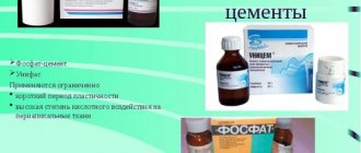
All cements are divided depending on their base - zinc phosphate, silicate, polycarboxylate, etc. Glass ionomer cements are considered the most modern and high-quality today.
Purpose and properties of the material
The brand of dental glass ionomer cements Fuji (Fuji) belongs to the company GC (Japan). Founded in 1921, GC is today a world-renowned manufacturer of dental products.
The company produces more than 600 types of materials, instruments and equipment for dental treatment. Due to their high quality, GC glass ionomer cements are in demand and are sold all over the world, including in Russia.
The basis of glass ionomer cements (GIC) is aluminofluorosilicate glass crushed to powder and polyacrylic acid.
The advantages of the material include high chemical adhesion to enamel and dentin, long-term release of fluoride, significant tensile and compressive strength, hypoallergenicity, and biocompatibility with human oral tissues.
One of the main disadvantages of GIC is its slow hardening. The initial hardness, which occurs 4-5 minutes after the start of mixing, is provided by calcium ions linking polyacrylic acid chains.
This process is unstable due to the solubility of calcium compounds in water or, on the contrary, dehydration. Final curing, provided by aluminum ions, occurs in a period of 2 weeks to 6 months.
Modification of glass ionomer cements with composites allows for faster curing due to photopolymerization of the composite, as well as giving the material greater hardness and aesthetics. Such cements are called “compomers” (a combination of the names “composite” and “ionomer”).
The GC company produces both unmodified glass ionomer cements (Fuji I (Fuji 1), Fuji II (Fuji 2), etc.), and modified (Fuji IX (Fuji 9), Fuji Plus Set, Fuji Plus-Powder, etc.) . The former are used mainly for fixing prosthetic structures, the latter – for dental restoration.
Provocateurs for the development of a cyst on the root of a tooth under the crown and methods for its treatment.
Come here to find out how much tooth depulpation costs before dentures.
This address https://www.vash-dentist.ru/protezirovanie/nesemnyie-p/koronki-np/kogda-trebuetsya-zamena-zubnyih.html is all about replacing dental crowns.
The use of glass ionomer cements from the GC Fuji line: theoretical and practical aspects
One of the latest trends in modern dentistry is working in accordance with the principles of the minimal intervention concept (MI Concept). The main objectives of this concept are control over caries disease, restoration of mineral balance and careful restoration of teeth, implying the preservation of as much dental tissue as possible, and not just “banal sealing of the defect” even with the most highly aesthetic material. It is impossible to cure a bacterial disease purely surgically, and secondary caries should be considered as a natural continuation of the disease [1, 5, 9].
Today, the highest priority is atraumatic dentistry, which meets the rules of minimal invasion and preservation of dental pulp. Organizing the work of a dentist according to these principles allows you to preserve the patient’s dental health for many years and reduce the risk of long-term complications.
Every year new restoration materials appear on the market. The most popular are composite materials in combination with adhesive systems due to their highly aesthetic properties and good strength characteristics in the field of direct occlusal loads. At the same time, composite materials are not perfect due to their inherent disadvantages [4, 8]:
- Polymerization shrinkage and stress.
- They do not have self-adhesion to tooth tissues.
- Lack of adhesion to tooth cement.
- Imposing high demands on cavity preparation (only a healthy surface of enamel and dentin is capable of forming a strong connection with the adhesive system).
- Lack of remineralizing effect on hard tooth tissues.
- Hydrophobic, labor intensive.
In view of this, there is a problem of existing complications (leakage of the filling, postoperative pain, irritation of the pulp, loss of tooth vitality, the presence of stresses in the tooth), and the durability of composite restorations remains far from ideal.
Glass ionomer cements (GIC) can be alternative materials in restorative therapy in a number of clinical situations. The range of currently produced GICs with improved and improved performance characteristics allows us to successfully solve a number of clinical problems that arise in the practice of a dentist. And, apparently, ignoring this group of restoration materials, giving preference only to composites, is incorrect, first of all, in relation to the patient’s dental health, even if we are talking about the fate of one tooth.
The main positive properties of GIC are [3, 6-8]:
- Physical and chemical affinity with hard dental tissues.
- Self-adhesion to tooth tissue (both intact and partially demineralized dentin; acid etching and absolute dryness of the surface are not required).
- Bioactivity, cariesstatic and antibacterial effect. GIC is the only material that solves the problems of treating demineralized dental tissues. Ion exchange adhesion ensures complete sealing of the cavity, the underlying demineralized layers will be isolated and will undergo remineralization, and the remaining bacteria will be cut off from receiving food. The cariesstatic effect is achieved largely due to the prolonged release of fluoride from the cement mass. This process begins immediately after filling and continues for at least one year. Diffusion of fluorine into surrounding tissues causes increased mineralization and promotes the formation of fluorapatites in enamel and dentin. This leads to increased acid resistance and decreased dentin permeability, worsening living conditions for pathogenic microorganisms and preventing the development of recurrent caries. The bacterial contamination of the surface of GIC fillings is significantly lower than that of composites and cements of other groups. GICs have a battery effect - they are capable of adsorbing fluoride ions from food and exogenous prophylaxis. When the environment surrounding the tooth becomes acidic (cariogenic situation), GIC releases fluoride into the adjacent tissues.
- The low modulus of elasticity provides high elasticity, which allows GIC to withstand occlusal loads under fillings and crowns, help compensate for polymerization shrinkage of composite materials, and also eliminate stresses that arise in the cervical region during microbending of the tooth during chewing.
- The coefficient of thermal expansion of GIC is close to the coefficient of thermal expansion of tooth tissue, which is important for ensuring long-term tightness at the filling/tooth tissue interface.
At the same time, “classical” (ordinary, traditional) GICs have a number of disadvantages (low strength characteristics, insufficient aesthetics, unsatisfactory performance characteristics, negative impact of mechanical and vibration factors in the process of “maturation” of the filling), limiting their clinical use and requiring the doctor to fulfillment of a number of conditions and technical techniques.
The advantages of modern (strengthened, packable) GIC are:
- Improved physical properties (high compressive strength, increased resistance to abrasion and cracking, wear resistance).
- The compactable viscosity of the cement mass allows it to easily condense into the cavity; the material does not stick to tools.
- Less sensitive to external influences during the “maturation” of the cement mass.
- Shortened curing time (up to 5 minutes); can be processed with abrasive tools within 3-7 minutes after application.
- The time for water loss after placing a filling (sensitivity to dehydration) has been reduced from 6 months to 2 weeks. The problem can be solved by using special varnishes.
- Good aesthetic characteristics.
GICs are the materials of choice in the following clinical situations [2, 3, 6, 11]:
- Indispensable for patients with low rates of caries resistance of hard dental tissues, with active and often recurrent caries, as an option for permanent and “delayed” filling.
- Poor oral hygiene (group of patients who are difficult to motivate).
- Subgingival carious lesions.
- Non-carious lesions of hard dental tissues (with this pathology, the structure of enamel and dentin changes, and adhesive systems of composite materials designed for the normal structure of these tissues may be ineffective).
- It is technically impossible to ensure complete isolation of the cavity from moisture.
- Treatment of children, adolescents, elderly patients.
- Low initial level of mineralization of hard dental tissues, treatment of initial caries, option of “transitional sealing” of immature fissures.
- As a necessary alternative to the adhesive technique in the sandwich technique.
The article presents the results of using various GICs from the existing Fuji GC line in a variety of clinical situations.
Using GIC with high fluoride release - GC Fuji Triage
This GIC can be used in conditions of high humidity and when it is impossible to control salivation; it has an increased level of fluoride release (6 times higher than other traditional GIC!). Its use is almost ideal in clinical situations requiring remineralizing treatment of dental hard tissues and stabilization of further carious destruction:
- In patients with multiple active carious lesions.
- In the technique of indirect pulpotherapy.
- To protect exposed areas of the tooth.
- In the treatment of hypersensitivity, erosions, caries in the spot stage.
Fluidity and insensitivity to moisture allow GC Fuji Triage to be used for sealing fissures of both temporary and permanent teeth with incomplete mineralization, even in conditions of incomplete eruption. The white shade of the material (Fuji Triage White) self-cures within 4 minutes, the use of a pink shade (Fuji Triage Pink - contains a light-absorbing dye) allows you to apply accelerated forced curing in 20-40 seconds, which is very convenient in children's practice and makes it easier visual inspection during repeated inspections.
CLINICAL CASE STUDY #1
Patient B., 6 years old. On examination: OHI-S=1.2; kpu+kpu=8 (subcompensated degree of caries). The fissures of the recently erupted tooth 4.6 are chalky. Immature fissures 4.6 were sealed using Fuji Triage (pink shade).
The effectiveness of using GC Fuji Triage as a sealant for primary and permanent molars for 1 year in 58 children aged 2 to 8 years using invasive and non-invasive sealing techniques was monitored. Repeated examinations were carried out after 6, 8, 12 months. When assessing the clinical effectiveness of the sealant, the safety of the material in the fissures, as well as the absence or presence of carious lesions, was taken into account. Analysis of the results showed that after 12 months, with the invasive method of sealing, complete preservation of the sealant was observed in 82.3±2.3% of cases, with the invasive method in 95.4±2.5% of cases. The appearance of carious lesions was not detected in any of the observed cases.
CLINICAL CASE STUDY #2
The parents of patient K., 7 years old, complained of a defect in the area of tooth 1.1, with which the daughter’s tooth had already erupted. On examination: OHI-S=0.8; kpu+kpu=10 (decompensated degree of caries). The use of GC Fuji Triage is dictated by the following anamnestic and clinical circumstances:
- Immaturity of hard dental tissues 1.1.
- Aplasia of enamel in the middle sector of the tooth crown.
- Hypocalcemia, hip dysplasia.
Fuji Triage White was used to seal the defect. The patient is under clinical observation, each erupting tooth is monitored (Fig. 1).
Rice. 1a. Clinical example No. 2: appearance of tooth 1.1 upon presentation. Rice. 1b. Clinical example No. 2: tooth 1.1 after filling with GC Fuji Triage (white shade).
Use of light-curing hybrid restorative cement - GC Fuji II LC Improved
This cement is multifunctional - it can be used for aesthetic restorations (mainly class III and V), as a base for sandwich technology, and also for restoring tooth cores. It has improved mechanical properties (a large amount of filler increases abrasion resistance), good aesthetics and polishability (smaller glass particle sizes; 11 shades on the Vita scale), as well as sufficient working time (3'45), which makes it possible to place several fillings at once.
CLINICAL CASE STUDY #3
The parents of patient M., 2 years old, complained of the destruction of the anterior teeth of the upper jaw in their son. On examination: the child is conditionally contactable; PLI=1; kpu=4. Diagnosis: early childhood caries; Dentin caries 5.1, 5.2, 6.1, 6.2. The capsule form used is GC Fuji II LC Improved, shade A2.
Due to his early age, it was necessary to work at a fast pace and restore all four teeth in one visit. All restoration work was completed 10 minutes after preparation of carious areas (damaged tissue was excavated, enamel edges were smoothed using a drill). The material was added in 2 stages, 2 capsules of cement were used (Fig. 2).
Rice. 2a. Clinical example No. 3: view of the anterior teeth of the upper jaw of patient M., 2 years old, upon presentation. Rice. 2b. Clinical example No. 3: b - view of the teeth after preparation. Rice. 2c. Clinical example No. 3: c — view of teeth after filling with GC Fuji II LC Improved.
CLINICAL CASE NO. 4
Patient A., 26 years old, complained of aesthetic defects in the area of the mandibular incisors. On examination: OHI-S=2.4; CPU=11. In the cervical area of teeth 3.1, 3.2, 3.3, 4.1, 4.2, carious cavities within the dentin were revealed. The enamel surrounding the defects shows signs of demineralization. After a series of motivational measures and removal of dental plaque, carious cavities were prepared. The restorations were made with GC Fuji II LC Improved, shade A3 (Fig. 3).
Rice. 3a. Clinical example No. 4: type of teeth 3.1, 3.2, 3.3, 4.1, 4.2 at presentation. Rice. 3b. Clinical example No. 4: immediately after filling with GC Fuji II LC Improved. Rice. 3c. Clinical example No. 4: 2 days after filling.
Use of self-curing reinforced GIC for esthetic restorations - GC Fuji VIII GP
Due to the content of specific resins, the material has sufficient light transmission for use in the area of the anterior group of teeth and is economical in use.
CLINICAL CASE NO. 5
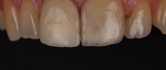
Patient M., 22 years old, came in on the insistent recommendation of his mother with complaints about the unsatisfactory appearance of the anterior teeth of the upper jaw. On examination: OHI-S=3.2; CPU=16. Viscous saliva. The lesions are located within the enamel and dentin, covering from 30 to 100% of the area of the vestibular surface 1.1, 1.2, 1.3, 2.1, 2.2, 2.3 with a greater depth of damage in the cervical region; filled with loose necrotic black-brown mass, easily removed with an excavator. The surrounding areas of enamel on the periphery are gray-white in color and lack shine. Abrasion of the cutting edges of the teeth.
To make a final diagnosis and determine the immediate causes of the development of this damage to the hard tissues of the teeth, anamnesis and dental health history were carefully collected. From the anamnesis: lesions of the anterior teeth of the upper jaw, the area of which increased over time, was noticed by the patient’s mother 4-5 years ago; did not go to the dentist; the teeth didn’t bother me; the patient has not been exposed to toxic or chemical substances during this period of time. According to the mother, it was found that from the age of 15, the son spends a significant part of his time, including night hours, in front of a computer screen. Currently, many hours of work on the computer continue to occur (about 10-12 hours a day).
Based on anamnestic and objective data, a diagnosis was made: computer necrosis of hard dental tissues in the area 1.1, 1.2, 1.3, 2.1, 2.2, 2.3 (according to ICD-10 - K03.81 - radiation (post-radiation) necrosis).
Necrotic tissue was removed down to dense dentin. The prepared cavities were medically treated with a 0.05% chlorhexidine solution and filled with GC Fuji VIII GP. An individual selection of oral hygiene products was carried out, calcium preparations and antioxidants were prescribed orally (Fig. 4).
Rice. 4a. Clinical example No. 5: view of the anterior group of teeth of the upper jaw upon presentation. Rice. 4b. Clinical example No. 5: teeth 1.3, 1.2, 1.1, 2.1, 2.2, 2.3 after preparation. Rice. 4c. Clinical example No. 5: teeth 2.1, 2.2, 2.3 immediately after filling with GC Fuji VIII GP. Rice. 4g. Clinical example No. 5: after G-Coat varnish. Rice. 4d. Clinical example No. 5: appearance of teeth a week after filling. Rice. 4e. Clinical example No. 5: appearance of teeth after a year. Rice. 4g. Clinical example No. 5: after polishing fillings.
The patient is registered at the dispensary. A follow-up visit was scheduled after 3 months. The patient did not show up at the appointed time. Active appearance on call took place after 10 months. There were no complaints. OHI-S=2.3. A slight brown staining was noted at the filling/tooth interface (due to the patient’s poor motivation, failure to maintain satisfactory oral hygiene, and the presence of a bad habit—smoking). Replacing glass ionomer fillings with composite ones is not indicated in this clinical case.
Ultra-strong, highly aesthetic glass ionomer restorative system for the chewing group of teeth - EQUIA
The EQUIA system includes EQUIA Fil, a next-generation glass ionomer material, and EQUIA Coat, a highly loaded polymer protective coating that takes glass ionomer technology to a whole new level.
The second component of the EQUIA Coat system is a nanofilled varnish, which not only prevents dehydration, but also protects the restoration from external influences, increases resistance to abrasion, and adds shine to the surface. EQUIA is recommended for permanent filling of load-bearing cavities of class I, as well as small cavities of class II [12], and is an alternative to amalgam and compomers (flexural strength 31.9 MPa; Vickers hardness 112 Hv).
CONCLUSION
- Composite materials and glass ionomer cements are complementary materials in restorative dentistry. The choice of material should be strictly based on clinical indications, taking into account the fact of the need for remineralizing treatment of dental tissues. The ultimate goal of restoration should be the restoration of the biological and functional usefulness of the tooth as an organ and the prevention of the spread of infection beyond the source. Using bioactive GIC in methods of “delayed” and permanent filling, as well as as a base “for composite material”, the doctor acts not just as a “reinsurer”, but in fact, based on an understanding of the biochemical mechanisms of resistance of hard dental tissues, performs this stage of therapy, which largely guarantees a favorable prognosis.
- The use of glass ionomer cements from the GC Fuji line, which have improved physical, biological and manipulation characteristics, is indispensable in a number of clinical situations encountered in the practice of a dentist, in full accordance with the concept of minimal intervention.
LITERATURE
- Artyushkevich A.S. Prevention and treatment of caries of varying degrees of activity / A.S. Artyushkevich, Z.R. Valeeva, G.V. Kuznetsova // Dental Journal. - 2007, No. 1. - P. 6-9.
- Aspects of the use of GIC in dental practice / I. K. Lutskaya [et al.]. - Mn.: BelCNMI, 2000. - P. 189-190.
- The use of glass ionomer cements in the treatment of dental caries in pediatric dentistry / V. P. Mikhailovskaya [et al.] // Dental Journal. - 2009, No. 4. - P. 304-308.
- Lobovkina L. A., Romanov A. M. Application of glass ionomers in therapeutic dentistry / L. A. Lobovkina, A. M. Romanov // Practitioner dentist. - 2011, No. 1. - P. 1-5.
- Mount G. Minimal intervention in dentistry. Classification of carious cavities / G. Mount // New in dentistry. - 2005, No. 2. - P. 73-77.
- Nikolaev A.I. Glass ionomer cements / A.I. Nikolaev, L.M. Tsepov, A.I. Bychkov // Institute of Dentistry. - 1999, No. 3. - P. 48-53.
- Clinical effectiveness of contemporary adhesives: A systematic review of current clinical trials / M. Peumans [et al.] // Dent Master. - 2005. - Vol. 21. - P. 864-881.
- Mount GJ Longevity in glass-ionomer restorations: review of a successful technique / GJ Mount // Quintessence Int. - 1997. - Vol. 28. - R. 643-650.
A complete list of references is in the editorial office.
ISO requirements
Requirements for dental cements are established by international standards ISO 9917-2-95 and 9917-91. The regulated parameters depend on the purpose of the cement. So compositions for filling dental canals should have:
- working time , max., min. – no more than 20;
- hardening time , hour – no more than 72;
- fluidity , min., mm – not less than 20;
- solubility ,% – no more than 3;
- film thickness , microns – no more than 50.
Restoration mixtures must meet the following requirements:
- compressive strength , MPa – not less than 130;
- erosion , mm per hour – no more than 0.05;
- hardening time , min – 2-6.
General requirements for dental cements include biocompatibility with the human body and tissues, uniform distribution of pigments, absence of foreign impurities, homogeneity of consistency and some others.
Cost of services
If treatment is started on the day of treatment, consultation and treatment plan are free
| Treatment of superficial caries with the installation of a light-curing filling | RUB 3,800 |
| Treatment of medium caries with light filling | 4,200 rub. |
| Treatment of deep caries with light filling | RUB 5,900 |
| Complex treatment of 1 tooth canal | 1,300 rub. |
| Filling 1 tooth canal | 1,400 rub. |
| Consultation | 550 rub. |
| Titanium anchor pin | 1,300 rub. |
| Anchor pin (carbohydrate, fiberglass) | RUB 1,850 |
Indications and contraindications
Indications for the use of Fuji cements are determined by the type of specific brand, and depend on its purpose (fixing, spacer, restoration). In particular, the last class of materials, which includes Fuji 9 and Fuji 8, are shown for:
- filling cavities of the 1st and 2nd classes of primary and permanent teeth (the latter without significant chewing load);
- filling cavities of the 1st and 2nd classes using the sandwich technique for restoration at any degree of load;
- class V carious cavities
- formation of a tooth stump for subsequent prosthetics.
Fixing compounds are used for fastening micro- and bridge-like prostheses made of various materials.
The main contraindication to the use of Fuji cements is the openness or thin thickness of the septum between the pulp and the filling. This obstacle is eliminated by placing calcium hydroxide pads on the pulp horns.
It is also possible that a particular patient’s body is hypersensitive to some component of the cement. In this case, a composition is selected that does not contain this element.
Indications for use:
- Fixation of metal or metal-ceramic crowns, bridge structures, inlays and onlays.
- Fixation of composite inlays, onlays, crowns and bridge structures.
- Fixation of all types of ceramic inlays.
- Fixation of metal-free crowns and bridges made of high-strength types of ceramics (zirconium oxide frameworks).
- Fixation of metal, ceramic and fiberglass pins.
Instructions for use
The use of Fuji cements includes a number of sequential stages, which include tooth preparation, preparation of the prosthetic structure, mixing of the material and its use (fixation of the prosthesis or tooth restoration).
Tooth and structure preparation
When preparing the outer surface of the tooth for a crown, standard techniques are used. The tooth is ground using rotary instruments, cleaned with water and pumice, and dried moderately with air or a swab (the tooth should retain its shine).
Apply conditioner to the prepared surface for 10-20 seconds, depending on the brand used. Wash it off and dry the tooth again. After this, it becomes ready to fix the prosthesis.
When preparing the internal cavities of a tooth for an inlay, onlay or filling, the main attention is paid to protecting the pulp from the filling material - if it is open or located at a short distance from the filling.
In this case, a calcium hydroxide liner is applied to the bottom of the cavity or exposed pulp. There is no need to create retention points on the walls of the cavity.
Preparing for mixing
Preparing cement components for mixing involves two steps: reducing the density of the powder, which may be increased due to long-term storage (which can lead to inaccuracy in measuring the powder), and removing air from the spout of the bottle with liquid.
The first is achieved by lightly tapping the bottle of powder on the palm of your hand. The second is to hold it for a few seconds after turning the bottle of liquid over so that the air from the spout can rise to the top of the bottle.
After selecting the components from the bottles, you must remember to immediately close them tightly with stoppers.
Kneading
The mixing procedure is determined by the amount of the mixture prepared and the type of cement used, and is carried out in strict accordance with the instructions for the material.
If a small amount of cement is mixed, all the powder and liquid are mixed at once within 20 seconds. If the quantity is significant, the powder is divided into two parts.
The first is mixed with the liquid for 5 seconds, then the second part is added and mixed for 15 seconds. So the total mixing time is 20 seconds . The mixture should be used immediately after preparation.
It is recommended to mix large quantities of cement on a cooled sheet. This allows you to increase working time without losing technological properties.
In addition, the GC line of cements includes the Fuji Plus EWT brand, which, thanks to additives, has an increased working time. It can be used for complex restoration operations that require more time than usual.
The video shows the process of mixing cement.
Fixation and restoration
When fixing prosthetic structures, only the inner part of the crowns or the internal cavity for onlays and inlays is covered with cement, after which fixation is immediately carried out with a moderate load. Excess squeezed out cement is removed.
When restoring the coronal part of a tooth, cement is introduced into the cavity and compacted using a suitable instrument. At this stage, it is important to prevent the formation of bubbles in the thickness of the material.
A matrix can be used to shape the outer surface of the filling. When applying a filling, it is necessary to keep within 2 minutes of working time.
An important point in restoration technology is to protect the filling from moisture and dehydration, which can disrupt the process of primary hardening of the material. This is done by coating the filling with varnish (Fuji Coat or Fuji Varnish).
The finishing treatment of the filling or tooth stump should begin 6 minutes after the start of mixing. After finishing the treatment, you need to re-coat the filling with a protective composition to prevent it from getting wet or losing moisture.
The restored tooth can be loaded moderately no earlier than an hour after the filling is completed.
Characteristics of combined stamped crowns and indications for their installation.
This publication contains all the most important things about metal-free dental crowns.
Here https://www.vash-dentist.ru/protezirovanie/nesemnyie-p/koronki-np/metallicheskuyu-s-napyileniem.html find out what spraying is done on metal crowns.
Advantages of installing Fuji fillings in Aristocrat-Dent dentistry
- Use of high quality cement with a long service life.
- Use of modern equipment and tools for filling.
- High professionalism and many years of experience of specialists.
The material has excellent light transmittance, so the filling will look as natural as possible. This is especially important when restoring the frontal area of the smile.
The cost of installing a Fuji filling in Moscow is affordable for most of our patients, and we accept payment in any convenient form. Our clinic guarantees the safety and effectiveness of the medical procedures performed. When visiting our dentist, adults and children feel as comfortable as possible.
We invite you to make an appointment by phone or online using a special form. The price of Fuji fillings is indicated on our website. We are waiting for your calls!
Release form, equipment and price
Typically, Fuji cements are sold in the form of two components - powder and liquid, located in different bottles. Less commonly used is the capsule form, in which the powder and liquid, separated by a septum, are in one capsule.
This form is convenient because it eliminates manual measuring and mixing. After activating and mixing the contents of the capsule (performed with certain tools), the cement is squeezed out of it into the prepared cavity.
The cement components (liquid, conditioner and powder) can also be purchased separately. Prices and equipment for some brands are shown in the table below.
| Material grade | Compound | Price, ₽ |
| Fuji I (Fuji 1) | Powder (35 g) Liquid (20 ml) | 5000 |
| Fuji IX (Fuji 9) | Powder (15 g) Liquid (6.4 ml) | 3500 |
| Fuji II (Fuji 2) Powder | Powder (15 g) | 4700 |
| Fuji II ( Fuji 2) Liquid | Liquid (6.8 ml) | 4500 |
Storage conditions and warnings
The shelf life of Fuji cements is 3 years if storage conditions are met: dark place, temperature 4-25 °C.
If components of the material accidentally come into contact with the oral mucosa or skin, they are immediately removed with a cotton swab soaked in alcohol, and the area is washed generously with water. If cement or its components get into your eyes, they should also be quickly rinsed with water and consult an ophthalmologist.
To prevent material from getting into the patient's oral mucosa, it is recommended to use a rubber dam.
Reviews
GC's glass ionomer cements have a reputation for quality materials that provide effective treatment. What does your personal experience say about this? If you have used Fuji brand materials, please tell us about the results of your treatment. You can leave your comment in the form at the bottom of this page.
If you find an error, please select a piece of text and press Ctrl+Enter.
Tags crowns fixed prosthetics
Did you like the article? stay tuned
Previous article
Orthodontic devices V.Yu. Kurlyandsky and their features
Next article
Reasons for installation and principle of use of the Begg orthodontic apparatus