body .attr-article__date{ background: none; padding: 0; }
NEAD EAD SEAD South Administrative District South-Western Administrative District CJSC Central Administrative District SZAO Northern Administrative District 01 02 03 05 06 07 08 09 1 0 1 1 1 2 14 18 15 16 17 Babushkinskaya Prospekt Mira Pervomaiskaya Baumanskaya Paveletskaya Teply Stan Shipilovskaya Prague Academic University Barrikadnaya River Station Oktyabrskoye Bratislava Taganskaya Academician Yangelya October Field
The call center is open 24 hours a day
Ambulance 24/7
Home Health from A to Z Useful information Vomiting blood
Date of publication: 04/01/2016
until January 31
You get 10% cashback when purchasing a gift certificate More details All promotions
Vomiting is a reflex eruption of the contents of the stomach and, in some cases, the duodenum through the mouth.
Vomit is usually formed from food debris, stomach acid and mucus, but it may also contain bile, blood and pus. Vomiting blood is a really serious symptom.
More about vomiting
Content
- Causes and routes of infection
- Symptoms
- Methods for diagnosing the disease
- Treatment
Cytomegalovirus is one of the types of herpes. Almost every person on earth is a carrier of the herpes virus, and most people become infected with it in childhood.
Most of the time the virus is in a “dormant” state. However, some of its manifestations can be hazardous to health.
Cytomegalovirus enters the body, like all other types of herpes. If a person’s immunity is normal, this virus may not manifest itself in any way for an unlimited amount of time.
What does vomiting blood look like?
The blood in the vomit may be scarlet or just coagulated. The presence of such blood in the vomit in significant quantities indicates heavy gastric bleeding. In small quantities, the presence of blood can be explained by Melory-Weiss syndrome, that is, rupture of the mucous membrane of the lower third of the esophagus with strong vomiting.
If blood remains in the stomach for any length of time, it reacts with gastric juice, oxidizes and turns brown; such blood in the vomit will resemble coffee grounds.
Causes and routes of infection
The main cause is the HCMV-5 virus, which is transmitted from person to person. In children older than several months and adults, infection occurs almost asymptomatically and for the most part does not pose a particular health hazard.
But a child can become infected with this virus even before birth, i.e. in this case we are talking about a congenital virus. And then cytomegalovirus can cause serious damage to the body. developmental defects , serious disorders in the nervous system and digestive system, in the cardiovascular and musculoskeletal systems may occur Smirnova, E.V. Rossikhina, N.S. Dyupina The role of cytomegaloviruses in obstetric pathology and neonatology // Vyatka Medical Bulletin, 2010.
The virus penetrates the placenta, so it affects children even at the stage of intrauterine development, and can be transmitted during childbirth and breastfeeding.
The main routes of infection, which with a 98% probability lead to the disease:
- Blood transfusion - infection occurs during: organ transplantation;
- blood transfusion;
- using non-sterile syringes for intramuscular and intravenous injections.
Hemorrhagic stroke: rehabilitation and treatment
Motor recovery
Rehabilitation of motor function also begins at the stage of inpatient treatment, after the provision of primary medical care.
The limbs are moved to improve blood circulation in them. This helps reduce tone, prevent congestion and pneumonia. The position of the patient's limbs can be corrected using a splint or special weights. It is customary to turn bedridden people over every 2 hours so that they do not develop bedsores. Already in the first days after an attack, you can do passive gymnastics, in which a relative or health worker helps the person move his limbs smoothly. Such procedures are recommended only if the patient does not experience pain during the process. Then, as the condition improves, the patient is allowed to first sit in bed and then try to get up. Before starting to walk, a person trains to change the emphasis from foot to foot, regaining the sense of his position in space. Then the patient learns to move with the help of various supports, walkers, and canes.
Movement restoration techniques
Physical therapy plays a major role in the rehabilitation of motor function: exercises for various muscle groups are developed for the patient, training on special simulators, and the use of devices to reduce tone are recommended.
Therapeutic massage is very useful. At the first stage, stroking the limbs with increased tone and rubbing muscles with decreased tone are used. To improve the effect, you can use a warm heating pad before the session. The massage can last from 5 to 20 minutes, depending on the patient's condition.
Among the methods of physiotherapy, oxygen baths, electrophoresis of neck vessels, and electrical stimulation of weakened muscles have proven themselves well.
Speech restoration
Work on returning the patient’s speech can begin even if more than a year has passed since the attack. You need to speak with patients after a stroke slowly, pronouncing words clearly, do not rush to answer, and ask questions that require monosyllabic answers.
It is recommended to work with a speech therapist who will help restore the functionality of the facial and oral muscles; exercises in front of a mirror may be useful. Gradually, as improvements occur, you can complicate the tasks and encourage the person to construct more complex phrases.
Restoration of breathing, swallowing
After a patient is admitted to the hospital, he is usually fed with a tube, then he most often has to learn the skill of independent feeding again. To simplify rehabilitation, you need to properly prepare food for a stroke victim. The food should be warm and not too hard or soft in consistency. It is important to choose aromatic dishes, they promote the production of saliva.
Patients should not be rushed while eating; food usually takes them at least half an hour. It is important to help them hold the utensil or spoon and motivate them. The strength of the swallowing muscles is restored with regular repetition of the feeding process.
Psychological recovery
Many patients after a hemorrhagic stroke experience depression and cognitive impairment. Medicinal therapy, courses of nootropics and neurometabolic drugs help restore thinking.
To correct the psychological state, it is recommended to work with a psychologist. Sessions of group and individual psychotherapy have proven themselves well. The support of the patient’s relatives and friends is of great importance, which can restore a person’s confidence in his own relevance and fortitude.
Kulikova Anna Aleksandrovna, neurologist
Symptoms
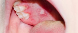
If the infection develops in children during the neonatal period, it may be accompanied by symptoms:
- enlarged lymph nodes;
- tonsillitis;
- labored breathing;
- impaired swallowing and sucking reflexes;
- prematurity;
- strabismus;
- jaundice of newborns. Source: R.Zh. Seisebaeva, A.E. Almaganbetova, F.N. Kasymbekova, E.S. Ataibekova, G.M. Abdrakhmanova Epidemiology of congenital cytomegalovirus infection // Bulletin of KazNMU, 2022, No. 1.
Cytomegalovirus can develop asymptomatically, which is the most dangerous. Due to the asymptomatic course of the infection, children may develop changes in the skull skeleton, lack of body weight, and mental development disorders.
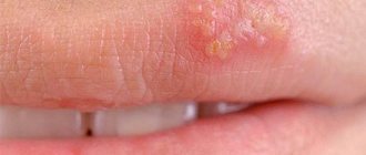
Acquired infection (when a child becomes infected at an older age) can occur with the following symptoms:
- enlarged lymph nodes (especially in the neck);
- lethargy, drowsiness;
- temperature increase;
- pain in muscles and joints.
Most often, the infection goes away without any specific treatment when infected at an older age than infants. But if the symptoms of infection do not go away after 2 months, consult a doctor immediately!
Causes of hemoptysis.
Most often, the presence of streaks of blood in the sputum during a strong paroxysmal cough during a cold in young people is not a dangerous symptom. It’s just that with a strong cough, the wall of the blood vessels can be damaged and a small amount of blood enters the lumen of the bronchi. Such hemoptysis occurs immediately after the inflammatory process in the bronchi subsides. However, even in this case, an examination by an ENT doctor and a chest x-ray are necessary to rule out another cause of hemoptysis, which we will now talk about.
- Quite often the cause of hemoptysis is diseases of the pharynx and paranasal sinuses. If blood appears in the sputum, an examination by an ENT doctor is necessary to rule out a tumor of the larynx, pharynx or inflammation of the sinuses (sinusitis, frontal sinusitis).
- In 20% of cases, the cause of hemoptysis is tumors of the bronchopulmonary system.
- Pulmonary embolism (damage to lung tissue due to a clot blocking the pulmonary artery supplying that section) can also cause hemoptysis.
- Severe heart disease: myocardial infarction, heart defects, cardiomyopathies can cause the appearance of blood streaks in the sputum. This occurs because high pressure in the pulmonary vessels, which develops in heart disease due to weakness of the heart muscle, leads to dilation of the pulmonary vessels and their damage.
- Sometimes the appearance of blood in the mouth due to diseases of the esophagus or stomach can be mistaken for hemoptysis.
Treatment
Drug therapy is used when a child is infected as a newborn or in severe cases of the disease. Treatment is not aimed at completely getting rid of the virus, which is physically impossible, but at reducing its activity. Antiherpetic drugs are most often used for treatment. To draw up a treatment plan, contact your pediatrician.
Sources:
- A.I. Smirnova, E.V. Rossikhina, N.S. Dupin. The role of cytomegaloviruses in obstetric pathology and neonatology // Vyatka Medical Bulletin, 2010.
- R.Zh. Seisebaeva, A.E. Almaganbetova, F.N. Kasymbekova, E.S. Ataibekova, G.M. Abdrakhmanova. Epidemiology of congenital cytomegalovirus infection // Bulletin of KazNMU, 2022, No. 1
The information in this article is provided for reference purposes and does not replace advice from a qualified professional. Don't self-medicate! At the first signs of illness, you should consult a doctor.
Infection caused by human herpes virus type 6
Human herpes virus type 6 (HHV-6) is a DNA virus of the Herpesviridae family of the Betaherpesvirinae subfamily of the Roseolavirus genus. HHV-6 was first isolated in 1986 from peripheral blood B lymphocytes of patients with non-Hodgkin lymphoma, which occurs in patients with HIV infection. The virus belongs to the subfamily of betaherpes viruses, is the closest genetic relative of CMV, there are two variants: HHV-6A and HHV-6B.
Replication of the virus in peripheral blood mononuclear cells occurs relatively slowly and is accompanied by lysis of the host cell. HHV-6, like other herpes viruses, is characterized by the ability to persist and latency in the body of an infected person. The virus exhibits tropism for a wide range of host cells: it was found in lymph nodes, peripheral blood lymphocytes, monocytes, macrophages, kidney cells, salivary glands, and brain. During an acute infection, the pathogen can be isolated from the blood. After infection, HHV-6 infection becomes latent. The place of virus latency has not been studied; it is assumed that the virus remains latent for some time in monocytes and macrophages. The virus infects the salivary glands and is released from them. Detection of the virus in the blood is typical only during the febrile stage of sudden exanthema and, probably, during reactivation of the virus and generalization of infection under conditions of immunosuppression. The pathogenesis of reactivation of infection is unclear.
HHV-6 infection is an anthroponosis. The source of infection is a person suffering from a manifest or latent form of infection, as well as virus carriers. Routes of transmission of infection: airborne droplets, household contact, parenteral, transplacental. Transmission factors are saliva, sputum, blood. The infection is characterized by universal susceptibility.
The high pathogenetic significance of HHV-6 has been shown: it can cause acute skin lesions in young children (sudden exanthema of newborns), fever in newborns with convulsive syndrome, chronic fatigue syndrome (at the same time, recent studies attach greater importance to HHV-7 in the development of this pathology ), mononucleosis-like syndrome; in immunocompromised individuals – cause fever, pneumonia, hepatitis, and central nervous system damage. It has been proven that the virus can also act as a cofactor for HIV. Along with the occurrence of a primary infection, reactivation of the virus is possible: in children intrauterinely infected with HIV-1, primary HHV-6 infection contributed to a more rapid development of clinical manifestations already during the first year of the child’s life. The presence of an active HHV-6 infection in an HIV-infected child may lead to more rapid progression of the underlying disease during the 1st year of life. Cases of pneumonia and encephalitis of HHV-6 etiology in patients with HIV infection have been described. HHV-6 DNA was determined in brain tissues of deceased patients at the stage of AIDS. In patients with HIV infection with severe immunosuppression, HHV-6 damage to the central nervous system, lungs and other organs is possible, but the clinical characteristics of damage to individual organs, the diagnostic sensitivity and specificity of various laboratory markers have not been precisely characterized.
Verification of the diagnosis of HHV-6 infection is carried out only with positive results of laboratory tests.
Differential diagnosis.
Entero- and adenovirus infection, measles, rubella, scarlet fever, pneumonia, otitis media, acute pyelonephritis, meningitis, pneumococcal bacteremia, allergic rashes.
Indications for examination
- Maculopapular rash (exanthema) in combination with lymphadenopathy after a short fever;
- enlargement of the occipital, posterior cervical and/or parotid lymph nodes;
- research after contact with a patient with sudden exanthema or other infection caused by HHV-6 or suspected of these nosological forms;
- differential diagnosis of exanthema diseases;
- immunodeficiency states;
- chronic fatigue and a decrease in performance by more than 50% with a duration of about 6 months in the absence of other diseases that cause similar symptoms;
- symptoms of congenital infection, developmental defects in newborns.
Etiological laboratory diagnostics include identification of the pathogen in cell culture, detection of viral DNA, determination of specific IgM, IgG antibodies to HHV-6 antigens.
Material for research
- Blood plasma, CSF, leukocyte fraction of blood, saliva - DNA isolation, identification of the pathogen in cell culture;
- blood serum - determination of AT.
Comparative characteristics of laboratory diagnostic methods.
Detection of the pathogen in cell culture is currently not used for routine diagnosis of infection caused by the HHV-6 virus due to the complexity, duration of execution and the need for certain research conditions.
The main method of differential diagnosis of infection is the detection and determination of the concentration of HHV-6 DNA by PCR. When testing whole blood to diagnose infection, DNA quantification is preferred to differentiate between latent and active infection, since the virus may be present in the white blood cells of healthy individuals. Detection of viral DNA in plasma but not in whole blood confirms the presence of an active infection. The results of determining HHV-6 DNA in a quantitative format allow for dynamic monitoring: based on the increase in concentration in peripheral blood, leukocytes, CSF, saliva, establish the activity of the infectious process, identify reactivation, and evaluate the effectiveness of the therapy.
To identify specific IgM and IgG antibodies to HHV-6 antigens, ELISA is mainly used. Determination of IgG antibodies can be performed in qualitative and quantitative format. Detection of IgM antibodies makes it possible to establish a diagnosis of a current primary HHV-6 infection; the results of determining IgG antibodies in a quantitative format allow for dynamic observation and assessment of the state of post-infectious immunity to HHV-6.
Indications for the use of various laboratory tests (herpes type 6 - analysis).
Indicators of active infection are the presence of HHV-6 DNA and IgM antibodies. IgM antibodies appear in the blood 4–7 days after the onset of the disease and persist for several months. IgG antibodies appear in the blood on days 7–10 of the disease and persist throughout life, therefore, to establish the fact of primary infection, quantitative determination of IgG antibodies over time is necessary. Determination of virus-specific IgG antibodies can be used in screening studies to determine the presence of immunity to HHV-6.
Features of interpretation of laboratory research results.
Detection of a specific fragment of HHV-6 DNA in patient biomaterial samples (blood plasma, CSF, scrapings from the mucous membrane of the oropharynx) allows one-time testing to confirm the fact of HHV type 6 infection.
The detection of specific IgM antibodies, markers of the acute phase of the disease, indicates a primary infection or reactivation of the infection. A single detection of IgG antibodies is not unambiguous evidence of a primary infection.
Prices
| Name of service (price list incomplete) | Price |
| Online opinion of a pediatrician (SPECIAL) | 0 rub. |
| Appointment (examination, consultation) with a pediatrician, primary, therapeutic and diagnostic, outpatient | 1750 rub. |
| Consultation (interpretation) with analyzes from third parties | 2250 rub. |
| Prescription of treatment regimen (for up to 1 month) | 1800 rub. |
| Consultation with a candidate of medical sciences | 2500 rub. |