Performing syndrome diagnosis
To properly check for the presence of a disease, it is necessary to perform a full range of various procedures. In particular, diagnosis of stylohyoid syndrome includes a number of actions and manipulations.
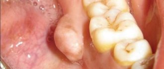
First of all, a professional examination of the patient is performed, as a result of which the doctor examines and identifies the compaction of the bone growth in the area of the anterior zone of the cervical spine of the patient. If you press on this part, the person should experience pain, and his health will deteriorate greatly.
Secondly, an X-ray of the facial skeleton, skull bones, and neck is taken.
At the time of diagnosis, you should carefully approach the procedures, because this disease can easily be confused with other similar pathologies, the symptoms of which are very similar. As an example, suppuration of the tonsils can be cited.
General information
Stylohyoid syndrome (elongated styloid process syndrome) was studied in detail and described in 1937 by the American otolaryngologist W. Eagle, therefore it also bears his name - Eagle syndrome. Anatomical changes in the styloid process occur in 18–30% of adults, but clinical manifestations develop in only 1–5%. Women are more likely to experience this disease; the average age of presentation is 50-60 years. Stylohyoid syndrome refers to multidisciplinary problems that combine neurology, dentistry, and otolaryngology.
Stylohyoid syndrome
Clinic
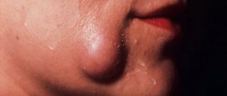
In the presence of an elongated and/or curved styloid process, or a calcified stylohyoid ligament, or a calcified stylohyoid fold, or elongated horns of the hyoid bone, excessive pressure of these structures occurs on the internal and external carotid arteries. Due to this, in the areas fed by the carotid arteries, many seemingly unrelated clinical symptoms appear, such as:
- sensation of a foreign body in the throat;
- chronic inflammation of the pharyngeal mucosa;
- pain in the maxillary joint;
- pain and noise in the ears;
- unilateral and bilateral orbital or headache;
- “shooting” pain when turning the head.
According to the literature, patients with stylohyoid syndrome most often complain of poorly localized pain with unilateral localization in the upper anterior part of the neck and irradiation to the pharynx, root of the tongue, and ear. In this case, pain can spread to the temporomandibular joint, lower jaw, temporal, buccal areas, and submandibular triangle. In some patients, pain occurs in the teeth of the upper and lower jaws. M. Kiely et al. (1995) described the spread of pain to the sternocleidomastoid muscle, and D. Savic (1987) - to the supraclavicular fossa, shoulder girdle and anterior chest wall. Patients usually characterize the pain as dull, constant with periods of intensification and weakening. Its intensity increases towards the end of the day, intensifying when turning or throwing back the head, after a long conversation or singing, or changes in weather conditions.
Patients usually complain of pain in only one organ, most often in the pharynx, ear, temporomandibular joint, and only a detailed survey allows us to clarify its localization and areas of distribution. Some patients develop glossopharyngeal neuralgia with its characteristic painful paroxysms. Dysphagia is a common symptom of the disease, and difficulty swallowing is usually associated with a significant increase in pain in the throat and ear. Some patients are bothered by the sensation of a foreign body in the throat, others are worried about a “constantly sore throat”; they may experience pharyngeal spasm, persistent dry cough without objective signs of inflammation in the upper respiratory tract.
Stylopharyngeal syndrome
As a rule, it is right-sided, since the right styloid process is normally longer than the left by an average of 0.5 cm. Pain occurs as a result of pressure, for example, by an elongated and inwardly curved styloid process, on the tissue in the area of the tonsillar fossa and irritation of the nerve endings of the glossopharyngeal nerve. The intensity of pain varies greatly - from minor pain or the sensation of a foreign body in the throat, especially when swallowing, to sharp, severe constant pain in the throat and tonsils, radiating to the ear. Some patients also note pain on the front surface of the neck, in the area of the hyoid bone. Occasionally, these sore throats are mistakenly associated with pathological changes in the tonsil and it is removed, and ongoing pain is explained by irritation of the nerve endings by a postoperative scar. However, stylopharyngeal syndrome should be differentiated not only from damage to the tonsils, for example by a chronic inflammatory process, but also from neuralgia of the glossopharyngeal nerve. Neuralgia of the latter is characterized by paroxysmal burning or shooting pains in the pharynx, tonsils, root of the tongue, radiating to the ear, which occur during talking, laughing, coughing, yawning and eating.
Styloid-carotid syndrome (“carotid artery syndrome”).
The pathogenesis consists of pressure from the tip of the elongated and outwardly deviated styloid process on the internal or external carotid artery near the bifurcation of the common carotid artery, irritation of the periarterial sympathetic plexus and pain receptors. When the internal carotid artery is irritated, the pain is constant and is felt in the frontal region, orbit, eyes, that is, in the area of branching and blood supply of the internal carotid artery or its branches, in particular the orbital artery. Due to the pressure of the appendage on the external carotid artery, pain radiates along its branches to the temple, crown, face (below the eye).
- Why does the jaw hurt on one side when chewing near the ear: what does pain on the right and left mean, how to treat it?
Publications in the media
Congenital syndrome characterized by a triad: • Congenital absence, insufficiency or hypoplasia of the abdominal wall muscles • Anomalies of the urinary tract (megacystis, dilation of the ureters and prostatic urethra) • Bilateral cryptorchidism.
Variants of this anomaly in girls and boys, for example, only isolated (without other defects) insufficiency of the abdominal muscles or this insufficiency in combination with either cryptorchidism or urinary tract ectasia, are designated by the term pseudoprune disorder.
Incidence : 1 in 35,000–50,000 births.
Genetic aspects • 100100 - plum belly syndrome • 264140 - plum belly syndrome, pulmonary stenosis, mental retardation, deafness.
Combined anomalies. Individual observations of the combination of this syndrome with hearing loss, anal atresia, skeletal malformations, anomalies of the gastrointestinal tract, heart, lungs, underdevelopment of the genital organs, open urachus, and malformations of skeletal muscles are described.
Clinical picture
• Abdominal wall. Aplasia of the muscles of the anterior abdominal wall, sometimes the lower medial sections of the abdominal wall consist only of skin, fat and deposits of fibrous tissue on the peritoneum •• The abdominal wall stretches in utero and acquires that typical wrinkled appearance in the newborn, which was the reason for denoting this pathology with the term “plum” stomach". The thinned and folded abdominal wall allows you to freely palpate any organs of the abdominal cavity. Directly under the skin, peristalsis of the intestines and ureters can be observed •• Children cannot sit up from a lying position. In order to sit down, they usually turn over on their stomach and stand up from this position, resting on their limbs •• The act of coughing is impaired, which leads to recurrent respiratory tract infections.
• The testes are not descended into the scrotum (cryptorchidism) •• Histologically, these testes are indistinguishable from normal gonads at an early stage of development •• Pathology of spermatogenesis (even after surgical correction) •• In adults, there are secondary sexual characteristics, libido and erection.
• The kidneys may be dysplastic. However, changes in the renal parenchyma usually do not correspond to those that occur with obstructive uropathy.
• Ureters •• Non-obstructive megaureter •• Vesicoureteral reflux •• Ascending infection and pyelonephritis.
• The bladder is enlarged and deformed •• There is a significant volume of residual urine •• Urachal cleft saves the life of a child with urethral atresia.
• The prostate gland is hypoplastic •• The prostatic part of the urethra is dilated, but there is no bladder outlet obstruction •• Atresia of the anterior urethra sometimes occurs.
• Gastrointestinal tract. There may be various anomalies •• Malrotation in combination with loose bowel •• Atresia of the large and small intestine •• Gastroschisis •• Torsion of the spleen •• Persistent cloaca •• Hernia of the umbilical cord.
• CVS (anomalies occur in 10% of cases) •• PDA •• VSD •• ASD •• Tetralogy of Fallot.
• Musculoskeletal anomalies of the limbs occur in 1/3–1/5 patients •• Clubfoot •• Scoliosis •• Congenital hip dislocation.
• Respiratory system. Pulmonary hypoplasia occurs in 30% of deceased newborns with plum belly syndrome, therefore, after the birth of the child, it is necessary to first examine the respiratory organs.
Diagnostics • Antenatal diagnosis is possible with the help of ultrasound •• However, the diagnostic accuracy and reliability of prenatal ultrasound continues to be controversial •• If plum belly syndrome is suspected in the fetus, it is necessary to perform repeated repeated ultrasounds •• After antenatal diagnosis, various management tactics are possible: termination of pregnancy, preparation for postpartum surgical correction. Intrauterine interventions are not recommended, with the exception of intrauterine decompression of the urinary system • Postnatal diagnosis •• The diagnosis of plum belly syndrome should be suspected in any child born with a flabby abdominal wall and absence of testicles in the scrotum •• Diagnosis is aimed at a complete examination of the urinary system and identification of associated malformations .
Early postnatal management • During the first days of life, the newborn is closely monitored with special attention to pulmonary function • Laboratory testing of renal function is necessary. Bacteriological examination of urine is periodically indicated. Ultrasound and voiding cystourethrography are also performed • To completely empty the bladder, it is recommended to perform palpatation of the suprapubic region • Fighting infection (prophylactic administration of antibiotics).
Treatment. There is no single point of view on the treatment of this defect; each child with plum belly syndrome requires a purely individual approach • The goal of any treatment method (conservative or surgical) is to prevent infection and preserve kidney function • Conservative tactics are most optimal •• Surgical intervention is indicated only in those cases , when prophylactic antibiotic therapy and follow-up are ineffective and do not allow preserving renal function and preventing persistent bacteriuria with or without bacteremia •• Possible surgical operations ••• Urinary diversion and drainage of the urinary tract (percutaneous nephrostomy, pyelostomy, ureterocutaneostomy or cystocutaneostomy) • •• Reduction cystoplasty (cystoplasty with reduction in the size of the bladder) ••• Corrective interventions on an enlarged ureter ••• Abdominal wall plastic surgery ••• Orchiopexy.
Prognosis • Mortality with this syndrome was 20% among patients of early infancy and 50% among children of the first two years of life • Early deaths were mainly due to pulmonary hypoplasia and respiratory complications. At a later age, among the causes of death, renal failure and sepsis, or both together, came to the fore • Currently, the tendency is for survival to increase over the years and for these patients to live longer.
Synonyms • Prune-belly syndrome • Eagle-Berrett syndrome • Abdominal muscle deficiency (AMD - abdominal musculature deficiency) • Triad syndrome • Flabby abdomen syndrome • Absence of muscles of the anterior abdominal wall syndrome.
ICD-10 • Q79.4 Plum navel syndrome
Diagnostics
To make a diagnosis, it is very important to carefully collect complaints and medical history. So, it is necessary to establish when the painful sensations appeared, what nature they are, what provokes an increase in pain, and what causes a weakening. It is necessary to accurately determine the areas where pain occurs and where it radiates. This is important for differential diagnosis with neuralgia of other nerves (occipital, glossopharyngeal, upper laryngeal, trigeminal, auriculotemporal).
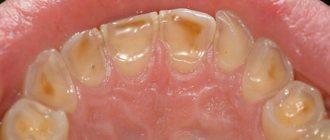
It is mandatory to examine the throat to rule out pharyngitis and tonsillitis.
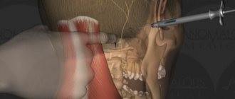
An additional examination method is skull radiography (or targeted radiography of the stylohyoid process).
Causes
Eagle syndrome develops as a result of changes in the structures of the stylohyoid complex (SPC), namely the styloid process of the temporal bone (STBS), stylohyoid ligament (SL) or hyoid bone. Anomalies of the SHOVK are associated with its elongation or incorrect location - deviation, curvature. Pathological changes in the ligament are caused by its calcification (incomplete/complete, unilateral or bilateral). Anatomical defects of the hyoid bone are represented by an increase in the lateral processes - horns.
The development of stylohyoid syndrome can be provoked by:
- fracture of the styloid process;
- pharynx injuries;
- dysfunction of the muscles attached to the process;
- tonsillectomy;
- Forestier's disease.
Diagnosis of stylohyoid syndrome in Pirogov
At the National Medical and Surgical Center named after N. I. Pirogov, the diagnosis is made by studying the anamnesis in conjunction with studies carried out in the clinic.
The disease is most often detected by the following method: palpation of the cervical spine or the fossa of the tonsillar part of the oral cavity occurs, resulting in pain.
Then the patient receives a referral for fluoroscopy, which confirms the diagnosis. There have been cases when an increase in the size of the stylohyoid process did not have a painful effect on patients. Before the pathology was discovered, they felt quite comfortable.
Classification
The clinical symptoms of Eagle syndrome depend on which anatomical structures are irritated by the hypertrophied stylohyoid complex. There are 2 variants of the syndrome:
- stylopharyngeal (classic) – associated with irritation of the branches of the glossopharyngeal nerve, characterized by persistent pain;
- styloid-carotid (carotid artery syndrome) - develops when the carotid arteries are involved in the pathological process, accompanied by cerebrovascular manifestations.
Prognosis and prevention
Stylohyoid syndrome belongs to diseases that significantly reduce the quality of life and ability to work of patients, and in some cases can lead to disabling consequences. Fast and accurate diagnosis, based on clinical and objective signs, allows you to choose the right (conservative, surgical) tactics and save the patient from suffering.
In terms of preventing stylohyoid syndrome, it is recommended to avoid injuries to the pharynx and SHOVK; in case of severe pain in the cervicofacial area, immediately contact a specialist. Greater awareness and vigilance of doctors regarding Eagle syndrome is necessary, which will avoid its underdiagnosis and improper treatment.
You can share your medical history, what helped you in the treatment of stylohyoid syndrome.
Complications
Almost all patients note a decrease in performance, loss of strength and mood, and sleep disturbances. The most dangerous complications of awl-carotid syndrome are transient ischemic attacks and stroke of ischemic type. They are often caused by dissection of the internal carotid artery with the formation of an intramural hematoma that occludes the lumen of the vessel. Compression of the internal jugular vein provokes intracranial hypertension. In some cases, sudden death may occur due to vagal arrhythmia or myocardial depression.