Publication date: 03/26/2020
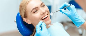
The Axioma Dental clinic has its own visiograph and orthopantomograph - this is digital X-ray equipment that allows you to achieve high efficiency in diagnosis and treatment. A visiograph is an ultra-sensitive technique for creating clear, accurate images of teeth. You no longer have to wait for the X-ray film to be developed; the image is instantly visualized on the screen. This saves the patient’s time without harming health.
An orthopantomograph is a safe alternative to X-ray machines. The technique takes a panoramic photograph of the patient’s entire dental system. The equipment is indispensable in any dental field for developing a treatment plan. The dentist can quickly make a diagnosis and obtain detailed information about the condition of the dental system.
The principle of operation of the visiograph
A dental visiograph is a system of equipment that includes an ultra-sensitive touch sensor, an X-ray, and a computer for digitizing data. The principle of operation of the technique is similar to x-rays, but it is a safe technique. If the film unit directs rays onto the film, then the visiograph imaging device sends data to an ultra-sensitive matrix, a CCD sensor. The microcircuit chip consists of photodiodes that can capture and visualize the smallest details.
The main advantage is that to create an image you need an ultra-low dose of X-ray radiation, only 2-3 μSv. This is safe for humans, and the doctor has the opportunity to reduce the time for diagnosis.
How to take pictures using a visiograph:
- The lead apron is protection for the patient, who wears it before the procedure.
- Placement of the sensor in the oral cavity - it can be wireless or wired. An individual protective cover is put on the sensor and placed in the oral cavity. The sensor is applied to the area where pathology is detected, the condition of the bone tissue is clarified for successful implantation, treatment of caries, pulpitis.
- The sensor of a dental touch imaging device is connected to a digital device for data visualization, digitization and analysis of results.
- Snapshot.
Within a few seconds, the doctor sees the full clinical picture on the PC screen, makes a diagnosis and quickly prescribes treatment.
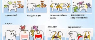
What can be found in a visual image?
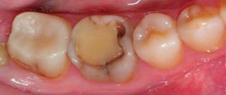
Radiovisiography is used in therapeutic dentistry, implantology and endodontics. The technique is indispensable in the treatment of dental diseases, filling canals and mechanical treatment of tooth roots. From the resulting image, a qualified dentist will be able to accurately determine the shape of the canal, its length and depth, and also select the correct size of the metal pin. Using the image, it is not difficult for a specialist to detect part of the chipped instrument and control the quality of the filling.
Visiographic image reveals:
- inflammatory foci (granulomas, cysts);
- caries (in the picture it will be visible as a dark, almost black area);
- loose, connective and bone tissue (gray areas in the image);
- dense structures such as pins, fillings and dentures (their presence will be indicated by white areas in the photo).
The extremely sensitive chip has a high level of detail. The end result is a sharp and clear photo. A high-quality image will help the doctor make a diagnosis and identify the smallest inflammatory and carious lesions. A visual image can provide the dentist with detailed information about the condition of the fillings.
The difference between a visiograph and an x-ray
A visiograph is a device that does not independently emit rays, unlike an X-ray machine. Its task is to receive the rays onto an ultra-sensitive sensor and convert them into an accurate image for data analysis. At the same time, modern equipment does not need to produce a wide beam of radiation. A radiovisiograph is a very thin targeted beam of light that is captured by a sensor.
The system’s sensor is able to recognize and capture the smallest particles of radiation in just a fraction of seconds, which reduces the procedure and risks for humans. Modern digital equipment differs from x-rays in the speed of obtaining an image; shutter speed:
- visiograph - 0.06 seconds.
- film apparatus - 0.6.
Conclusion - innovative technology allows you to achieve maximum safety for the patient and reduce procedure time.
Commissioning, repair and maintenance of medical equipment
In our company, you can enter into an agreement for regular maintenance of medical equipment, as well as order equipment repair or commissioning services. We work with equipment of any complexity and always cope with the assigned tasks 100%.
Leave an online application on the website, and our specialist will contact you as soon as possible to clarify all important details.
Advantages and disadvantages of a visiograph
Pros of sensory equipment:
- Minimum radiation dose.
- High image accuracy.
- Maximum safety for the patient.
- The procedure can be performed for pregnant women and children - consultation with a doctor is required.
- Compact device, able to work in a small office.
- Instantly visualize your photo without the need to develop film.
- The doctor gets the opportunity to see each tooth - multiple magnification of the image, storage of data on a PC, transmission via e-mail, local network.
- The manipulation takes several minutes.
- Editing a picture - the dentist uses different filters to improve the visual perception of the picture.
- The procedure is painless and does not require special preparation.
The disadvantage is the high price of equipment and procedures. But in modern dentistry, the priority is accurate diagnosis, effective treatment, and patient safety. Therefore, reputable clinics invest money in purchasing equipment.
The essence of the X-ray machine
During treatment, prosthetics or implantation, it is necessary to take dental x-rays many times: before, during and after the intervention. The devices familiar to many caused irreparable harm to the patient’s body, because the radiation dose in them is very high. This was necessary so that the reflected X-rays could be imprinted on the film, thereby creating an image.
“Classic” X-ray also required a staff member who knew how to develop images, as well as an entire darkroom. And patients had to wait a long time for examination results.
Features of the orthopantomograph
Dental orthopantomograph is equipment for creating high-resolution panoramic images. Devices of various types have an overview imaging system, which makes it possible to visualize the entire jaw, the patient’s teeth, and jaw joints. The principle of operation is that the X-ray emitter and sensor in digital modifications rotate around the head, forming a clear three-dimensional image of the jaws.
Digital orthopantomographs make shooting as safe as possible for humans - the radiation level is 2-3 times less than with film units. Innovative technology does not require laboratories to develop images; the picture will be ready quickly and visualized on the computer screen without waiting for results.
If the orthopantomograph has a cephalostat, the doctor can take a frontal profile photograph of the skull, which is needed to build orthodontic structures.
Innovative technology works with different shooting programs:
- Standard overview.
- Separately upper and lower jaw.
- Photo of the maxillary sinuses.
- Children's photography.
- Image enlargement mode.
- Other programs for different clinical cases.
Safety, speed, high-resolution image quality, and information content are the characteristics of equipment used by clinics all over the world.
How dangerous is this?
The most common question regarding a visiograph is how dangerous it is. In fact, due to its design, it is no more dangerous than a regular digital camera or office scanner. The radiation dose received by the patient during radiography of the teeth of the lower jaw will be 2 microsieverts and 5 μSv for the upper jaw. This amount of radiation is extremely safe for both the patient and the doctor. But only if the doctor follows safety standards and correct positioning. At the same time, individual forced ventilation must operate in the office, which will disperse ionized air, since it is hazardous to health.
Here is an example for comparison: when working with Soviet installations, the average radiation dose ranges from 20 to 30 μSv, and when using the 5D1 device, the values can reach up to 80 μSv.
Modern dentistry with visiograph and orthopantomograph
The technical equipment of the dental clinic is a guarantee of accurate diagnosis, effective treatment, and error-free diagnosis. Without the latest generation equipment, a medical institution cannot guarantee the patient a high level of service.
Axioma Dental Clinic is a center that invests money in purchasing innovative equipment. Doctors have a Sirona orthopantomograph and a visiograph at their disposal. This provides benefits to our patients:
- Examinations in one clinic - no need to waste time, sign up in advance for x-rays at clinics.
- Safety - there is no risk of receiving a high dose of X-rays.
- Treatment of children, adults, pregnant women - innovative equipment does not cause harm to health.
- Instant results - the photo will be ready in a few seconds.
The Axioma Dental clinic is the right approach to treating teeth, gums, correcting row defects, and implantation using safe equipment.
Sources:
- Yarnova E.A. Possibilities of radiation diagnostic methods in visualization of the temporomandibular joints in cases of dentofacial anomalies
- Goncharov I.Yu. Planning the surgical stage of dental implantation in the treatment of patients with various types of missing teeth, defects and deformations of the jaws, 2009
- Robustova Tatyana Grigorievna. Dental implantation: surgical aspects, 2003
Expert author:
Makanina Lina Viktorovna
Dentist-therapist
The information presented in this article is provided for reference purposes and does not replace the advice of a qualified specialist.
At the first signs of illness, you should consult a doctor.
Tell us about us:
Popular methods of radiation diagnostics
Today, the most common and popular method of radiation examination in outpatient practice is intraoral radiography of teeth, or intraoral photograph of the tooth. Sometimes intraoral photographs of teeth are called targeted, which is incorrect. A targeted photograph is a photograph taken outside the standard positioning, and standardized studies are named according to the positioning method.
At a therapeutic appointment during endodontic treatment, at least three intraoral photographs of each tooth under examination must be taken:
- A diagnostic image is necessary to assess the condition of periodontal tissues at the time of examination, make a diagnosis, determine the number and shape of roots, the direction of canals, and choose treatment tactics.
- measuring photograph - a photograph of a tooth at the treatment stage with endodontic instruments inserted into the canals with a fixed stopper length of the working part or verifiers after instrumental treatment of the canals. If the orthogonal projection is performed correctly, provided that the visiograph program is accurately calibrated and there is no projection distortion for incisors and premolars, some measurements can be taken from the diagnostic radiogram. For multi-rooted teeth, it is preferable to measure the length of the canals using endodontic instruments (Fig. 1), an apex locator or from a three-dimensional photograph.
- a control photograph is taken immediately after the end of endodontic treatment in order to determine how well the root canals are filled, as well as after a certain specified time, in order to verify the absence or detect the presence of complications (Fig. 2). When examining multi-rooted teeth and in cases where there is an additional canal, in an image taken with the orthoradial direction of the beam (direct projection), the root canals often overlap each other, which significantly complicates diagnosis and can lead to errors in the treatment process. To obtain a separate image of the root canals, radiography with an oblique (eccentric) direction of the central beam is used (Fig. 1). For each specific case, the mesial or distal inclination (angulation) of the tube in the horizontal plane is selected (for more details, see: Rogatskin D.V., Ginali N.V. The Art of Dental Radiography, 2007).
Ideally, maximum information about the topography of the roots and the condition of periodontal tissues can be obtained by performing polypositional radiography. In this case, for diagnostic purposes, three photographs are taken - one in a straight line, with an orthoradial direction of the beam, and two in an oblique projection - with a distal-eccentric (Fig. 1) and mesial-eccentric direction of the beam (respectively, straight, posterior oblique and anterior oblique projections).
The most important aspects of successful intraoral radiography are standardization and consistent manipulation. Standardization of manipulations means the ability of a specialist conducting a radiation examination to choose the optimal method for each case and take a series of identical images, regardless of the position, condition of the patient and the time separating one examination from another. That is, if a diagnostic or measurement image is considered to be of high quality, each subsequent clarifying and control image must be made with the same spatial and technical settings, and each subsequent image must be identical to the previous one (Fig. 1, 2).