An orthodontist makes such a diagnosis with code K07.3 according to ICD-10 (International Classification of Diseases, 10th revision) if the tooth has erupted with an inclination or displacement, or has completely appeared outside the dental arch. This mainly happens to the lower eighth molars, incisors and canines.
A companion to dystopia can be other anomalies in the position of the teeth - crowding, displaced or open bite, as well as retention.
Dystopic wisdom tooth: what is it?
A dystopic wisdom tooth is a third molar, also known as a “figure eight”, which is positioned incorrectly relative to the rest of the dentition. Such a tooth almost always needs to be removed.
A special case of a dystopic wisdom tooth is an unerupted, but fully formed figure eight, which turns out to be turned parallel to the gum. In this case, the tooth is there, although it is not visible, and it is in an incorrect position, so we can talk about dystopia.
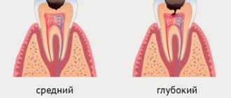
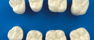
Two examples of dental dystopia
Clinical manifestations
A clinically unerupted tooth may not manifest itself in any way, and the person will not even know about the presence of a problem. Sometimes this state of affairs persists for life, and treatment is not required. But in almost 80% of cases, an impacted tooth causes a disorder, and then the person is forced to seek dental help.
As a rule, problems arise when wisdom teeth erupt incompletely. Then a hood forms over the crown - a gum overhang over the tooth, where food gets clogged and plaque accumulates. Over time, this causes inflammation - pericoronitis, during which the soft tissues become red, swollen and cause pain of varying degrees of intensity. A colorless or slightly cloudy liquid may be released from under the hood [11, 23].
If pericoronitis is limited to the catarrhal phase, the patient’s well-being practically does not suffer. But if the situation worsens and pericoronitis becomes purulent, problems appear with opening the mouth due to significant swelling of the mucous membrane surrounding the causative tooth, pain appears with shooting in the temporal, ear or occipital region on the affected side. The tonsil on the side of the impacted tooth can also become inflamed, and drops of pus are released from under the overhanging gum. General well-being is also affected - body temperature can rise to 37.8 - 38 degrees, regional lymph nodes enlarge, weakness and headache appear [5, 19].
Repeated repetitions of acute pericoronitis, interspersed with periods of good health, indicate the transition of the disease to the chronic stage. In this case, the pain when closing the jaws or chewing is moderate, and the formation of ulcers in the hood area comes to the fore. They appear due to the biting of swollen gums by antagonist teeth. A small amount of purulent contents is usually released from under the hood. General health is usually satisfactory, despite the fact that pericoronitis causes frequent exacerbations [4, 7].
If an impacted tooth is located in the frontal zone, a person may complain about the unattractive appearance of the dentition and reduced aesthetics of the smile. However, there may be no other complaints [14].
Causes of dystopic wisdom teeth
Very often, dental dystopia is associated with genetic reasons. A predisposition to shifting dentition is inherited along with small jaw size and other aspects of individual appearance that can cause teeth to shift. Doctors call other factors that provoke improper growth of molars, incisors and fangs:
- atypical formation of the tooth germ in the embryonic period;
- macrodentia, partial adentia, supernumerary teeth and other diseases;
- improper eruption (can be caused by early removal of the primary bite);
- small jaw size;
- mechanical damage, dental trauma;
- bad habits such as chewing foreign objects, thumb sucking (affect the development of malocclusion);
- late eruption of canines, for which there is no longer room in the dentition.
Modern dentistry successfully cures the patient, regardless of the causes of tooth dystopia and its position in the gum. The main thing is to seek help in time.
ICD-10 (Dentistry)
The article presents the international classification of diseases, tenth revision, relating to the dental profile.
K00—K14 Diseases of the oral cavity, salivary glands and jaws (click on the appropriate block to expand subcategories)
K00 Disorders of development and eruption of teeth
Excluding: impacted and impacted teeth (K01)
| K00.0 | Edentia
|
| K00.1 | Supernumerary teeth
|
| K00.2 | Anomalies in the size and shape of teeth
Excluding: Carabelli tubercular anomaly, considered as a normal variant and subject to coding |
| K00.3 | Mottled teeth
Excluding: deposits (growths) on teeth (K03.6) |
| K00.4 | Tooth formation disorders
Excludes: Hutchinson's incisors and mulberry-shaped molars in congenital syphilis (A50.5), mottled teeth (K00.3) |
| K00.5 | Hereditary disorders of dental structure, not classified elsewhere
|
| K00.6 | Teething disorders
|
| K00.7 | Teething syndrome |
| K00.8 | Other dental development disorders
|
| K00.9 | Dental development disorder, unspecified
|
K01 Impacted and impacted teeth
Excluding: impacted and impacted teeth with malposition of them or adjacent teeth (K07.3)
| K01.0 | Impacted teeth An impacted tooth is a tooth that has changed its position during eruption without obstruction from an adjacent tooth. |
| K01.1 | Impact teeth An impact tooth is a tooth that has changed its position during eruption due to an obstacle from an adjacent tooth. |
K02 Dental caries
| K02.0 | Enamel caries
|
| K02.1 | Dentin caries |
| K02.2 | Cement caries |
| K02.3 | Suspended dental caries |
| K02.4 | Odontoclasia
|
| K02.8 | Other dental caries |
| K02.9 | Dental caries, unspecified |
K03 Other diseases of hard dental tissues
Excluding: bruxism, teeth grinding NOS (not otherwise specified) (F45.8), dental caries (K02)
| K03.0 | Increased tooth wear
|
| K03.1 | Grinding of teeth
|
| K03.2 | Tooth erosion
|
| K03.3 | Pathological tooth resorption
|
| K03.4 | Hypercementosis
|
| K03.5 | Ankylosis of teeth |
| K03.6 | Deposits (growths) on teeth
|
| K03.7 | Change in color of hard tissues of teeth after eruption Excluding: deposits (growths) on teeth (K03.6) |
| K03.8 | Other specified diseases of dental hard tissues
If it is necessary to identify the radiation that caused the injury, use an additional code of external causes (class XX). |
| K03.9 | Disease of hard dental tissues, unspecified |
K04 Diseases of the pulp and periapical tissues
| K04.0 | Pulpitis
|
| K04.1 | Pulp necrosis
|
| K04.2 | Pulp degeneration
|
| K04.3 | Improper formation of hard tissue in the pulp
|
| K04.4 | Acute apical periodontitis of pulpal origin
|
| K04.5 | Chronic apical periodontitis
|
| K04.6 | Periapical abscess with cavity
|
| K04.7 | Periapical abscess without cavity
|
| K04.8 | Root cyst
Excludes: periodontal lateral cyst (K09.0) |
| K04.9 | Other and unspecified diseases of the pulp and periapical tissues |
K05 Gingivitis and periodontal diseases
| K05.0 | Acute gingivitis Excludes: acute necrotizing ulcerative gingivitis (A69.1), gingivostomatitis caused by herpes simplex virus (B00.2) |
| K05.1 | Chronic gingivitis
|
| K05.2 | Acute periodontitis
Excluding:
|
| K05.3 | Chronic periodontitis
|
| K05.4 | Periodontal disease
|
| K05.5 | Other periodontal diseases |
| K05.6 | Periodontal disease, unspecified |
K06 Other changes in the gingiva and edentulous alveolar margin
Excluding: atrophy of the edentulous alveolar margin (K08.2), gingivitis: acute (K05.0), chronic, NOS (not otherwise specified) (K05.1)
| K06.0 | Gum recession
|
| K06.1 | Gingival hypertrophy
|
| K06.2 | Lesions of the gums and edentulous alveolar margin caused by trauma. If necessary, identify the cause, use an additional code for external causes (class XX) |
| K06.8 | Other specified changes in the gingiva and edentulous alveolar margin
|
| K06.9 | Changes in the gingiva and edentulous alveolar margin, unspecified |
K07 Maxillofacial anomalies (including malocclusions)
Excludes: atrophy and hypertrophy of the half of the face (Q67.4), unilateral condylar hyperplasia or hypoplasia (K10.8)
| K07.0 | Main anomalies in jaw size
Excludes: acromegaly (E22.0), Robin's syndrome (Q87.0) |
| K07.1 | Anomalies of maxillo-cranial relationships
|
| K07.2 | Anomalies of dental arch relationships
|
| K07.3 | Anomalies of teeth position
Excludes: impacted and impacted teeth with normal position (K01) |
| K07.4 | Malocclusion, unspecified |
| K07.5 | Maxillofacial anomalies of functional origin
Excludes: bruxism, teeth grinding NOS (not otherwise specified) (F45.8) |
| K07.6 | Temporomandibular joint diseases
Excludes: current case of jaw dislocation (S03.0), sprain and strain of jaw joint(s) (S03.4) |
| K07.8 | Other maxillofacial anomalies |
| K07.9 | Maxillofacial anomaly, unspecified |
K08 Other changes in teeth and their supporting apparatus
| K08.0 | Exfoliation of teeth due to systemic disorders |
| K08.1 | Loss of teeth due to accident, extraction or localized periodontal disease |
| K08.2 | Atrophy of the edentulous alveolar margin |
| K08.3 | Delayed tooth root (retentive root) |
| K08.8 | Other specified changes in teeth and their supporting apparatus
|
| K08.9 | Changes in teeth and their supporting apparatus, unspecified |
K09 Cysts of the oral region, not elsewhere classified
Including: lesions with histological features of an aneurysmal cyst and other fibro-osseous lesion Excluding: radicular cyst (K04.8)
| K09.0 | Cysts formed during the formation of teeth
|
| K09.1 | Growth (non-odontogenic) cysts of the mouth area
|
| K09.2 | Other jaw cysts
Excludes: occult bone cyst of the jaw, Stafne cyst (K10.0) |
| K09.8 | Other specified cysts of the oral area, not classified elsewhere
|
| K09.9 | Oral cyst, unspecified |
K10 Other jaw diseases
| K10.0 | Jaw development disorders
|
| K10.1 | Giant cell granuloma, central
Excludes: peripheral giant cell granuloma (K06.8) |
| K10.2 | Inflammatory diseases of the jaws
If necessary, identify the radiation that caused the injury, use an additional code of external causes (class XX) |
| K10.3 | Alveolitis of the jaws
|
| K10.8 | Other specified diseases of the jaws
|
| K10.9 | Disease of the jaw, unspecified |
K11 Diseases of the salivary glands
| K11.0 | Salivary gland atrophy |
| K11.1 | Salivary gland hypertrophy |
| K11.2 | Sialadenitis Excludes: mumps (B26), Hereford uveoparotid fever (D86.8) |
| K11.3 | Salivary gland abscess |
| K11.4 | Salivary gland fistula Ex: congenital salivary gland fistula (Q38.4) |
| K11.5 | Sialolithiasis
|
| K11.6 | Salivary gland mucocele
|
| K11.7 | Disorders of the secretion of the salivary glands
Excludes: dry mouth NOS (R68.2) |
| K11.8 | Other diseases of the salivary glands
Excludes: sicca syndrome (Sjögren's disease) (M35.0) |
| K11.9 | Salivary gland disease, unspecified
|
K12 Stomatitis and related lesions
Excluding:
- decaying mouth ulcer, gangrenous stomatitis, noma (A69.0)
- cheilitis (K13.0)
- gingivostomatitis caused by herpes simplex virus (B00.2)
| K12.0 | Recurrent oral aphthae
|
| K12.1 | Other forms of stomatitis
|
| K12.2 | Cellulitis and oral abscess
Excluding:
|
K13 Other diseases of the lips and oral mucosa
Including: changes in the epithelium of the tongue Excluding:
- some changes in the gingiva and edentulous alveolar margin (K05-K06)
- cysts of the mouth area (K09)
- tongue diseases (K14)
- stomatitis and related lesions (K12)
| K13.0 | Lip diseases
Excluding: |
| K13.1 | Biting cheeks and lips |
| K13.2 | Leukoplakia and other changes in the oral epithelium, including the tongue
Excludes: hairy leukoplakia (K13.3) |
| K13.3 | Hairy leukoplakia |
| K13.4 | Granuloma and granuloma-like lesions of the oral mucosa
|
| K13.5 | Submucosal fibrosis of the oral cavity
|
| K13.6 | Hyperplasia of the oral mucosa due to irritation Excludes: hyperplasia of the edentulous alveolar margin due to irritation (denture hyperplasia) (K06.2) |
| K13.7 | Other and unspecified lesions of the oral mucosa
|
K14 Diseases of the tongue
Excluding:
- erythroplakia, focal epithelial hyperplasia, leukedema, leukoplakia of the tongue (K13.2)
- hairy leukoplakia (K13.3)
- congenital macroglossia (Q38.2)
- submucosal fibrosis of the tongue (K13.5)
| K14.0 | Glossitis
Excludes: atrophic glossitis (K14.4) |
| K14.1 | "Geographical" language
|
| K14.2 | Median rhomboid glossitis |
| K14.3 | Hypertrophy of the tongue papillae
|
| K14.4 | Atrophy of the tongue papillae
|
| K14.5 | Folded tongue
Excludes: congenital cleft tongue (Q38.3) |
| K14.6 | Glossodynia
|
| K14.8 | Other tongue diseases
|
| K14.9 | Tongue disease, unspecified
|
Dental diseases from other sections.
| A69.0 | Necrotizing ulcerative stomatitis
|
| A69.1 | Other Vincent infections - Fusospirochetous pharyngitis - Necrotizing ulcerative (acute): • gingivitis • gingivostomatitis - Spirochetal stomatitis - Vincent's ulcerative film sore throat: • tonsillitis • gingivitis |
| B00.2 | Herpetic gingivostomatitis and pharyngotonsillitis |
| B26 | Parotitis:
|
| B37.8 | Candidiasis of other localizations Candidiasis: • cheilitis • enteritis |
| D86.8 | Sarcoidosis of other specified and combined localizations:
|
| E22.0 | Acromegaly and pituitary gigantism Excluded:
|
| E53.0 | Riboflavin deficiency
|
| F45.8 | Any other disturbances in sensation, function, and behavior that are not associated with physical disorders and that are not mediated through the autonomic nervous system are limited to specific systems or parts of the body and are closely related in time to stressful events or problems. Psychogenic:
|
| J36 | Peritonsillar abscess
Excluded:
|
| L55—L59 | Diseases of the skin and subcutaneous tissue associated with exposure to radiation
|
| M35.0 | Sicca Sjögren's syndrome Sjögren's syndrome with:
|
| Q38.2 | Macroglossia |
| Q38.3 | Other congenital abnormalities of the tongue
|
| Q38.4 | Congenital anomalies of the salivary glands and ducts:
|
| Q67.4 | Other congenital deformities of the skull, face and jaw |
| Q87.0 | Syndromes of congenital anomalies affecting primarily the appearance of the face
|
| R68.2 | Dry mouth, unspecified Excluded: decreased secretion of salivary glands (K11.7) dry mouth caused by:
|
| S03.0 | Jaw dislocation:
|
| S03.4 | Sprain and strain of the joint (ligaments) of the jaw Temporomandibular joint (ligament) |
| class XX | External causes of morbidity and mortality:
|
Types of wisdom teeth dystopia
Wisdom tooth dystopia is classified depending on which direction the crown is shifted. The following types of disease are distinguished:
- vestibular (forward displacement);
- oral (teeth located behind the entire row);
- mesial (forward bend);
- distal (backward tilt);
- with supraposition (above the dentition);
- with infraposition (below the dentition);
- with tortoposition (the tooth is rotated around its axis);
- with transposition (the tooth is in the place of another tooth).
In the most severe cases, you can observe the appearance of a tooth outside the oral cavity: for example, inside the nose. This is very dangerous and requires immediate treatment.
Procedure for providing assistance to children with dental damage
Immediately after receiving an injury, the patient is provided with primary medical care, which includes a general assessment of the child’s condition, pain relief, prescription of antibiotics, anesthetics and other medications, as well as a preliminary diagnosis. After this, the patient’s parents are given a recommendation to make an appointment with a pediatric dentist (therapist), who will carry out full dental treatment. The doctor providing specialized care carries out:
- registration of a medical history (including legal and social aspects of the case);
- collecting anamnesis (origin of injury, presence of concussion, local symptoms);
- clinical examination of the injury (visual examination, percussion, palpation, temperature tests of the pulp, instrumental methods);
- additional diagnostics (electroodontodiagnostics (EDD), transillumination, targeted radiography, occlusal radiography, computed tomography);
- making a diagnosis based on examination, clinical and other studies;
- choice of treatment tactics taking into account the nature of the damage, possible risks, benefits and costs.
The period of rehabilitation of a child after an injury, starting from the moment of emergency assistance, can take 1-3 or more days. Given the complexity of treatment, the period of complete restoration of the integrity and functions of damaged teeth often takes a longer period - several months or even years.
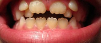
Symptoms of dystopia
Symptoms of dental dystopia are most often visual and easily noticeable. It is enough to look at the position of all the elements of the dentition to see that some of them are in the wrong places or are simply growing unevenly. Other symptoms:
- there is no tooth in the bite, and the period of eruption has already passed;
- inflammation and pain are felt at the site of the wisdom tooth;
- The wisdom tooth did not appear on time
It is interesting that in a number of cases there is no wisdom tooth at all - it does not form and does not erupt. Dentists call this option a variation of the norm.
Ways to stimulate the eruption of an impacted tooth
If the x-ray shows that the unit that has not erupted is located vertically or is anatomically correct, but its roots are not yet developed, it is possible to carry out physical procedures aimed at stimulating natural eruption:
- vacuum massage of gingival tissues, increasing the tone and strengthening of the mucous membrane;
- finger massage and rubbing of the gums using tinctures of eucalyptus, calendula or sage;
- electrical stimulation with galvanic or pulsed current using vegetotropic drugs;
- electrophoresis with Lidase, Adrenaline and humic acids;
- ultraphonophoresis with calcium chloride;
- vibration vacuum massage;
- low frequency ultrasound therapy;
- stimulation with laser currents of infrared and low-intensity types;
- selection and wearing of removable dentures with irritating effects - regional therapy.
Consequences of dystopia
Although dental dystopia does not seem like a terrible disease, it can create many problems. This is why wisdom teeth that have grown incorrectly are most often removed. Among the most unpleasant consequences of the disease:
- disruption of the entire dentition, because due to one incorrect element, the rest move or cannot erupt correctly;
- injury to the tissues of the cheeks, tongue, lips, which can cause ulcers and even provoke the development of tumors;
- complication of oral hygiene, which leads to inflammation, caries, both on the dystopic and adjacent teeth;
- the appearance of gingival pockets where bacteria accumulate, leading to pericoronitis;
- impaired chewing function, which in turn reduces the quality of food and can lead to deterioration of the gastrointestinal tract;
- difficulty pronouncing words, poor diction;
- violation of the facial skeleton, unaesthetic appearance of the lower part of the face.
Almost always, dystopia leads to unpleasant aesthetic consequences, which causes complexes and rejection of one’s appearance in the child, and then in the adult. Of course, this can be worked out with a psychologist, but it is much wiser and more correct to deal with the cause than to deal with the consequences.
Prevalence
Data on the prevalence of pathological resorption have significant discrepancies. Thus, E. Harris [31] in his study determined the frequency of apical root resorption in the permanent dentition in patients who were not treated orthodontically, and found that from 7 to 10% of 306 patients showed obvious apical resorption. F. Vier and J. Figueardo [68] showed that the prevalence of teeth with periapical lesions with resorption is more than 82%. A report by M. Haapasalo and U. Endal [29] estimated the prevalence of internal inflammatory root resorption to be between 0.1 and 1%, confirming that the estimate is quite rough and may be incorrect.
Differences in study methods, variability in pre- and post-treatment radiographic images, misdiagnosis, and undetected lesions are challenges in determining the overall prevalence of pathologic root resorption.
In some cases, it is difficult to determine what is causing the violation of the tooth structure: due to developmental disorders or resorption mechanisms. [52]
Diagnosis of dystopia
An orthodontist or even a dental therapist can easily determine tooth dystopia during a routine examination if the anomaly is not complex. In some cases, when an element of the dentition remains in the gum, ends up in the palate or another place, during the examination one can only suspect a problem.
If the doctor has not found one of the teeth that should have already erupted in a child (or an adult, if we are talking about eights), he will refer the patient for an x-ray examination. The picture clearly shows all the irregularities and you can see the formed teeth in the soft tissues. To clarify the position and for subsequent treatment, arthopantomography, creation of plaster models of the jaws, and teleradiography are used.
Signs and symptoms, how to diagnose
Now let's move on to the question of how to understand that there is a problem. Sometimes complete retention occurs with virtually no symptoms, and then the dentist can detect it at an appointment or using an x-ray. If the tooth does erupt partially, you can notice it yourself at home. It is necessary to carefully examine the problem area, try to feel the crown growing under the gum with your finger, but without unnecessary zeal. In dentistry, to make an accurate diagnosis, the patient is sent for radiography, and in some cases, a computed tomography scan is required.
X-ray examination is an important part in diagnosing the problem
Often the pathology is accompanied by inflammation of surrounding tissues, swelling and redness. It causes discomfort when eating, when trying to open the mouth wide. It creates a threat of rapid spread of carious processes, development of pulpitis, and chronic periodontitis. Another common sign of the problem is the formation of follicular cysts. Such neoplasms can provoke abscesses, sinusitis and even purulent-necrotic processes in the jaw.
When is the best time to treat dental dystopia?
Like any dental disorder, dystopia should be treated immediately after the problem is discovered. The easiest time to change the position of teeth is during childhood and adolescence (up to about 18 years of age). This is due to the time of formation of the jaw bones. Until adulthood, the bone is still quite soft, which makes it easy to align the bite.
Sometimes, although rarely, trainers are used to treat dystopia. Read more about them in the article: Trainers for teeth straightening: description, varieties, tips for use
If we are talking about dystopic molars , then they are almost always removed. The third molars themselves are considered an atavism and an optional element in the dentition; moreover, their treatment is associated with problems and difficulties. Therefore, doctors do not try to save these teeth.
Sometimes treatment is associated not with removal, but with grinding of the tooth. This decision is made if the dystopic element does not interfere with the chewing function and does not violate the aesthetics of the oral cavity. In this case, it is recommended to be constantly monitored by a doctor, since such elements of the dentition are easily affected by caries. Moreover, the consequences are much more serious than in ordinary cases: incorrect tooth position stimulates the development of bacteria, inflammation, and complications.
Installation of braces for the treatment of canine dystopia
Bruised tooth
A tooth bruise is a fairly common dental injury in patients’ lives. Damage to the tooth, disruption of its fixing (support-retaining apparatus), but most importantly - without displacement of the tooth (that is, the tooth was in the alveolus and remains) - all this describes the picture of a tooth bruise.
Symptoms of a tooth bruise
A patient with a tooth bruise may complain:
- For constant, aching pain,
- for pain when touching a tooth,
- for pain when chewing food,
- when the tongue touches the surface of the tooth.
Also, when a tooth is bruised, its mobility is noted. Because the periodontium of the tooth suffers the most.
Tooth Bruise Clinic
Tooth bruise clinic. When a tooth is bruised, a dentist determines painful percussion and mobility.
Diagnosis of tooth bruise
Diagnosis of a tooth bruise: there are no changes on the x-ray. But the dental pulp may not respond to temperature and electrical tests for several weeks, but over time its sensitivity may be restored, therefore, for a high-quality diagnosis of a tooth bruise, it is necessary to conduct repeated tests.
Treatment of tooth bruise
The main goal of treating a tooth bruise is to provide rest to the tooth for a period of one month. Of course, first you need to explain to the patient that eating nuts or gnawing apples will not work. If a tooth is bruised, it is recommended to remove the causative tooth from contact with the antagonist. This can be done by grinding, or you can use special orthodontic aligners. When the pain subsides, you need to re-test the vitality of the pulp. In case of necrosis, it is necessary to carry out endodontic treatment, offer the patient bleaching or orthopedic treatment.
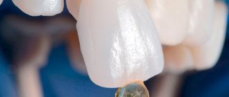
Removal of dystopic wisdom tooth
Removing a dystopic wisdom tooth is a fairly serious operation that requires professionalism and care from the doctor. Incorrect actions can lead to dislocation of adjacent teeth, lower jaw, injury to the mandibular canal and other problems. Therefore, it is very important to contact experienced specialists who are familiar with the specifics of removing complex teeth.
Indications for removing a dystopic tooth
Clear indications for removal:
- detection of a jaw cyst;
- traumatic dystopia;
- diseases of the jaw caused by this tooth;
- difficulties in treating caries in adjacent teeth;
- the appearance of pulpitis and periodontitis.
The practice is that the decision to remove a child is made taking into account all these factors. In an adult, it is more likely to decide whether to keep a tooth or not – removal is considered a universal solution, the simplest and safest. They resort to it in almost all cases.
Stages of removing a dystopic wisdom tooth
The removal operation is carried out in several stages. Compliance with technology allows you to protect the patient and simplify the operation for the doctor. Also, before starting work, detailed studies must be carried out: a full x-ray of the jaw is taken, all available caries is cured, etc.
During tooth extraction, the surgeon performs the following actions:
- Introduction of anesthetic. This may be local anesthesia or general anesthesia if several teeth are to be removed at once.
- Complete exposure of the tooth. To do this, a flap of mucous membrane and periosteum are peeled off.
- I sawed off the walls using a drill. This is necessary to align the element and make it easier to remove.
- Tooth extraction. The entire molar is removed using forceps. If the tooth was previously destroyed, its fragments are removed. It is important that the doctor removes all the fragments, otherwise inflammation will occur.
- Application of antiseptics to prevent the development of wound infection.
- Returning the mucosal flap to its place.
- Applying seams.
Once the wound is stitched, the operation is over, but the treatment is not. After a week, you should definitely see a doctor to have the stitches removed. It is also advisable to be observed by a specialist for 1-2 months to make sure that the removal had no consequences.
Removal of a dystopic tooth in pictures
Postoperative care
After extraction of an impacted tooth, it is recommended to adhere to the following rules:
- For 20 minutes after removal, you need to keep the tampon in your mouth, pressing it against the wound to stop bleeding.
- If the bleeding has not stopped and is going too intensely, you need to contact a specialist again and immediately.
- It is important not to eat anything for at least 4 hours after extraction; you are allowed to drink only clean water. After this time, you can eat only soft and warm food until the wound heals.
- If the mucous membrane is swollen, it is recommended to apply a cold compress to the cheek for 10–15 minutes.
- In case of inflammation in the area of the extracted tooth, it is important not to heat the hole and consult a doctor.
- A blood clot that has formed in the wound cannot be removed, as it protects it from bacteria.
- From the second day after removal, you can rinse your mouth with a weak solution of Furacilin, a decoction of sage or chamomile to prevent infection.
- Severe pain can be relieved with analgesics.
On a note!
The tissue of the hole after extraction of an impacted tooth heals on average in 3 to 4 weeks. During this period, they monitor the healing process and consult a doctor if concomitant dental problems develop.
Forecast of dystopia
The appearance of a dystopic tooth in a child is not such a serious problem (especially if there is only one). Before a person’s facial skeleton stops growing, the position of the main teeth can be normalized quite quickly. Therefore, it is recommended to begin treatment immediately after identifying the problem. In this case, all the teeth will most likely fall into place and in the future, in adulthood, will no longer cause concern.
If correction of dystopia in an adult is required, this almost always causes many difficulties. After 18-20 years, teeth are no longer so mobile, so correcting their position requires many preparatory interventions. It is much easier to remove a problematic tooth than to try to put it back in place.
Being cured in a timely manner, dystopia has virtually no negative impact on a person’s future life. Most often, the correct bite is maintained for a long time and does not require supervision by a doctor.
Bottom line
If it is determined that tooth retention has no prospects, it is removed. There is no prevention in this case. Considering that elements that have not erupted are potentially dangerous for the development of complications, the decision on their extraction or preservation is made by a specialist individually.
Sources
- https://ultrasmile.ru/retinirovannye-i-distopirovannye-zuby/
- https://www.KrasotaiMedicina.ru/diseases/zabolevanija_stomatology/impacted-tooth
- https://guide-dental.com.ua/retinirovannyj_zub/
- https://DentConsult.ru/polost-rta/retinirovannye-zuby.html
- https://CreateSmile.ru/retinirovannyj-zub/
- https://mildent.ru/retinirovannyy-klyk
- https://jsmiles.ru/ortodontiya/15-anomalii/261-retinirovannyi/
Prevention of dystopia
Dentists are confident that if you carefully monitor your child’s dental health from early childhood, you can avoid most bite problems. Dystopic teeth are no exception, because their appearance is not always associated with hereditary factors. To prevent dystopia, doctors recommend the following:
- proper nutrition, adherence to the daily routine of a pregnant mother;
- prevention of jaw injuries from an early age;
- regular visits to the dentist, wearing devices to correct malocclusion;
- giving up bad habits (including weaning off the pacifier after a year and from thumb sucking in sleep), etc.
If you and your child regularly visit a dentist, he will give recommendations on what exactly to do in your case.
How is the impacted segment extracted?
Removing an impacted tooth is a complex surgical procedure. You can trust its implementation only to an experienced dental surgeon. The cost of removing an impacted tooth exceeds the cost of removing normally erupted teeth, and the patient also needs to take this fact into account. After the operation, the patient has to deal with pain for some time, and there is a high probability of developing complications after removal of the impacted segments. To minimize all the troubles after surgery, the patient must follow dental recommendations and properly care for the oral cavity.
The operation to remove the impacted segment proceeds according to the following plan:
- Diagnosis of the problem and sanitation of the oral cavity. If necessary, a few days before the operation the patient is prescribed vitamins and sedatives.
- Anesthesia. Local or general anesthesia may be used.
- Cutting through the gum and removing soft tissue to expose the bone. Work with gum tissue is carried out using a laser or scalpel. If the doctor uses a laser, the gums tolerate the intervention better, but the cost of the procedure increases.
- Preparation of bone tissue with a bur and opening access to the segment to be removed.
- Extraction of the entire dental unit using special forceps. In cases where the tooth cannot be removed immediately, the doctor has to saw it with a bur and remove it piece by piece.
- Plastic surgery of hard/soft tissues, suturing (if necessary), treating the operated area with antiseptics and anti-inflammatory drugs.
How is complex removal performed?
Extraction of impacted molars is a procedure with increased trauma and is classified as complex. You will have to peel off the mucoperiosteal flap and separate the tooth from the bone using a bur. Then you need to dislocate it with forceps or an elevator, and after removing it, apply sutures. If exposure of adjacent roots is observed, resection of their apex is performed, followed by retrograde filling2. After the procedure, painkillers and antibiotics, and oral baths with an antibacterial solution may be prescribed. Recovery will take a little longer than after simple removal - up to a week.
Removing such a tooth is a full-fledged operation
The duration of the procedure itself can take from 20 minutes to an hour. For pain relief, local anesthesia is used, including in combination with sedation. In some cases, namely if it is necessary to extract several impacted molars at once or if the patient has insurmountable dental phobia, the operation can be performed under general anesthesia. To do this, the clinic must have the appropriate license, a qualified anesthesiologist, as well as all the necessary equipment to monitor the condition of the patient’s body and provide an emergency response in case of unexpected reactions from his body.
This is interesting: Dangers of radicular dental cyst and methods of its treatment
If the crown has erupted almost completely, sometimes it can be removed without cutting the mucosa. It is enough to grab the visible part with forceps, unscrew it and, after dislocation, carefully remove the tooth from the socket. It should also be noted that the denser structure of the bone tissue of the lower jaw somewhat complicates the procedure and often requires more powerful anesthesia with complete blocking of the conduction of the mandibular nerve.
Contraindications
Surgery to remove an impacted tooth is contraindicated in the following cases:
- state of hypertensive crisis;
- acute stages of heart disease or diseases of the nervous system;
- existing viral or infectious diseases:
- existing diseases of the blood and hematopoietic system;
- in the last phase of menstruation in women;
- if less than 14 days have passed since the operation to terminate the pregnancy.
Conclusions. Expert advice
Dystopia is the incorrect position of a tooth in the jaw: its displacement forward or backward, down or up, as well as rotation around its axis or tilt. The appearance of such an anomaly is associated both with hereditary factors and with bad habits or mechanical damage to the jaw. The sooner dystopia is detected, the easier it is to cure. In adults, the wisdom tooth most often found to be dystopic is the third molar, because there may simply not be enough space in the jaw for it to erupt.
The consequences of such anomalies include general malocclusion, frequent inflammatory diseases, impaired chewing function, and much more. Depending on the complexity of the case, dystopia is treated either by tooth extraction or with the help of braces. If the decision is made to remove it, it is important to carry out the operation carefully and correctly. If treatment is started on time, the prognosis for the disease is positive.
Classification of dental trauma
Let's look at several classifications of dental trauma.
According to the International Classification of Dental Diseases based on ICD-10 (WHO, Geneva, 1997), the following dental injuries are distinguished:
- S02.5 Tooth fracture.
- S02.50 Fracture of tooth enamel only;
- S02.51 Fracture of the crown without damage to the pulp;
- S02.52 Fracture of the tooth crown with damage to the pulp;
- S02.53 Fracture of tooth root;
- S02.54 Fracture of the crown and root of the tooth;
- S02.57 Multiple fractures of teeth;
- S02.59 Tooth fracture, unspecified;
- S02.2 Tooth dislocation;
- S02.20 Tooth luxation;
- S02.21 Tooth intrusion or tooth extrusion;
- S02.22 Tooth dislocation (disarticulation).
If we talk about the possible causes of dental trauma, then the following causes of dental trauma are distinguished - the etiological classification of dental trauma:
- Domestic or domestic dental trauma;
- Road or automobile dental trauma;
- Sports dental injury.
Domestic trauma occurs in 60% of patients who complain of dental trauma. And among these 60%, more than half are due to trauma to the central incisors of the upper jaw. Also, men use dental herbs more often than women J
Classification of dental trauma by time of occurrence can be:
- Acute tooth trauma
- Chronic tooth injury
Acute tooth trauma
Acute tooth trauma most often occurs as a result of a strong, simultaneous impact. The cause of acute dental trauma may be
- hit,
- falling face down
- falling on your back
- chin strike and much more;
Chronic tooth injury
Chronic tooth trauma is caused by constant mechanical impact. That is, there is a constant thinning of the tooth enamel. The most common causes of chronic tooth trauma are:
- Bad habits such as biting threads, biting nails, gnawing seeds and nuts, etc.
- Incorrect installation of the intracanal pin,
- Orthodontic treatment,
- Occlusal trauma,
- As a complication of fluorosis, enamel hypoplasia, cysts and tumors in the jaw.