Molars (6, 7 and 8 teeth) – where they are located, what structural features they have and nuances in treatment
A healthy adult should normally have 32 teeth - 16 each in the upper and lower jaws.
Moreover, they all differ in shape and structure from each other, depending on the location and their immediate tasks. Molars are the sixth, seventh and eighth teeth located on both sides of both jaws. Their main purpose is to ensure thorough chewing of food. They must be strong and hardy, and these requirements partly explain their special structure. Further in this article, read more about molars – what kind of teeth they are, when they “come out” to the surface in children, and whether there are any nuances associated with their treatment.
Upper molars
For the upper row of the jaw, the molars have the following anatomical surfaces:
- The buccal part is convex, with a prominent groove that is most susceptible to caries. When performing hygienic cleaning, it is recommended to pay special attention to this area.
- The chewing side has some differences and a large number of tubercles, which increases the role of this group in performing the chewing function. One of these features is the triangular trigon, which unites the following types of tubercles - protocone, metacone, paracone.
- The palatal part is distinguished by such a characteristic feature as the Carabelli tubercle, located on the border of the surface from the side of the palate and the medial area. In rare cases it has a separate root.
- The mesial part has a convex or straight line between the cementum and enamel. The root system is complex and can be barrel-shaped, divergent, or cylindrical.
- The distal one resembles the medial one and requires special attention when cleaning (dental floss is recommended to completely remove any remaining food).
Where are molars located in the mouth?
The central teeth and those located on the sides belong to the incisors. They are largely responsible for how a smile looks. These are followed by canines and 2 more premolars on each side on both jaws. The last ones in the row are called molars - there are 8 or 12 of them in total, if together with wisdom teeth (eights). They are the sixth, seventh and eighth elements in the series.
Molars are the largest in the mouth, characterized by a wide chewing surface with pronounced tubercles and depressions in the center. They are also covered with an enamel layer, but, unlike the front incisors, they can withstand pressure weighing up to 75 kg1. Durable and wear-resistant, they are designed for constant mechanical impact, namely for thoroughly crushing food. Below is more information about where they are located and what features they are characterized by.
Molars are the largest in the mouth and have a wide chewing surface.
Location on the upper jaw
They are somewhat larger in size than their antagonists, only there are usually 4 tubercles on their chewing surface, not 2. This feature allows them to grind food better. Most often, they have 3 roots, and in about 10% of cases, as many as 4 of them are formed in the second molars. Eights have from 1 to 5 roots, but in the vast majority of cases there are only 2-3 of them.
The structure of the elements on the lower jaw
The first and second ones are almost identical in structure. Only the surface of the first is more ribbed; it can have up to 6 tubercles. But the second one has 4 of them according to the standard. The third ones, they are also eights, like on the upper jaw, are underdeveloped, more often they are only half-cut and initially lie in the wrong position. Their root system is very tangled, and the canals are almost always impassable.
The photo shows the numbering of teeth
Location of premolars
If the fangs are involved in tearing off pieces from the whole product, then the premolars are the rudiments of the chewing group of teeth. They are called small radical elements. They are located behind the fangs in two units, and outwardly they vaguely resemble them. The premolar units have 2 cusps on the chewing surface. This promotes good food grip, and the wide crown successfully chews food. The peculiarity of these rudiments is that they are absent in the primary dentition, but in the permanent dentition there are eight of them. In addition to grinding food, small radical units are capable of crushing it and roughly grinding it.
A characteristic feature of these masticatory organs is the presence of 2 tubercles on top of the crown. They are called buccal and lingual. They have a single root. The first premolar rudiments on both jaws are key stable units, while the second are variable organs that often undergo reductional changes. Some medical scientists view premolars as canines connected to each other. The upper and lower premolar units are structurally different.
Features of the lower premolars
The premolars of the lower jaw have a specific anatomy, a more rounded shape, similar to a barrel. Its crown is significantly inclined towards the tongue. The organ is stable, solid, and has excellent resistance. It is able to withstand high loads and considerable pressure when chewing. Units have one root. Characteristic features of premolar rudiments:
- Weak expression of the lingual tubercle, which is part of the median ridge.
- The furrow between the elevations is weakly expressed.
- The lingual tubercle, which looks like a semicircular belt, is slightly separated.
- Its convexity faces the tongue.
- The sharp tip is located on that tubercle that stands out well.
- The groove divides the lingual tubercle into 2 parts.
The length of the first unit is on average 22 millimeters. Usually she has one channel. It is very rare that two canals that converge are diagnosed. The largest cavity is in the area below the neck. The canal has an oval shape and ends in a narrowing. The root is characterized by a distal deviation. The length of the second rudiments is from 20 to 24 millimeters. In most cases there is only one root. There are organs with two roots. One root can have 2 independent channels. There is one channel inside 1 root. Near the top it can be divided into 2 separate passages.
Features of the upper premolars
The premolars of the upper jaw always have crowns with pronounced angles. Their edges are clearly aligned, and the main elements of the crowns are well defined. The first premolar organ has 2 roots, as well as:
- The average length is 21 millimeters.
- Most often there are 2 roots and two canals.
- There can be one root with one channel.
- Sometimes there are two canals in one root.
- It is rare that a unit has 3 roots and three canals.
One third of the roots have a distal bend. The direction of the organ cavity is buccal-palatal. It is located quite deep, on the same level with the neck of the rudiment. Its covering is a thick layer of dentin. The shape of the canal mouths is funnel-shaped. This allows you to freely enter the canal when opening the cavity. Second units:
- The average length is from 2 to 2.4 centimeters.
- Often such organs have 1 root and one canal.
- There may be 2 roots and two canals.
- Rudiments with 3 roots and three canals are very rare.
Typically, the canals of such teeth have two orifices. The canals are connected and also opened by a single apical foramen. 20 percent of units have 2 holes. The cavity of the chewing organ is located at the level of the neck itself. The shape of the channel is slit-like.
Features and nuances that need to be taken into account during treatment
On the chewing surface of the molars there are tubercles and, accordingly, grooves - the latter are called fissures. They are cone-shaped, teardrop-shaped and grooved - they provide the ability to thoroughly crush and grind incoming food. Plaque settles in the fissures, food debris gets stuck, so they are especially susceptible to caries. More often in children's, but also in adult dentistry, a sealing procedure is carried out to prevent carious processes.
The photo shows chewing teeth
On a note! Many people call all chewing teeth “molars,” but this is not entirely true. In fact, premolars (small) and molars (large) are molars - their roots do not dissolve when the bite changes.
The first ones on the upper jaw are sixes
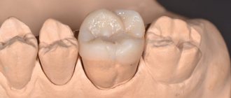
The first ones on top are considered the largest in the mouth. Their length can reach up to 2.5 cm. They have a diamond-shaped chewing surface on which there are four tubercles - two on the side of the cheeks and tongue. Moreover, the former have a conical shape, while the latter have a rounded shape.
On one of the anterior tubercles there is also an additional tubercle, and on the lateral surface on both sides there is a small groove. The surface on the lingual side is more convex. When a primary dentition changes, these sixth teeth are usually the first to grow. The process usually begins at the age of 5-8 years.
The photo shows the structure of the first upper molar
Second permanent molar – upper seventh
Slightly smaller than sixes in size, the second upper molars usually grow to 1.9-2.3 cm. The chewing surface is diamond-shaped or triangular with smoothed edges, 3-4 cusps, but without additional ones. In most people (about 57%), the upper second have 3 roots and 3-4 canals. In rare cases, there are 4 roots or the two front ones grow together.
The photo shows the structure of the second upper molar
Third or eight on the upper jaw
The eighth teeth in the top row, also known as wisdom teeth, are distinguished by their diamond-shaped or spike-shaped crown part. There can be from 2-4 to 6 tubercles on the chewing surface. The root system is often very twisted and tangled. Usually the top eights have 2-3 roots, less often their number varies from 1 to 4-5 pieces.
It should be noted that in about 15% of all people, wisdom teeth do not grow at all. It is believed that this is not an anomaly, but a natural evolutionary process. In fact, these are rudiments, the need for which a person lost quite a long time ago, as he developed and abandoned raw hard food in favor of heat-processed food. But to this day, for most of us, by the age of 20-25, eights begin to emerge - this process is usually difficult and painful, often accompanied by inflammation and in most cases requires complete removal of the problematic element.
The photo shows the structure of the third upper molar
Another difficulty is that the canals in the root system of third molars are almost always deformed and impassable. These teeth, as a rule, remain impacted and dystopic, that is, they do not erupt completely and initially grow in the wrong direction. When the inflammatory process occurs, tissue infection occurs, caries or pulpitis develops. It is almost impossible to cure a tooth with such a complex and difficult-to-reach root system. However, if the eights are completely fine, doctors leave them. Subsequently, they can become a good support for the prosthesis.
Sixths on the lower jaw
These are the largest molar teeth on the lower jaw, from 1.9 to 2.4 cm long. The diamond-shaped chewing part is covered with tubercles - 2 on the lingual side and 3 on the cheek side. They are separated by grooves - fissures. The crown is slightly inclined inward, the surface on the side of the cheek is voluminous, but not as convex as on the upper antagonist. There are usually 2 roots and 3-4 canals, in rare cases there may be 2.
The photo shows the structure of the first lower molar
Second lower molars and their features - sevens
The shape is almost identical to the neighboring sixes, but slightly smaller in size. Usually they have 4 tubercles, sometimes with a small additional one. They are also separated by fissures by longitudinal and transverse pits. They usually have 2 roots, but in some cases they grow together. There are usually 3 to 4 root canals.
The photo shows the structure of the second lower molar
Eights – wisdom teeth of the lower jaw
The crown of the figure eight can have a different shape, but there are usually 4-5 tubercles on the chewing surface. Most often there are 2 roots, but there are cases when there is only one. As on the upper jaw, on the lower jaw they grow dystopic or remain impacted, have a complex and difficult-to-reach root structure, and therefore are most often subject to removal.
The photo shows the structure of the third lower molar
Molar placement
The molar large units are located on the back of the jaw. Each jaw has six of them. They decrease in size from front to back. That is:
- the first rudiment is the largest;
- the second one is smaller;
- the third tooth is small;
The molar units are located most posteriorly in the arc. They are also called extreme. The number eight, that is, the wise tooth, grows in the period from 18 to 35 years. It happens that he does not appear at all. Row anomalies and bite defects can form in the absence of individual units or groups of rudiments. Often this concerns the end organs. Remember that normally there should be 28 units without eights, and with them 32 organs. The primary dentition has eight outermost units, the permanent dentition has twelve. In the molar group, the sixth units are key, and the 7s are variable, the most variable being the eights.
If caries treatment is carried out in a timely manner, the chewing organs will last a long time. The health of the whole body, the functioning of the gastrointestinal tract, liver and kidneys depends on this. Therapy should begin at the white spot stage, while the cavity has not yet formed. Prevention of caries is of great importance. To do this, you need to visit the dentist at least once every 6 months, carefully care for your oral cavity, and follow the dentist’s recommendations. Modern medicine has technologies the use of which can save even a dilapidated organ. Therefore, the key to health is timely treatment after a complete diagnosis and correct diagnosis.
It is important from childhood to teach a child to brush his teeth and treat the oral cavity. Children should understand that the main cause of caries is pathogenic bacteria living in dirty plaque. Therefore, trips to the hygienist will not allow the active proliferation of pathogenic microbes, subject to regular preventive cleanings.
Structural features
Those who looked at their teeth in the mirror noticed that they all had a different design. The structure of the crowns depends on:
- from the arrangement of organs in a row;
- depending on the shape of the unit;
- depending on the size of the rudiment;
The anatomical structure of the mammary organs is basically the same as the permanent units. True, temporary rudiments have distinctive features.
- They are smaller in size;
- The width of their crown prevails over the height;
- In the cervical area, the enamel layer is thickened;
- The shade of the enamel is bluish;
- The roots are widely spaced and short;
In addition, in the alveolar arch the temporal units are arranged steeply vertically. This is influenced by the fact that behind the roots there are already rudiments of permanent teeth. The upper units have 3 roots, one of them is lingual and 2 are buccal. The crowns have a cuboid shape, on the chewing surface of which there are 3 or more tubercles. The groove that separates the tubercles has two parallel lines and one perpendicular. The crowns of the rudiments on the lower jaw are somewhat elongated, and the fissures are located almost cross-shaped. These chewing organs have 2 roots. In eights, the roots often merge, the shape of such a root is conical. It happens that wise teeth grow with 4-5 roots. Since the upper and lower eights are close to each other, occasionally they can grow together.
To keep the chewing organs healthy, dentists may prescribe dental remineralization. This procedure restores the enamel layer well due to its saturation with deficient elements. The enamel ceases to be porous, acids do not penetrate its pores and do not contribute to the leaching of useful minerals. This event prevents and eliminates caries at an early stage, improves the aesthetics of crowns with fluorosis, reduces the sensitivity of units, brightens crowns and normalizes the microflora of the oral cavity.
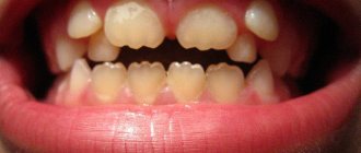
When do permanent molars appear in children?
When chewing teeth come in, as in the case of all others, the child may exhibit characteristic symptoms, only in this case they are more pronounced. There may be a headache, and the temperature may rise to 38 ⁰C. If several climb at once, the immune system weakens and becomes susceptible to viruses. During this period, many children have a red throat, runny nose and enlarged submandibular nodes. About how long it takes molars to cut and how many of them erupt in the end, the information is below:
- up to 3 years – the child grows only incisors, canines and 2 premolars on both sides,
- 6-8 years - the first molars are cut, and with them 4 more elements appear in the rows. Some of them are permanent, others are milky, but soon they will change,
- 10-12 years – milk fours and fives begin to change,
- 12-13 years old - sevens come out from below and above,
- 16-35 years and older – third molars or wisdom teeth may emerge.
The photo shows a diagram of the eruption of permanent teeth.
“I remember my eldest son had a headache at night, he was 6 then. I cried a lot, my temperature rose, but not much. And no cough, no sore throat. I gave him Nurofen, drank it, and fell asleep. In the morning I wanted to take a look at your throat, and here on you - two teeth appeared on your lower jaw, right after the A’s.”
Olechka_K, from correspondence on the woman.ru forum
Molars for children are completely new teeth that grow on the farthest parts of the jaws. And permanent premolars appear after the loss of milk fours and fives.
Treatment methods for chewing teeth in adults and children
The whole difficulty lies in the difficult passage of the channels of the masticatory elements. When processing them, there is a serious risk of injury to the pulp, which can provoke pulpitis. In this case, two treatment options can be distinguished:
- depulpation – partial or complete removal of the pulp with further endodontic treatment, that is, cleaning and filling the canals. This method is more often used in adult dentistry,
Surgical method of pulp treatment - biological method - a medicine, usually an antibiotic, is applied to the affected area of the pulp, after which a temporary filling is placed on top. The active substances of the drug destroy pathogenic microorganisms and stop the inflammatory process. It must be admitted that pulpitis on baby teeth progresses much faster than on adults, so quite often the doctor has no choice and removes the affected temporary element. If necessary, prosthetics can be performed before a permanent one appears.
This method involves killing the pulp with medication.
Molars are directly involved in the process of chewing food. Like other teeth, they require timely diagnosis and treatment. And in case of their early loss, it is imperative to take care of their correct replacement - the best option for this would be implantation.
Regular cleaning is the basis of hygiene. But even if we follow this rule responsibly, we cannot completely clean out all food debris and bacteria in one such procedure. Subsequently, they transform into plaque and hard stone, which causes gums to become inflamed, teeth to decay and rot. Therefore, it is so important to regularly visit the dentist for a preventive examination and professional cleaning of plaque and deposits. It is also important not to forget to rinse your mouth after each meal, use floss and, preferably, an irrigator. Proper care of your oral health will be the best prevention of any dental diseases.
1Dmitrienko, CB Anatomy of human teeth, 2000.
Structure and functions
The structure of molars includes two parts:
- pulp, that is, soft tissue;
- group of hard tissues - dentin layer, enamel, cement.
Anatomically, the following areas are also distinguished:
- crown part protruding above the gum tissue;
- neck in the form of a border between the crown and root system, clinical and anatomical parts are distinguished;
- root system that provides fixation, located deep in the bone structure.
Eights belong to this group of teeth, but they may not be present in everyone. They do not carry any special functional load; if problems arise during development or eruption, removal of wisdom teeth is recommended.