Removing a tooth is the last thing a dentist will suggest. Due to a number of subjective and objective reasons, it is impossible to maintain the dentition in ideal condition until old age.
Dental defects are the loss of one or more dental units, leading to a violation of the integrity of the dentition, deviation in the development of physiological occlusion, and an abnormal arrangement of individual teeth.
Reasons contributing to the development of dental defects
Dental defects arise due to:
- untimely treatment of caries;
- inflammatory processes in the oral cavity;
- presence of malocclusion;
- genetic conditioning;
- diseases leading to changes in metabolic processes;
- damage to the jaw apparatus;
- various neoplasms.
Other causes of dental deformities
In addition to deformations of the development of the skull, other reasons can lead to changes in the dentition:
- Poor posture. When posture is impaired, the body begins to adapt. Not only the position of the spine changes, but also other parts of the musculoskeletal system. These adaptive changes affect the dentofacial apparatus. A defect in the dentition develops.
- Bad habits in a child. This may involve prolonged pacifier or finger sucking. Such habits greatly affect the formation of the dentition and can lead to tooth movement and changes in bite.
- Intrauterine pathologies. They may be due to various reasons. For example, diseases suffered by the mother during pregnancy. As a result, deformations of the jaws and tooth buds may develop.
- Periodontal diseases. Weakened tissues cannot hold teeth in their normal position. They begin to shift, the dentition becomes deformed.
- Partial edentia. When one or more units are lost, the rest begin to shift. As a result, a dental defect develops.
Pigmentation
If this defect appears, the color of the tooth changes. It may turn brown, gray or almost black, pink or red, yellow. The shade depends on what dye is applied to the crown. For example, if it is tea or coffee, the color will be brown, and if it is nicotine tar, it will be yellow or grayish. If the color of the crown changes unevenly and clearly visible pigmented areas appear, this indicates enamel defects. It absorbs dyes better where there is demineralization and the structure of the fabric is damaged. Pigmentation itself is not dangerous, but it can be associated with other dental health problems. It often appears in the presence of chips, poor hygiene, and some concomitant diseases. To restore pigmented areas, professional teeth cleaning and remineralization are performed. Sometimes whitening, restoration of crowns or installation of veneers may be required.
Types of dental deformities
Teeth can move from their normal position in different ways. Depending on the type of displacement, types of dental deformations are distinguished:
- Rotation of a tooth relative to its axis.
- Tilt towards the palate, cheek, tongue.
- Vertical displacement of teeth, leading to their lengthening.
- The distal displacement of one unit back in relation to the rest of the dentition.
- Mesial displacement is the opposite of distal displacement, since the deformed tooth moves forward.
- Crowding is characteristic of an underdeveloped small jaw. There is not enough space for normal development of the dentition. The teeth move in different directions, overlapping each other.
- Gaps and crevices in the dentition develop if the jaw size is large. Visible gaps (diastemas, tremata) form between adjacent dental units.
In complex cases, a combined displacement can be observed, combining different anomalies. Visually, such defects look like uneven teeth.
Prevention
A set of measures aimed at preventing the development of dental anomalies in children includes:
- balanced nutrition of a woman during pregnancy in order to provide the fetus with the substances necessary for its skeletal system;
- inclusion in the diet of a growing child of vitamins and microelements necessary for the proper formation and mineralization of teeth;
- preventing the development of bad habits in children that cause occlusion disorders (prolonged use of a pacifier, finger sucking);
- timely treatment of dental, infectious, endocrine and other somatic diseases;
- regular monitoring of the condition of the dental system.
Diagnosis of dental deformations
Dental deformities are not an independent diagnosis. Treating them only with traditional methods used to correct the bite is not always effective. If the cause that caused the deformation of the dentofacial apparatus is not identified and eliminated, there is a risk of relapse.
To make a diagnosis and select treatment options for dental deformation, doctors at our clinic use:
- Visual examination of the oral cavity. The doctor will definitely ask you when the problem began. This will help determine the causes of the development of malocclusion.
- Paraclinical studies. After a visual examination and medical history, you will be prescribed tests: x-rays of the teeth and alveolar process, x-rays of the temporomandibular joint, tomography, etc.
Examination of the dentition gives the dentist a lot of information about the bite. With its help, you can determine the nature of the deformation, the condition of the periodontium, and roots. Instrumental diagnostic methods are used to diagnose functional and morphological disorders.
With the help of examination and questioning, you can obtain other data on the condition of the dentofacial apparatus:
- Position of teeth in the jaw. The position of the dentition is assessed in relation to the sagittal plane, focusing on the line between the incisors. If this line is displaced, a more precise occlusion study is necessary. The doctor determines the size of the incisal overlap, the position of individual units, and the nature of their occlusal surface.
- Features of the movement of the lower jaw during opening and closing of the mouth. Zigzag movement of the jaw may indicate pathologies of the TMJ and masticatory muscles.
Impaired closure of teeth and their rotation around their axis. Movement of molars and premolars is easily diagnosed during examination.
Instrumental diagnosis of dental anomalies allows an accurate diagnosis.
Classification of anomalies of the dentofacial system
Bite defects develop for various reasons and cannot be ignored. This problem concerns not only aesthetics, but also general health. The pathological arrangement of teeth affects the functionality of the jaw. Due to poor quality of chewing food, various problems with the digestive organs arise.
Modern orthodontics offers effective treatment methods for children and adults with defects in the dental system. But, first of all, you need to make a correct diagnosis and determine the degree of complexity of the anomaly, which will allow the specialist to choose the right treatment in each specific case.
Let's start with the main thing - the classification of anomalies of the dental system, displayed in the diagnosis. We propose to consider the division of anomalies according to etiological and morphological characteristics (Kalvelis classification , orthodontics
).
Types of anomalies:
1. Anomalies in the number of teeth - supernumerary teeth, partial or complete hypodontia.
2. Anomaly of shape and size – ugly shape, spiky, oversized teeth.
3. Defects in the structure of hard dental tissues.
4. Premature or delayed teething in children.
5. Anomalies in the position of individual teeth in a row: distal, palatoglossal, mesial, labiobuccal, etc.
6. Diastema (disproportionately large spaces between teeth).
7. Crowding of teeth in a row.
8. Anomalies in the shape of the dentition (asymmetrical arrangement, V-shaped, etc.).
9. Sagittal malocclusion.
10. Vertical malocclusion (deep, open).
11. Transverse malocclusion (narrowed rows, disturbed relationship of lateral teeth, etc.).
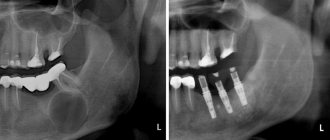
Anomalies of the dental system can be genetic, congenital or acquired. For example, intrusion is quite common in orthodontics.
, in which partial or complete immersion of the tooth crown into the alveolar process occurs, and the root moves into the jaw bone. This anomaly is acquired in nature and occurs as a result of tooth trauma due to a strong blow or fall.
Treatment
Treatment methods for dental deformities are chosen depending on the type of pathology. Most deformities not associated with the loss of antagonist teeth develop in childhood. Therefore, the problem needs to be resolved as early as possible. In orthodontics they use:
- Plates. Can be used immediately after baby teeth are replaced by permanent teeth. The plate puts pressure on the dentition, as a result it is aligned. Installation of plates is indicated for patients aged 7-12 years. At this time, the child’s bones are growing rapidly, so the technique will be effective.
- Trainers. They can be used in early childhood. The trainer is a silicone structure that resembles a mouth guard. They are worn for several hours. The elastic trainer protects the dentition from excessive pressure from the tongue and cheeks and prevents teeth from moving.
- Braces. Can be installed on children over 12 years of age. The orthodontist determines the need to use braces based on the results of the study.
These are the most common options for orthodontic treatment of dental deformities. Specialists at the Zuub.rf dental clinic believe that the approach to eliminating this problem should be comprehensive. It is important not only to visually straighten the dentition and correct the bite, but also to eliminate the cause that led to the development of the anomaly.
Hyperesthesia
This is a condition in which the enamel thins and becomes overly sensitive. Sometimes this can cause the shade of the crown to change, but more often hyperesthesia is manifested by acute aching pain when the teeth come into contact with irritants. The reaction can occur to drinks that are too cold or hot, sour or sweet. Hyperesthesia can have several causes, including too aggressive whitening, poor daily hygiene, regular damage to the enamel when brushing your teeth or due to contact with sour or sweet foods and drinks. It also occurs with calcium deficiency, malocclusion (if occlusion is disturbed) and as a complication of bruxism (teeth grinding during sleep). To treat hyperesthesia, you need to eliminate the cause of thinning enamel (for example, adjusting your daily dental care, correcting your bite, or using mouth guards for bruxism). To remove high sensitivity, remotherapy is performed. To do this, the teeth are coated with a remineralizing composition and fluoride varnish. While the enamel remains highly sensitive, it is better to rinse your mouth with clean water after each meal.
VI. GENERAL ISSUES
Partial absence of teeth (partial secondary edentia) is one of the most common diseases: according to the World Health Organization, it affects up to 75% of the population in various regions of the globe [13].
In our country, in the general structure of medical care for patients in dental treatment institutions, this disease makes up from 40 to 75% and occurs in all age groups of patients [9, 13, 19].
Partial absence of teeth (partial secondary adentia) directly affects the patient’s quality of life. Partial absence of teeth (partial secondary adentia) causes disruption, up to complete loss, of a vital function of the body - chewing food, which affects the processes of digestion and the intake of necessary nutrients into the body, and is also often the cause of the development of diseases of the gastrointestinal tract of an inflammatory nature .
No less serious are the consequences of partial absence of teeth (partial secondary edentia) for the social status of patients: disorders of articulation and diction affect the patient’s communication abilities; these disorders, along with changes in appearance due to loss of teeth and developing atrophy of the masticatory muscles, can cause changes in the psycho-emotional state, up to mental disorders.
Partial absence of teeth (partial secondary adentia) is also one of the reasons for the development of specific complications in the maxillofacial area, such as the Popov-Godon phenomenon, dysfunction of the temporomandibular joints and the corresponding pain syndrome.
Untimely restoration of the integrity of the dentition in the case of their partial absence (partial secondary adentia) causes the development of such functional disorders as overload of the periodontium of the remaining teeth, the development of pathological abrasion, and disturbances in the biomechanics of the dentoalveolar system.
Untimely and/or poor-quality treatment of partial absence of teeth (partial secondary adentia) leads to the development of diseases of the dental system such as periodontal disease, and in the long term - to complete loss of teeth - complete secondary adentia of both jaws.
The concept of “tooth loss due to an accident, tooth extraction or localized periodontitis” (K08.1 according to ICD-C - International Classification of Dental Diseases based on ICD-10) and terms such as “partial secondary edentia” and “partial absence of teeth” ( in contrast to edentia - a disorder of the development and eruption of teeth - K 00.0), are essentially synonymous and are applied both to each of the jaws and to both jaws. A synonym for the terms “partial absence of teeth” and “partial secondary edentia” is also the concept of a dentition defect, meaning the absence of one or more teeth.
Partial absence of teeth (partial secondary adentia) should be distinguished from primary adentia, in which a defect in the dentition has developed due to the absence or death of the rudiments of permanent teeth. Partial absence of teeth (partial secondary adentia) is a consequence of caries and its complications, tooth extraction and/or loss due to an accident (trauma), periodontal disease, etc.
Caries in our country is one of the most common diseases. Its prevalence in adults aged 35 years and older is 98-99%. The rates of caries complications are also high: the percentage of extractions in the age group over 35-44 years is 5.5, and in the next age group - 17.29% [33]. In the structure of dental care in terms of visits, patients with pulpitis, which, as a rule, is a consequence of untreated caries, make up 28-30% [32].
The incidence of periodontal disease in the age group 35–44 years is 86% [31].
These diseases, if untimely and poorly treated, can lead to spontaneous loss of teeth due to pathological processes in periodontal tissues of an inflammatory and/or dystrophic nature, to the removal of teeth that cannot be treated and/or their roots due to deep caries, pulpitis and periodontitis.
Untimely orthopedic treatment of partial secondary adentia, in turn, causes the development of complications in the maxillofacial area and the temporomandibular joint, and also aggravates the process of tooth loss.
The main sign of partial absence of teeth (partial secondary adentia) is the absence in the dentition of one to fifteen teeth on one of the jaws [8, 35].
The clinical picture is characterized by the absence of one or more teeth in the presence of one or more natural teeth or their roots. Manifestations of partial absence of teeth (partial secondary adentia) depend on the topography of the defects and the number of missing teeth and are varied.
A feature of this pathology is the absence of pain syndrome in patients. In the absence of one or two, and sometimes several teeth, patients often do not feel discomfort and do not consult a doctor.
The partial absence of even one tooth in any functionally oriented group of teeth can lead to the development of the Popov-Hodon phenomenon, direct or reflected traumatic nodes, resulting in inflammation in the gingival margin, destruction of bone tissue and the development of pathological pockets, primarily in the area of the teeth , limiting the defect.
In the absence of one or more frontal teeth in the upper jaw, the clinical picture is characterized by the symptom of “retraction” of the upper lip. With a significant absence of lateral teeth on the soft tissues of the cheeks and lips.
In the absence of even one frontal tooth in the upper and/or lower jaw, diction may be impaired.
Partial absence of teeth in both jaws without preservation of antagonizing pairs of teeth in each functionally oriented group of teeth leads to a decrease in the height of the lower part of the face, often to the development of angular cheilitis (“jams”), pathology of the temporomandibular joint, changes in facial configuration, expressed nasolabial and chin folds, drooping corners of the mouth.
The partial absence of chewing teeth causes dysfunction of chewing; patients complain of poor chewing of food. Sometimes significant partial adentia is accompanied by habitual subluxation or dislocation of the temporomandibular joint.
After the loss or removal of teeth, atrophy of the periodontal ligaments occurs in the corresponding areas of the jaws; with the loss of more than two teeth, atrophy of the alveolar processes themselves gradually develops, progressing over time.
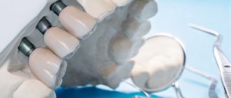
Partial absence of teeth (partial secondary edentia) is an irreversible process. Restoring the integrity of the dentition is possible only by orthopedic treatment methods using fixed and/or removable denture structures.
CLASSIFICATION OF PARTIAL ABSENCE OF TEETH (PARTIAL SECONDARY EDENTIA)
In clinical practice, partial absence of teeth (partial secondary adentia) of the upper jaw and lower jaw is not distinguished. The classification principles are the same for both jaws. The Kennedy classification of partial secondary adentia (dentition defects) has become most widespread and practically used [8, 14, 17].
This classification distinguishes four classes: 1. Bilateral distally unlimited defect (end defect). 2. Unilateral distally unlimited defect (end defect). 3. Unilateral distally limited defect (included defect). 4.Lack of front teeth (defect in the frontal region) (included defect).
Each class has a number of subclasses. In the clinical application of the Kennedy classification, the doctor rarely encounters “pure” classes; variants of subclasses and/or a combination of defects of various classes and subclasses are much more often observed.
Another well-known classification of dentition defects is the classification of E.I. Gavrilova [8]. It distinguishes four groups of defects: 1. End one-sided and two-sided. 2. Included (lateral - one-sided, two-sided and front). 3. Combined. 4. Jaws with single teeth preserved.
Close to this classification is the Wild’s classification of defects [17, 54], which distinguishes the following main categories (classes) of partial secondary edentia: 1. Unilateral or bilateral terminal defect of the dentition. 2. One or more included defects. 3. Combination of terminal (terminal) and included (included) defects of the dentition.
In recent years, due to the importance of assessing the functional state of the dentition in partial secondary edentia, modifications according to Wild are increasingly used [17, 54].
When determining patient models, taking into account the functional state of the dentition and the possibility of restoring lost functions, which depends on the topography and the number of remaining teeth, it is more convenient to take as a basis the principle laid down in the classifications of E.I. Gavrilova and Wilda.
GENERAL APPROACHES TO DIAGNOSIS OF PARTIAL ABSENCE OF TEETH (PARTIAL SECONDARY EDENTIA)
Diagnosis of partial absence of teeth (partial secondary adentia) is made through a clinical examination, history taking and clinical examination. Diagnostics is aimed at eliminating factors that prevent the immediate start of prosthetics. Such factors may include the presence of:
- unsanitized teeth; — not removed roots under the mucous membrane; - exostoses; — tumor-like diseases; - inflammatory processes; - diseases and lesions of the oral mucosa.
When diagnosing, it is necessary to take into account the results of clinical and X-ray examinations of existing teeth, especially those planned for support, including their periodontal status, as well as the general and functional state of the dental system.
GENERAL APPROACHES TO TREATMENT OF PARTIAL ABSENCE OF TEETH (PARTIAL SECONDARY EDENTIA)
Basic principles of orthopedic treatment of partial secondary adentia:
1. When planning orthopedic treatment, the priority should be to preserve the remaining teeth.
2. Each tooth planned to support a prosthesis must be assessed from the perspective of the condition of hard tissues, pulp, periapical tissues, and periodontium. Depending on the results of this assessment, the support is determined as reliable, questionable or unsatisfactory. First of all, reliable teeth should be used for supports. The preservation of a tooth largely depends on its strategic importance as a support for the prosthesis, as well as on the ratio of labor intensity and cost of treatment measures necessary to preserve it and achieve results.
3. You cannot begin prosthetics without preparatory measures, if necessary.
4. Not every dental defect requires prosthetics. Prosthetics up to the complete set of dentition is not mandatory. The individual characteristics of the patient’s dental system play a decisive role.
5. Orthopedic structures should provide opportunities for optimal oral hygiene.
6. When making fixed bridges, structures of short length are preferred. Long structures that connect several functionally oriented groups of teeth into a single block should be avoided. Expanding the scope of prosthetics is justified only in conditions where this solution is the only opportunity to ensure optimal individual functioning of the dentofacial system.
7. Poor oral hygiene of the patient is a relative contraindication to fixed prosthetics.
8. The worse the patient follows medical recommendations and cooperates with the doctor, the simpler the orthopedic design should be [51].
The goal of treating patients with partial secondary adentia includes simultaneous solution of several problems:
— restoration of sufficient functional capacity of the dental system;
— prevention of the development of pathological processes and complications; — improving the quality of life of patients;
— prevention or elimination of negative psycho-emotional consequences associated with missing teeth.
Denture fabrication is not indicated if the existing denture is still functional or if its function can be restored (eg, repair, relining).
The manufacture of a prosthesis includes: examination, planning, preparation for prosthetics and all activities for the manufacture and fixation of the prosthesis, including the elimination of deficiencies and control. This also includes instructing and teaching the patient how to care for the denture and the oral cavity.
The orthopedic dentist must determine the features of prosthetics depending on the anatomical (taking into account the topography of defects in the dentition), physiological, pathological and hygienic state of the patient’s dental system.
When choosing between equally effective types of prostheses, the doctor should be guided by cost-effectiveness indicators. In cases where it is impossible to immediately begin and complete treatment as planned, the use of temporary prostheses, including removable or fixed immediate prostheses, is indicated. You can use only those materials, instruments, equipment, systems (for example, implantation), means of prevention and treatment that are approved for use by the Ministry of Health of Russia, clinically tested, the safety of which has been proven and confirmed by clinical experience. If there is a confirmed allergic reaction of oral tissues to the prosthetic material, tests should be carried out and the material that has proven to be tolerable should be selected.
When planning and conducting orthopedic treatment, it is necessary to take into account the health status, somatic status, and chronic diseases of the patient.
The most important stage of treatment is preparing the dental system for prosthetics.
Prosthetics should be carried out after the following measures:
— complete sanitation of the oral cavity should be carried out (pay attention to teeth with increased sensitivity);
— the feasibility of preserving teeth affected by caries and other diseases (x-ray and electroodontometric control), including filled ones, teeth with periodontal lesions, etc., should be checked when planning them as supporting ones;
— pulpless teeth must have roots sealed to the apex (x-ray control);
— the necessary treatment must be carried out for diseases of the periodontium and oral mucosa;
— if pathological processes in the teeth and jaw bones are suspected, it is necessary to conduct an X-ray examination;
- teeth and roots that cannot be preserved must be removed.
If a pathological process is detected on an x-ray, it should be eliminated before the fabrication of a permanent orthopedic structure. Any treatment aimed at eliminating oral diseases that prevent permanent dentures must be completed in full.
If it is impossible to completely eliminate pathological processes, primarily in the periapical tissues, prosthetics should take into account the possibility of subsequent surgical intervention. In such cases, radiological monitoring is necessary, no later than after 9 months [38].
Making a prosthesis for the jaw with partial secondary edentia includes: preparation of teeth, casts (impressions) from both jaws, production of diagnostic and working models, determination of the central relationship of the jaws, checking the design of the prosthesis, application, fitting, fitting, installation, fixation, remote control and corrections .
In the treatment of partial absence of teeth (partial secondary edentia), fixed bridges, cantilever fixed dentures, single crowns on teeth, partial removable plate and clasp dentures are used.
Bridges are usually indicated if:
- up to 4 incisors are missing, but chewing function is provided by natural teeth or existing bridges;
- in the area of lateral teeth on one side of the jaw no more than 3 teeth are missing and the dentition can be restored using a bridge with supports on both sides;
— the bridge will serve to fix the removable denture [51]. Thus, bridges are made with support on natural teeth on both sides (with the exception of cantilever ones). Bridges are not indicated:
- with insufficient ability of the periodontium to withstand loads and such general somatic diseases that adversely affect periodontal tissue;
- if an x-ray of the supporting tooth indicates a pathological process that cannot be stopped.
When replacing missing molars, the body of the bridge should be made with a wide wash (about 1 mm), not adjacent to the mucosa. In other areas of the jaws, the body of the bridge should not be adjacent to the mucous membrane (the tip of the dental probe should pass freely under the body of the bridge). It should be noted that the concept of a “tangential” intermediate part of a bridge prosthesis refers to a visual impression; in fact, a distance between the body of the prosthesis and the mucosa must be ensured, sufficient for free rinsing.
When using solid-cast metal-ceramic and metal-plastic bridges and crowns, a “garland” is always made from the oral side. Crowns with veneers and facets in bridges on the upper jaw are made only up to the 5th tooth inclusive, on the lower jaw - up to the 4th inclusive. The lining of the chewing surfaces of the lateral teeth is not shown in principle.
Crowns are indicated: - to preserve the tooth, if this cannot be achieved by other methods for a long time; - to protect the tooth from damage by the prosthesis; - for supporting the prosthesis; — to change the relationship of the jaws during prosthetics [51].
Artificial stamped and solid crowns for partial secondary edentia can be used to cover abutment teeth in the manufacture of removable partial laminar dentures and clasp dentures.
In the manufacture of artificial solid-cast crowns, four types of preparation are used, each of which has its own advantages and disadvantages, as well as recommended indications (Table 1).
Table 1
Clinical features of various forms of preparation of tooth stumps for crowns
| Preparation form | Advantages | Flaws | The appropriate type of prosthesis |
| Tangential (no shoulder) | Maximum preservation of hard tooth tissues, ease of implementation, smaller gap between the edge of the crown and the stump of the tooth in case of inaccuracy in the preparation form or errors during cementation | Lack of a clear preparation boundary, risk of excessive grinding of hard tissues, greater risk of trauma to the gingival margin | Solid crowns (without veneer), metal-ceramic and metal-plastic crowns with oral and vestibular garland |
| With a semicircular ledge | Clear preparation margin good distribution of impression, modeling and fixation materials relatively gentle method for hard tissues | The relative difficulty of solving the problem of prosthesis retention with a short clinical crown is the risk of complications (gingival recession) with an excessively convex veneer in the area of the gingival margin | Solid metal-plastic and metal-ceramic crowns |
| Preparation with a rectangular circular shoulder | Clear preparation margin Possibility to achieve optimal aesthetics due to sufficient space Low risk of excessive grinding in the cervical area | Large loss of hard tissues, danger of pulp damage, large cemented gap due to inaccurate preparation form or errors during cementation | Porcelain (jacket) crowns and half-crowns, metal-ceramic crowns with fired shoulder mass |
| Preparation with a shoulder-bevel at an angle of 135° | Clear preparation boundary, low risk of excessive grinding in the cervical area, smaller gap between the crown edge and the tooth stump in case of inaccurate preparation shape or cementing errors | Difficulty in performing, loss of hard tissue | Metal-ceramic and metal-plastic crowns, especially for front teeth |
In the case of partial absence of teeth and the impossibility of making a fixed bridge structure, as a rule, one-piece clasp dentures should be made. In this case, it is necessary to take into account the condition of periodontal tissues and the principles of hygiene.
As teeth are lost and the length of the toothless portion of the alveolar process (dentition defect) increases, the indications for the use of removable laminar dentures expand.
In the case of partial absence of teeth and the impossibility of redistributing the load on the periodontium of the supporting teeth, as a rule, partial removable lamellar dentures made of plastic without complex support-retaining elements are indicated.
The use of combined (combined) dentures is indicated if, using the necessary connecting elements and a sufficient number of preserved supporting teeth, it is possible to achieve functionally more favorable fixation and stabilization than using a clasp denture with clasp fixation or a partial removable laminar denture.
Connecting elements such as rod (beam) systems, telescopic crowns and attachments can only be used if the load is evenly distributed on the remaining abutment teeth.
In the absence of strict specific indications for prosthetics using dental implants, this type of treatment can only be used at the insistence of the patient on the basis of an appropriate agreement.