According to antiplagiat.ru, the uniqueness of the text as of October 16, 2018 is 99.9%.
Key words, tags: X-rays, tooth extraction, orthopantomogram, periodontitis
A dental cyst belongs to the group of jaw cysts. Jaw cyst is a general concept that unites many pathologies. The formation of a cystic cavity in the jaw can be caused by an inflammatory process, or in rare cases, by a growing tumor. It can be in a “dormant” state and not manifest itself in any way, or, on the contrary, be in an active, acute phase, get sick, affect neighboring healthy tissues, and cause various diseases. In any case, the presence of cysts in the jaw and, in particular, the presence of a dental cyst, requires attention and serious consideration.
Tooth cyst: historical background
The word “cyst” is of Greek origin (“kystis”) and literally means “bubble.” A dental cyst is a cavity-like tumor-like formation. Previously, when X-rays were not taken, it was extremely difficult to see such formations and, as a rule, dentists encountered them during the extraction of teeth from patients. Thanks to the advent of X-ray diagnostics at the end of the 19th century (1895), it became possible to plan the treatment of dental cysts. And today, if a patient regularly visits the dentist’s office, we can successfully diagnose and eliminate dental cysts at those stages when they do not pose a danger.
Causes of development of lateral neck cyst
A lateral neck cyst or branchiogenic tumor (the name of the disease comes from the Greek word “kystis” - bubble) refers to congenital benign tumor formations, which is a consequence of pathological embryonic development of the fetus. Long-term studies indicate a direct connection with abnormal intrauterine development of the fetus in the early stages of pregnancy.
Already from the fourth week of pregnancy, the formation of the gill apparatus in the embryo occurs, consisting of 5 pairs of cavities or “gill pockets”, gill slits and arches connecting them, involved in the development and formation of the structures of the child’s head and neck. With normal anatomical development of the fetus, the gill pouches gradually become overgrown, but in some cases, they persist and form various types of cysts (lateral, median).
Tooth cyst: anatomy
A dental cyst is a benign cavity tumor-like formation that has a membrane and an internal epithelial lining, the cells of which produce fluid. Modern medicine still cannot accurately answer the question of how and where an incipient cyst takes epithelial tissue from. However, most scientists are inclined to believe that it comes from the remnants of the dental epithelium, the so-called islands of Malasse-Astakhov. And it develops as a result of chronic odontogenic inflammation, when microbes enter the jaw bone through the tooth canal. Further development of the process can go in two directions, the result of which are pathologies: an extensive destructive process that does not have a shell - a non-shell formation (such a process is not a cyst), or a dental cyst itself - a shell-like formation.
Types of dental cysts
It is customary to distinguish between 2 types of dental cysts - follicular and radicular. Follicular cysts are less common than radicular cysts. In the presence of certain factors, it grows from the dental follicle - the membrane around the growing tooth. When it is large in size and involves adjacent teeth, it is called a follicular dentigerous cyst.
Radicular (odontogenic inflammatory) cysts form on the roots of teeth as a result of pathological processes spreading beyond the root apex. By location, radicular cysts can be apical (root), lateral (lateral), apicolateral and interradicular.
Diagnosis of lateral neck cyst
To confirm the diagnosis of “ lateral neck cyst ,” a differential approach to medical examination is required using modern diagnostic equipment - computed tomography, ultrasound, and, if necessary, puncture.
As a rule, the diagnosis of a lateral neck cyst is performed if the patient has a complication in which a noticeable increase in the size of the cyst, phlegmon or a characteristic abscess is already observed. When diagnosing a neck cyst, it is very important to exclude other diseases in the neck area that have similar manifestations and similar symptoms. A branchiogenic cyst can be differentiated from the following neck diseases: neck lipoma, lymphosarcoma, lymphangioma, neck teratoma, lymphadenitis, including the nonspecific tuberculous form, glomus tumor (vagus nerve), branchiogenic carcinoma, metastases from thyroid cancer.
Diagnosis of a lateral neck cyst consists of the following steps:
- complete collection of medical history, including hereditary factors; — external examination with palpation of the neck and lymph nodes; — computed tomography of the neck with contrast to clarify the size of the tumor, its location, type of fistula, contents of the cavity; — Ultrasound of the neck; — cyst puncture (according to indications); - fistulogram or staining of the fistula tract.
The mechanism of formation and growth of a dental cyst
The cystic cavity, located in the jaw, has external resistance from the surrounding jaw bone. But the epithelial cells of the lining begin to produce fluid, which gradually fills the cavity, creating excess pressure.
This pressure acts on the surrounding bone tissue, causing its gradual peripheral resorption, giving the cyst the opportunity to grow even larger, increase fluid secretion, and therefore put even more pressure on the walls. This is why cysts can grow to very large sizes, sometimes asymptomatically, if not accompanied by periodic inflammation. This may be due to the patient’s good immune status, low pathogenicity of the microflora in the lesion and other factors.
Is there a way to prevent branchiogenic cervical cysts?
Unfortunately, there are no preventive measures to prevent the development of this disease due to its nature of abnormal intrauterine conception. Most likely, this is a question for geneticists dealing with the problems of the etiology and pathogenesis of congenital anomalies of embryonic development. But, if a lateral cyst is nevertheless identified, then dynamic observation is recommended for children up to three years of age with periodic examinations every 3 months. Children should undergo regular medical examinations with a mandatory examination by an ENT doctor who can monitor the development of the tumor and eliminate various risks and complications.
Possible complications of a dental cyst
As the cyst grows, it can “push aside” nearby anatomical formations, such as the canal of the inferior alveolar nerve, and can disrupt the external contours of the jaw bones (plastic toy syndrome), thereby changing the contours of the face, that is, causing facial asymmetry. It may also involve other neighboring tissues in the pathological process. So, often, retrograde periodontitis develops in the teeth located next to the cyst. And if the cyst grows into the maxillary sinuses, chronic odontogenic sinusitis is formed, which can be asymptomatic for a long time, but, nevertheless, have a detrimental effect on health.
Reasons for education
Mandibular cyst
A jaw cyst (ICD code – K09.2-K10.0) is a hollow benign formation in the bone formed from fibrous tissue. Tumor cells are located in the capsule. They do not metastasize, but if not treated in a timely manner, they can provoke unpleasant pathologies, including an abscess and purulent inflammation that turns into sepsis. In some cases, a capsule filled with contents opens on its own, forming a fistula through the gum or into the maxillary sinuses, which can lead to intoxication of the body and infection of the oral and nasal cavities.
A cyst in the area of the lower and upper jaw is not always a consequence of a congenital malformation of the facial part of the skull, as dentists previously believed. The impetus for its appearance can also be:
- Caries, especially in advanced stages, when gum inflammation, periodontitis, and pulpitis occur. With prolonged inflammation, the formation of cystic cavities is possible.
- Chronic stomatitis, characterized by a prolonged inflammatory process against the background of the constant presence of pathogenic flora in the oral cavity.
- Injuries to chewing and front teeth. A split, dislocation or crack causes damage to tooth enamel, as a result of which pathogenic microorganisms enter the bone tissue.
The development of the pathological process is facilitated by a weakening of the immune system caused by smoking, poor nutrition, living under stress, and hypothermia. Cysts in the upper jaw most often grow due to pathologies of the maxillary sinuses. The lower jaw is no less susceptible to inflammatory processes, especially when hygiene rules are violated, stone formation, caries, or previous injuries.
The main reason for the systematic formation of a jaw cyst is the spread of infection into the deep layers of the tooth through the root canals.
Diagnosis of a dental cyst
The choice of treatment method for a dental cyst is based on a multifaceted diagnosis, which may include:
— examination of the patient; - “targeted” x-ray of the tooth, which the patient indicates as the causative tooth; - orthopantomogram (panoramic image of the jaw bones), which allows you to see pathologies and the most likely places of their development in the upper and lower jaws, in the maxillary sinuses, in the canals of the nerve trunks; - in some cases - a computed tomogram, which allows you to see the facial skeleton and skull bones, joints, maxillary sinuses in three projections (frontal, sagittal and horizontal) and in a three-dimensional volumetric image.
Classification and types of cysts
The classification of cysts of the upper and lower jaws is formed depending on the origin of the pathology, the location and size of the problem area, the internal contents, and the nature of the replaced tissues. They are of odontogenic and non-odontogenic type. Odontogenic ones include radicular, follicular, adental, and epidermoid formations. Nonodontogenic are divided into nasolabial, nasolabial, and globular-maxillary. The most common:
Primordial (primary, keratocyst)
Develops in any tooth germs in the form of multiple and single formations. Mainly diagnosed in the angles of the lower jaw or malaria. It can form at the site of a missing tooth. Characterized by frequent relapses.
Follicular cyst of the jaw (dental)
Follicular dental cyst
Occurs during the eruption and growth of a permanent tooth, as well as as a result of a malformation of dental epithelial tissues. It grows slowly. Most often occurs in children at the stage of replacing baby teeth. It is diagnosed exclusively by x-ray and has no specific manifestations in the early stages.
Retromolar
Appears in the molar area during teething, causing injury to the gums and mucous membranes. When an infection enters a problem area, local inflammation occurs, gradually leading to the formation of a cyst.
Radicular (root)
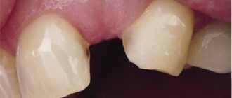
It occurs in most patients and is easily diagnosed, without requiring the use of additional examination methods. It is enough to run your tongue or finger over the formation, press on it and hear a specific crunch. It is most often observed in the upper jaw and is caused by advanced caries and tooth damage. Often, a radicular cyst of the upper jaw forms near the maxillary sinuses, contributing to the development of sinusitis.
Aneurysmal
A small growth containing hemorrhagic fluid inside the capsule (sometimes mixed with pus). Occurs in children aged 13-14 years. It looks like a painless lump that does not crunch when pressed. Chewing for a long time can cause pain. Prolonged course of the disease can lead to deformation of bone tissue.
Residual
A residual cyst grows at the site of an extracted tooth.
It grows on the lower and upper jaw at the site of an extracted tooth. Capable of reaching impressive sizes. It has unclear boundaries and is visible on an x-ray as a semicircle with a cut facing the bone dental bed.
Traumatic
Develops due to bruise, blow, damage to the gums and mucous membranes with a hard object. More often seen from below. It is distinguished by the absence of epithelial and connective tissues. In case of complications, the patient risks losing one or more healthy teeth.
Nasoalveolar (nasolabial)
A formation growing on the front wall of the upper jaw. It can destroy the outer shell of bones and grow into the roots, which can lead to loosening and loss of teeth. It has a round shape, clear boundaries, and limited mobility. Does not connect to skin.
Removal of a dental cyst. Historically significant and modern methods of surgery for removing dental cysts
Initially, the most common method of treating cysts was their removal. For this purpose, 2 fundamentally different methods were used: cystotomy and cystectomy.
Cystotomy (or the PARCH-I method) has historical significance in the development and establishment of medicine. Today this method is practically not used, but previously it was indispensable for removing large cysts. To avoid complications and its severe consequences, a wide channel was created between the cyst cavity and the vestibule of the oral cavity by suturing the edges of the cyst shell with the oral mucosa.
Cystectomy (or PARCH-II method) involves complete excision/removal of the cyst. This surgical intervention is often simultaneously accompanied by the removal of the root apex, the source of the formation of the cyst containing infected apical deltas. Today this method is the most popular.
The main goal of cystectomy is complete sanitation of the cyst cavity, which is not possible without resection of the root apex. The fact is that the root pulp in its apical third has an apical delta with an unclear structure and very fine branching of the canals, even in single-rooted teeth. If such canals can be cleaned during tooth treatment, then during retreatment this is almost impossible, especially considering the narrowness of the lateral deltas. Removing the root apex, in addition, allows you to most effectively clean the cyst cavity behind the root. This is especially true for teeth 12 and 22, which have a more curved root system. The operation is carried out as follows: using a small incision on the gum, the surgeon very carefully reaches the surface of the bone located above the cystic cavity, removes the wall with a trephine or bur, making sure that the neighboring teeth are not damaged, and then removes the cyst shell and performs a resection of the root apex . Next, sanitation of the cystic cavity is carried out and its subsequent revision for residual cyst particles. After this, in some cases, retrograde filling is performed. The final step in the operation to remove cystic formations is filling the resulting cavity with osteoplastic material and suturing the wound.
In the case of the formation of a large cyst, amputation of the apex of the tooth root can be performed, or half of the tooth and damaged root can be removed (hemessection), and in the case of an interradicular cyst, this can be done by corono-radicular separation.
Retention cyst of the lower lip
It is a common pathology of the oral mucosa. It is a benign neoplasm in the shape of a ball. The pathology usually develops due to blockage of the duct of the minor salivary gland. The reason for this may be the most common injury or inflammatory process.
The cyst occurs equally in both women and men. Its appearance does not depend on age. The cyst is characterized by the ability to very quickly increase in size, which affects a person’s daily lifestyle. That is why doctors recommend removing it.
The main reasons for the appearance of cysts
The neoplasm most often forms as a result of mechanical damage and trauma to the lip. These include burns, blows and biting. As a result of constant trauma to the gland, the excretory canal becomes clogged, the secretion begins to linger, and a small tubercle forms. Gradually it fills with liquid and increases in size. The inflammatory process after injury can also lead to the formation of pathology. A retention cyst of the lower lip can occur in a person at any age. The neoplasm is often a consequence of the congenital development of the germinal components of the so-called glandular cells.
In addition to traumatic effects, the cause of a cyst can be atrophy of the excretory ducts. Typically, such a violation is caused by a tumor that directly compresses the duct, or by a scar. The latter narrows the canal, and the accumulated secretion gradually expands the glandular lobe.
How to recognize pathology?
A retention cyst of the lower lip is a capsule of connective tissue with viscous contents inside. Outwardly, it resembles a small ball. The formation is painless, but may cause discomfort while talking or eating. The cyst tends to grow rapidly and can reach up to 2 cm in diameter. The mucous membrane above it usually does not undergo significant changes. Sometimes it takes on a bluish tint, which is explained by the accumulation of contents. The ball is covered with connective tissue, and inside it there is a clear liquid resembling saliva. On palpation the formation is soft. When eating food, the capsule is easily damaged, causing its internal contents to spill out, but after that the cyst fills up again. As a rule, the formation consists of one chamber, but cases of multi-chamber cysts are known.
Diagnosis confirmation
Recognizing a retention cyst is not difficult for a qualified specialist. When pressed with a finger, the formation disappears, but after a while it appears again. A more accurate diagnosis can be made after an ultrasound examination. During the procedure, the doctor evaluates the structure of the cyst, its size and contents. By probing the canals, the width of the duct and the presence of salivary stone are determined. Only after a complete examination can a specialist recommend treatment for a retention cyst of the lower lip.
The need for differential diagnosis In order to accurately make a diagnosis and subsequently prescribe competent treatment, it is important to be able to recognize this pathology among other neoplasms of a benign nature. The following types of salivary gland cysts are distinguished: ranula; submandibular; parotid; minor salivary gland. The ranula is localized above the hyoid-maxillary muscle. There are known cases of penetration into the submandibular region. This formation is large in size. It can displace the frenulum of the tongue, thereby preventing a person from fully eating and having conversations. The submandibular gland cyst is characterized by slow development. During palpation it is easy to detect a round formation. When this cyst grows, it can involve the upper areas of the mouth. In such a situation, bulging of the formation into the sublingual part is usually observed. Pathology of the parotid gland is quite rare. The main reasons for its formation include mechanical damage, inflammatory processes and blockage of ducts. It is the same factors that provoke the formation of another anomaly of the oral cavity called a retention cyst of the lower lip (photo presented in this article). The development of pathology at the initial stage is not accompanied by pronounced symptoms. As a result of mechanical damage, a so-called extravasal cyst can form. This formation is distinguished by the fact that granulation tissue is formed around it.
What treatment is required?
This pathology is treated by a dentist. After confirming the diagnosis, there are two possible solutions to the problem. The specialist may send the patient home, believing that the formation will resolve on its own. The second option involves surgical removal of the retention cyst of the lower lip.
The operation itself lasts no more than 30 minutes and involves the use of local anesthesia. The physician assistant twists his lower lip and holds it tightly. The dentist makes several cuts along the formation, frees the cyst from its internal contents and applies sutures. According to many patients, the operation itself, under the supervision of an experienced surgeon, is almost painless. The main troubles begin during the rehabilitation period, when the retention cyst of the lower lip begins to gradually heal. The operation can be performed using a laser. However, its help is used extremely rarely due to severe bleeding and a high risk of perforation of the gland membrane.
Postoperative period
After surgery, patients are advised to treat the affected area daily with a special antiseptic solution and monitor their condition. Some note that the first day after surgery is the most difficult. Talking, eating - all these simple actions cause painful discomfort, but after about a month everything returns to normal. It should be noted that the duration of the rehabilitation period directly depends on the size of the formation. Many patients report visual distortion of the lip and slight numbness even months after surgery. To avoid negative consequences, it is recommended to remove the pathology at the initial stage of its formation.
Quality criteria for dental cyst removal
Despite the fact that cystectomy is a surgical intervention, the main indicator of the quality of treatment will be the sanitation of the root canals, including retrograde filling. Currently, in addition to eliminating the cyst shell itself, the unspoken rule has become the filling of its cavity with one or another osteoplastic materials, because this completely eliminates the possibility of blood clot suppuration, promotes rapid healing of the bone defect and improves the mechanical stability of the tooth.
Prognosis for treatment of lateral neck cyst
The prognosis of a lateral neck cyst depends on the following factors:
- general health of the patient; - age; - the size of the cyst and its localization in relation to important organs and large vessels; — duration of cyst development; - forms of cysts - inflammation, suppuration; - contents of the cavity - pus or exudate; - type of fistula; - presence or absence of chronic diseases.
In general, the prognosis for treatment of a lateral neck cyst can be considered favorable. The risk of a lateral cyst degenerating into a malignant form is extremely low.
Indications for tooth extraction with a cyst
As a rule, conservative treatment carried out on time or surgery to remove the cyst allows you to save the tooth. But this is not always possible. A tooth must be removed in the following cases:
— the presence of severe pain symptoms when drug treatment is ineffective; - purulent inflammatory process when it is impossible to drain it; — fracture of the coronal part of the tooth, without the possibility of restoration with anchor pins and/or stump inlays; — obstruction of root canals; — the presence of multiple damage to the roots of the tooth or large damage; the tooth is almost completely destroyed and orthopedic restoration is impossible; - no need for dental treatment due to the presence of a prosthetic plan agreed upon between the doctor and the patient.
Features of the development of cervical cysts and treatment in childhood
Lateral neck cysts in newborns and young children occur in a latent form and are not expressed by clinical symptoms until a provoking factor appears - trauma to the child, the appearance of a respiratory infection or other inflammatory processes. Sometimes hormonal changes in the body can become such a factor.
Clinical practice shows that in children the formation of a lateral cyst is always preceded by acute respiratory infections, influenza and other diseases of the upper respiratory tract. There is a connection between the tumor and the lymphatic tract, during which microbes of a pathogenic nature penetrate into the internal cavity of the cystic formation, causing suppuration.
In childhood, surgical treatment of a lateral cyst is carried out from three years of age outside the acute stage. But in case of serious complications leading to a threat to life, surgical intervention is indicated at an early age of the child.
Price
The cost of treatment or removal of a dental cyst consists, like many other surgical interventions, of a number of factors. The complex of diagnostic procedures performed, types and methods of treatment, conservative preparation of canals, preoperative preparation, the operation itself, the osteoplastic materials used, postoperative observation - all this affects the final cost of treatment in these clinical situations.
A dental cyst is a serious pathology. In all cases, even in cases of complete absence of any symptoms, it poses a health hazard. With regular visits to the dentist, you can be sure: a dental cyst, if there is one, will be detected in time, and subsequent treatment will allow it to be eliminated without leaving a trace, preserving the health of the tooth.
According to antiplagiat.ru, the uniqueness of the text as of October 16, 2018 is 99.9%.
Key words, tags: X-rays, tooth extraction, orthopantomogram, periodontitis