Author of the article:
Soldatova Lyudmila Nikolaevna
Candidate of Medical Sciences, Professor of the Department of Clinical Dentistry of the St. Petersburg Medical and Social Institute, Chief Physician of the Alfa-Dent Dental Clinic, St. Petersburg
Medium caries is a stage of the carious process that occurs after the initial one and precedes the deep one. It is characterized not only by the destruction of enamel, but also by disruption of the dentin structure. Often the disease is practically asymptomatic, so the patient cannot independently identify the presence of this disease. The only method of self-diagnosis is to pay attention to changes in the appearance of the tooth when examining the oral cavity in a mirror (presence of holes) or to the appearance of sensitivity while eating.
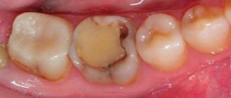
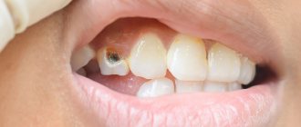
If you look at a photo of average caries, you will notice pronounced changes in the structure and color of the affected area. Most often, the disease develops on the contact (chewing) part of the tooth. In the photo you can see dark brown or almost black spots, small or large holes, which can be located on any part of the tooth. These holes are a carious cavity in which dentin gradually softens and food debris accumulates.
With chronic average caries, the destruction of the enamel and dentinal layer occurs faster - carious cavities can appear as early as five months after the initial changes in the tooth enamel. Most often, young children suffer from the chronic form of the disease.
Causes of the disease
There are several factors that lead to the occurrence of this disease:
- Cariogenic microflora of the oral cavity.
- Increased carbohydrate content in the diet.
- Reduced resistance of hard tissues.
- Lack of oral hygiene.
- Malocclusion.
- Genetic predisposition to the disease.
- Plaque or tartar.
- Weakening of the immune system.
- Bleeding gums.
- Changes in the composition of saliva.
- Lack of calcium.
- Fluoride deficiency.
- Poor diet.
- Lack of vitamins.
- Abuse of carbonated drinks.
The main cause of average caries is late diagnosis of the disease at an early stage.
Classification
There are acute and chronic forms of the disease. The form determines how quickly the process will develop and what the consequences will be.
Signs of an acute process:
- Rapid development. In a short period, pulpitis may develop, and the tooth will be completely destroyed.
- Dentin is soft and easily removed with dental instruments.
- The color of damaged tissue is light.
- The edges of the cavity are sharp, the walls are steep.
In a chronic process:
- Caries can take years to develop.
- The walls of the cavity are dark, almost black.
- Dentin is hard and cannot be removed with dental instruments.
- The edges of the cavity are smooth, the walls are flat.
Acute caries often develops on a child’s baby teeth and must be treated immediately. This development of the process is possible in adults.
Symptoms of the disease
The disease can exclusively affect the upper layers of dentin. The process of development of medium-sized caries is accompanied by destruction of the enamel layer and a violation of the boundary between enamel and dentin. As a result, patients complain of the following unpleasant sensations:
- The presence of a carious cavity, which is easily detected using the tongue. In some cases, it may have sharp edges and cause injury.
- Food getting into the carious hole if it is located on the contact part of the tooth.
- Short-term painful sensations from exposure to hot or cold food, liquids, from the chewing process and from mechanical impact on the diseased tooth. Once the cause of the pain is eliminated, the unpleasant symptoms quickly disappear.
What are the differences from other forms of the disease?
Deep caries is very similar to some forms of the disease, but there are still visible and invisible differences.
Difference from pulpitis
Pulpitis is a complication of advanced caries. The forms of the disease can be distinguished from each other by their characteristic symptoms:
- Defeat. With pulpitis, the nerve is affected; with caries, the nerve remains unharmed.
- Feelings of pain. With pulpitis, pain can occur spontaneously, even without exposure to irritants on the tooth. With caries, pain disappears along with the disappearance of irritants.
- Night sleep. With pulpitis, sleep can be disturbed due to severe pain; with caries, the tooth does not hurt at night.
Difference from average caries
The similarity between the two forms of the disease is that the patient in both forms feels pain from cold, sour or sweet foods. However, with average caries they go away within 1-2 minutes, with deep caries they last longer.
Medium caries is characterized by the fact that in this form the carious lesion does not affect the tooth pulp and does not cause inflammation of the nerve, as in deep caries.
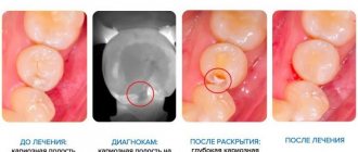
Diagnosis of the disease
To prescribe effective treatment for the disease and exclude the presence of other diseases, it is necessary to diagnose secondary caries, which is carried out by the treating dentist.
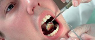
In most cases, an experienced specialist only needs an instrumental examination to make a diagnosis. It is carried out using a probe and a mirror. As a result of the examination, a carious cavity is discovered, inside of which there is softened dentin. Mild pain is present only when probing the bottom of the cavity, since there are exposed nerve endings there. The walls of the carious cavity are insensitive.
During diagnosis, the doctor pays attention to the following factors:
- Density of the cavity bottom.
- Localization of the carious hole.
- The color of the damaged tooth.
- The presence and nature of pain in the patient during the study.
A soft bottom indicates moderate dental caries, and its density may indicate the presence of a wedge-shaped defect. The tooth becomes gray if the patient has developed chronic periodontitis. The short duration of painful sensations indicates superficial caries, and if the pain lasts longer than ten seconds, the disease has acquired a deep form.
If the affected area is in a hard-to-reach place, the doctor may prescribe an X-ray examination for the patient. In some cases, the dentist recommends undergoing electroodontic diagnostics or thermal testing.
Is it possible to treat deep caries of a wisdom tooth?
The patient can make the decision to remove a wisdom tooth if pain occurs on his own. But only a highly qualified dentist can make a verdict on its treatment.
Treatment of a carious "eight" is considered rational if it has a partial covering in the form of a hood of gum, as well as an antagonist - the third molar located on the opposite side.
Removal shown:
- with a horizontal tooth position;
- presence or absence of 1st and 2nd molars;
- impossibility of treatment due to incomplete opening of the mouth;
- displacement of teeth in a row due to wisdom teeth.
Treatment of average caries
After diagnosis, it is necessary to urgently treat the disease to prevent the process from spreading to the deep layers of dentin. Otherwise, inflammation of the pulp may occur.
Treatment is carried out by preparing the damaged areas. It goes through several stages:
- The overhanging edges of the carious hole are carefully removed.
- Dentin, which has undergone a process of softening and pigmentation, is removed from the cavity.
- The bottom and walls of the damaged tooth are cleaned of the remains of destroyed tissue.
- A cavity is formed for subsequent filling.
During each stage, the dentist uses a specific type of bur, which differs in the shape and diameter of the tip. Preparation is carried out in bright light. At the end of this process, the cleaned cavity is treated with antiseptics, degreased, and then thoroughly dried.
The walls and bottom of the hole are coated with a special “glue”, and then the cavity is filled with filling material, which undergoes a curing process using chemical or light radiation.
The finished filling is corrected using diamond burs, as well as using various grinding discs, pastes and brushes. Correction can be made several times. The purpose of this procedure is to achieve maximum comfort in the patient when clenching the jaw. The restored surface should not cause even the slightest discomfort.
Publications in the media
Dental caries is a localized lesion of the hard tissues of teeth, manifested by demineralization and progressive destruction of enamel and dentin with the formation of a defect in the form of a cavity.
Epidemiology • Caries occurs at all ages; the prevalence of caries reaches 100%, which, according to WHO experts, makes it possible to classify it as a pandemic • Dental caries damage is assessed by the intensity indicator: “KPU index” - for permanent teeth, “KP index” - for temporary teeth, “KPU+kp index "- in a mixed dentition (the index is the sum of the number of teeth affected by caries [K], filled [P] and removed [U]).
Risk factors • Pathological pregnancy • Acute infectious and chronic systemic diseases • Sensitization of the body • Anti-infectious vaccinations and other conditions accompanied by decreased immunity • Excessive consumption of carbohydrates • Insufficient intake of mineral components and trace elements, in particular fluoride • Inadequate oral hygiene.
Etiology. Dental caries is an infectious disease associated with associated microflora (lactobacillus, veyonella, etc.), where the leading role is played by streptococci (S. mutans, S. sanguis, S. mitis, S. salivarius). The most cariogenic is S. mutans, which produces lactic acid only from glucose.
Pathogenesis. Development is determined by genetic resistance (caries resistance) or predisposition to the disease.
• A decrease in resistance to caries is associated with a decrease in the calcium content in the surface layer of enamel, more precisely with a decrease in the molar ratio “calcium/phosphorus” in hydroxyapatites below the minimum level (1.30).
• Immune disorders are associated with a decrease in the content of secretory IgA in saliva, which normally, by preventing the adhesion of microorganisms to the enamel surface, prevents the formation of soft plaque.
• Microorganisms form dental plaque (soft plaque), which is fixed on the surface of the tooth on the polysaccharide stroma.
• The microflora of the plaque damages the organic basis of the enamel, in which, with the loss of the ability to fix the mineral substrate, the process of incipient demineralization begins to predominate. In the resulting carious defect, microorganisms undergo enzymatic breakdown of the organic substances of the tooth.
Classification
• Based on the depth of the lesion, they distinguish •• initial caries •• superficial caries •• medium caries •• deep caries.
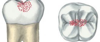
• Based on localization, they distinguish •• fissure caries - damage to grooves and natural depressions •• approximal caries - damage to contact surfaces •• cervical caries - damage to a tooth near its neck •• carious defects in other areas are considered atypical.
• According to the course, caries is distinguished as acute, acute, chronic and suspended.
Clinical manifestations. The clinical picture is very diverse and is determined by the nature of the process and the depth of damage to the hard tissues of the tooth.
• Acute forms (acute and acute) of caries are characterized by rapid development of the process, when within a short time (1–1.5 years) most of the teeth are affected; Often two or several foci are found in them. A carious defect is characterized by slight destruction of the enamel and extensive destruction of the underlying dentin. The walls and bottom of the cavity are formed by softened, low-pigmented dentin. When cold, sour or sweet food gets into the cavity, severe pain occurs. Plaque forms quickly. The viscosity of saliva increases, the activity of salivary blood lysozyme decreases, and the content of secretory IgA decreases. Impairments in the functional state of the autonomic nervous system may be detected.
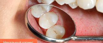
• Chronic dental caries develops unnoticed when one, or less often 2-3, teeth are sporadically affected, and such damage can go unnoticed for a long time. The carious focus is localized on typical surfaces, affecting molars, premolars and rarely the upper incisors •• Initial caries is a focus of demineralization of the enamel in the form of a spot on the labial and cheek surfaces of the teeth, closer to the neck, small in size, matte, whitish or dirty gray in color. Its contours are uneven, but clearly visible after staining with methylene blue solution. The probe glides easily over the surface without detecting a defect. There are no unpleasant or painful sensations. The teeth are usually abundantly covered with soft plaque •• Superficial caries is an enamel defect that does not cause unpleasant subjective sensations and is easily detected. Has clear spherical outlines; no changes are usually noted in the enamel surrounding the defect •• Middle caries is a cavity defect affecting the enamel-dentin junction and dentin layers. It has clear edges, the bottom and walls are dense and pigmented to varying degrees. There are no complaints or food gets stuck •• Deep caries is a cavity defect extending to the area of deep (suprapulpar) dentin: large in size, usually filled with decaying contents of a dirty gray color. There may be complaints of quickly passing pain when cold or sour food gets into the cavity. Cold stimulus causes pain. Probing the cavity floor is sensitive. The electrical excitability of the pulp is reduced to 20 μA.
Diagnosis of caries is based on complaints and the time period during which damage to several teeth occurs.
• An objective examination includes examination, probing and mandatory assessment of the hygienic condition of the oral cavity.
• Additional methods include thermometry (using water at a temperature below 18 °C), vital staining with 2% aqueous methylene blue or electroodontodiagnostics, radiography.
• To identify hidden and initial forms, transillumination, luminescent examination of the enamel surface and determination of the electrical conductivity of hard dental tissues can be used.
• To identify predisposition to caries, the acid resistance of enamel and its ability to remineralize are determined.
Differential diagnosis • Complicated stages of caries • Dental diseases of non-carious nature.
TREATMENT
Diet. In acute forms of caries, a diet rich in proteins and mineral salts is prescribed; vitamins A, C, D, B1, B6.
Drug treatment • General treatment is indicated for acute forms of caries. Combined calcium preparations are prescribed (calcinova, calcium-D3-nycomed); sodium fluoride orally 4–6 mg/day • Local non-operative treatment of initial caries consists of professional hygiene (removal of dental plaque) and remineralization of the affected enamel, which is carried out using fluorides in the form of solutions, pastes, gels, varnishes, often in combination with solutions of calcium salts, or remineralizing drugs (Remodent).
Surgery. Operative and restorative treatment of caries consists of anesthesia, instrumental treatment (necrectomy) of the tissues forming the bottom and walls of the carious cavity, and its subsequent filling with filling material or inlay.
Non-drug therapy. For initial caries, application or physical treatment methods are used: electrophoresis, ultraphonophoresis. For acute caries, UV irradiation is recommended.
Forecast. Without treatment, caries, with the exception of paused ones, necessarily develops into complicated forms.
Prevention. Aimed at increasing the resistance of teeth to caries and combating plaque formation.
• A balanced diet with mandatory consumption of milk and dairy products, sufficient intake of vitamins, limitation of easily digestible carbohydrates (sucrose, glucose). It is extremely important to introduce fluoride preparations into the body with drinking water (1 mg/l) or other fluoridated products (milk, salt).
• Treatment of acute infectious and concomitant diseases, carrying out general health measures.
• Regular and high-quality oral hygiene plays an essential role: twice-daily brushing of teeth using anti-caries pastes is recommended.
ICD-10 • K02 Dental caries
Prognosis and consequences of the disease
Treatment of this disease, if the dentist has sufficient experience, is successful - all unpleasant sensations in the sealed cavity completely disappear, and the tooth again acquires aesthetic appeal. If an untreated area remains under the filling, the disease will progress further. For this reason, it is necessary to carefully consider the choice of clinic and attending physician.
If average caries is not treated in a timely manner, the disease will enter an advanced stage, which is fraught with severe pain, the development of pulpitis and periodontitis, and then tooth loss.
Where to treat?
You can undergo treatment for deep caries at the Good Hands dental clinic. Our specialists will make every effort to save the tooth and carry out its restoration, maintaining its appearance and functionality. It is very important to seek professional dental care in a timely manner in order to prevent the development of pulpitis and periodontitis, which can lead to tooth loss. Our specialists use effective anesthetics and modern equipment in their work. Thanks to this, the treatment will be comfortable and painless. Book a free consultation with us by filling out the simple form on this page or calling us!
Disease prevention
After each meal, food particles remain in the oral cavity, which contribute to the development of decay processes and create a favorable environment for bacteria. To prevent the spread of harmful microorganisms, it is necessary to brush your teeth regularly.
If a person has a tendency to develop the disease, the dentist prescribes special toothpastes that can reduce the risk of the disease. Thus, we recommend ASEPTA PLUS REMINERALIZATION paste, which strengthens the enamel and prevents caries on teeth.
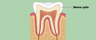
Tooth structure and types of carious lesions
A tooth consists of several parts. On top it is covered with hard mineralized enamel, underneath there is dentin, and even deeper - the root of the tooth and pulp. Caries is a pathological process that is accompanied by demineralization and softening of hard dental tissues with the subsequent formation of a carious cavity. The process can affect any part of the tooth; depending on this, caries of enamel, dentin or cement is distinguished.
Carious lesions of the enamel are characterized by symptoms such as softening, the appearance of a white or brown spot, roughness, and sometimes the carious cavity is located in the enamel. If the lesion affects the bone tissue of the tooth, which makes up its bulk, in this case we speak of dentin caries. This form of lesion is characterized by the formation of a carious cavity directly in the dentin.
Cement caries affects the root of the tooth and contributes to its exposure. It often occurs with gum disease, when there is no close contact between the tooth and the gum.
Clinical researches
Clinical studies have proven that regular use of professional toothpaste ASEPTA REMINERALIZATION improved the condition of the enamel by 64% and reduced tooth sensitivity by 66% after just 4 weeks.
Sources:
- Report on the determination/confirmation of the preventive properties of personal oral hygiene products “ASEPTA PLUS” Remineralization doctor-researcher A.A. Leontyev, head Department of Preventive Dentistry, Doctor of Medical Sciences, Professor S.B. Ulitovsky First St. Petersburg State Medical University named after. acad. I.P. Pavlova, Department of Preventive Dentistry
- Clinical experience in using the Asepta series of products Fuchs Elena Ivanovna Assistant of the Department of Therapeutic and Pediatric Dentistry State Budgetary Educational Institution of Higher Professional Education Ryazan State Medical University named after Academician I.P. Pavlova of the Ministry of Health and Social Development of the Russian Federation (GBOU VPO RyazSMU Ministry of Health and Social Development of Russia)
- Evaluation of the clinical effectiveness of a combined drug of local etiotropic action in the treatment of inflammatory periodontal diseases (E.L. Kalichkina, E.A. Tyo, Z.Z. Abubakarova) E.L. Kalichkina, candidate of medical sciences, assistant of the department of therapeutic dentistry, Kemerovo State Medical Academy of Roszdrav, Kemerovo E.A. Tyo, MD, professor, head of the department of therapeutic dentistry, Kemerovo State Medical Academy of Roszdrav, Kemerovo Abubakarova, assistant of the department of therapeutic dentistry, Kemerovo State Medical Academy of Roszdrav, Kemerovo GOU HPE Kemerovo State Medical Academy of Roszdrav , Kemerovo
Prevention of dentine caries in permanent and baby teeth
Measures to prevent dentin caries are similar to the prevention of caries in other parts of the tooth.
These include:
- Proper nutrition with a minimum amount of sugars and fast carbohydrates, rich in fluoride, calcium, phosphorus. Avoiding frequent snacking.
- Thorough daily brushing of teeth twice a day using a toothbrush, paste, and floss. The additional use of an irrigator to clean hard-to-reach places and interdental spaces, as well as mouth rinses, helps reduce the risk of caries.
- Schedule a visit to the dentist twice a year, as well as immediately contact a doctor at the first sign of caries.
- Professional teeth cleaning using special equipment twice a year.
- If necessary and as prescribed by a doctor, use vitamin-mineral complexes, which contain components beneficial for dental health.
- Drinking fluoridated drinking water, which is enriched with minerals.
Compliance with these preventive measures allows you to maintain the health and beauty of your teeth and reduces the risk of developing caries and other dental diseases.
The effectiveness of non-invasive treatment methods
If the damage caused to the tooth is insignificant, and the enamel is sufficiently dense, treatment of enamel caries in the white spot stage can be carried out without preparation and filling. In this case, the affected area is ground and polished, which avoids the recurrence of the disease. After this, the tooth is locally fluoridated, and the process of restoring the enamel from the inside begins. This treatment method is called remineralization. Fluoridation can also be done at home - using special preparations - but only after consulting a doctor.
Also, modern methods of non-invasive treatment of enamel caries include: chemical-mechanical treatment (infiltration), air-abrasive treatment and ozone treatment. All these methods allow you to do without a drill and carry out treatment with virtually no discomfort for the patient. At the same time, it is possible to preserve healthy tooth tissue to the maximum - the microhardness of the enamel is damaged during enamel caries, and all of the above methods allow you to act only on softened tissue, without affecting the healthy parts of the tooth.
Regardless of the chosen technique, in order for the treatment of dental caries to be effective and the disease not to recur, the participation of the patient himself is important. Only a conscientious approach to hygiene will help maintain oral health.