At the Clinical Hospital on Yauza you can:
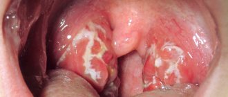
- quickly and efficiently examine the condition of the tonsils (perform pharyngoscopy, laboratory tests - bacteriological, immunological, etc.);
- decide on the choice of effective treatment for tonsillitis (inflammation of the palatine and pharyngeal tonsils): conservative - general and local, surgical.
We use the following methods of low-traumatic surgical treatment:
- tonsillotomy - partial removal of tonsils (tonsils) in children while maintaining their protective functions (all operations are performed under anesthesia);
- tonsillectomy - a low-traumatic operation to remove the tonsils;
- suturing wounds and using a bipolar coagulator to prevent blood loss;
- the use of innovations that accelerate and facilitate the healing process.
Timely diagnosed tonsillitis has a positive prognosis with conservative treatment without transition to the chronic stage. Radical treatment of chronic tonsillitis - tonsillectomy - prevents the development of systemic complications, often leading to disability.
Symptoms of tonsil cancer
- Dysphagia (difficulty swallowing) – the tumor interferes with swallowing, causing difficulty swallowing.
- Odynophagia (pain when swallowing) – the tumor grows, leads to the formation of ulcers, grows into the nerves, which leads to pain.
- Lump – spread of cancer to the lymph nodes makes the tumor clinically noticeable: the patient may notice a firm, palpable mass in the neck.
- Otalgia (ear pain) - the innervation of the oropharynx and ear has some connection, which means that the brain can “replace” pain in the oropharynx with pain in the ear (referred pain). The most alarming is pain in the ear on one side, without any other ear problems, not going away, unexplained by other diseases. It is important to understand that most causes of ear pain are associated with middle ear infection, eustachian tube dysfunction and other diseases of the ENT organs. But a combination of several symptoms may indeed indicate an oncological process.
- Trismus (spasm of the pterygoid muscles, causing difficulty opening the mouth).
- Feeling of a lump in the throat.
- A sore throat.
- Bleeding from the mouth.
- Change in speech (muffled voice).
- Weight loss.
Medial pterygoid muscle. getbodysmart.com
Consequences of tonsil removal
One of the most unpleasant consequences is weakening of the immune system throughout the body. As mentioned above, the tonsils are a natural protective barrier that blocks the passage of many bacteria and viruses into the lungs. After their removal, infectious diseases, flu and colds may occur more often. The same new coronavirus will have easier access to your respiratory tract.
The rehabilitation itself, even with the most painless surgical methods, can become an unpleasant surprise: swelling in the bronchi, soreness, crusting and the sensation of a foreign body in the throat. No doctor can answer for sure how long this will last - everything is too individual.
Please do not resort to surgical intervention unless absolutely necessary and clearly prescribed by a doctor. The tonsils are an important organ in our body, and its presence protects us from many unpleasant diseases. Contact only trusted ENT specialists and start treatment as early as possible.
Take care of your health!
Diagnostics
- Anamnesis collection, visual examination and palpation of the mucous membranes of the floor of the mouth, the anterior two-thirds of the tongue, the tonsils and base of the tongue, the mucous membrane of the palate, cheeks and gums, and the posterior wall of the pharynx.
- Diagnosis of HPV infection: immunohistochemical determination of the surrogate marker p16 in tumor tissue.
- Computed tomography of the chest and abdominal cavity to exclude metastatic lesions.
- Biopsy – cancer can be diagnosed only by taking a tissue sample from a growth that is suspicious for a malignant process.
- MRI of the soft tissues of the neck provides more accurate detail of the soft tissues compared to CT. Performed to assess the prevalence of the process.
- Ultrasound of the lymph nodes of the neck to determine their structure (if there is metastasis in the lymph nodes, they may not change their size, but the structure of the lymph node changes).
- PET-CT may be ordered if occult distant metastases are suspected in some situations.
- Fibercolonoscopy, fibrogastroduodenoscopy, laryngoscopy and some other studies may also be prescribed.
HPV-associated squamous cell carcinoma of the right tonsil (A) demonstrating p16 overexpression by immunohistochemistry (B) and evidence of high-risk HPV by in situ hybridization (C). Patients with HPV-related tonsil squamous cell carcinoma rarely harbor oncogenic HPV infection at other pharyngeal sites, SelvamThavaraj et all., ELSEVIER April 2014
Determination of stage
Determination of the stage of cancer is carried out according to the TNM system (T - tumor, N - nodulus, M - metastasis), including the size of the primary tumor, the spread of tumor cells to regional (located next to the primary tumor) lymph nodes and to distant areas (lungs, liver) . Today, the Russian Federation has adopted the seventh classification of tumors (the newest is the eighth). The connection between tonsil cancer and HPV infection is also taken into account during staging (but only for the eighth classification) - with similar morphological data of the tumor and its distribution, the stage of tonsil cancer associated with HPV infection is often lower, in contrast to cancer associated with other factors . In general, the prognosis for tonsil cancer associated with HPV infection is more favorable.
Tonsil cancer staging based on tumor size is “T”. headandneckcancerguide.org
Treatment
Early tonsil cancer can be treated with radiation therapy or surgery: small tumors confined to normal tonsil tissue can be cured by removing the tonsil (tonsillectomy). If the tumor extends beyond the tonsil itself, then the soft palate and the wall of the pharynx may need to be removed. Depending on the presence of tumor cells in the lymph nodes, lymphatic dissection may be performed - removal of regional lymph nodes (for head and neck tumors, they are located in the neck). Otherwise, the tumor can be treated with radiation without the need for surgery. The effectiveness of radiation therapy and surgery is similar.
So, for the treatment of early tonsil cancer there are two approaches:
- surgical treatment, which, depending on the results of pathomorphological examination, may be continued with a course of radiation therapy (adjuvant radiation therapy) either alone or simultaneously with chemotherapy;
- radiation therapy – alone or simultaneously with chemotherapy (simultaneous chemoradiotherapy).
Late cancer of the tonsils, growing into the peripharyngeal space, penetrating into the bones, base of the skull, and medial pterygoid muscle may be accompanied by symptoms such as temporal pain and trismus (spasm of the masticatory muscles, leading to difficulty opening the mouth). Treatment of advanced cancer is often accompanied by chemoradiotherapy. An option is surgical treatment of advanced cancer followed by chemoradiotherapy.
When performing surgical intervention, the decision on the need for subsequent treatment is made on the basis of a pathomorphological examination of postoperative material. Spread of the tumor beyond the capsule of the lymph node, removal of the tumor outside of healthy tissue (the tumor is not removed radically, that is, not completely), significant spread of the tumor to the lymph nodes and a large size of the primary lesion, as well as tumor invasion of nerves, lymphatic and blood vessels - all these characteristics are predictors of an unfavorable course of the tumor process and may require additional postoperative treatment in the form of reoperation (if the tumor is not completely removed), chemotherapy and/or radiation therapy.
For the treatment of metastatic tonsil cancer (M1), surgical treatment and radiation therapy are not indicated. In this case, the main approach is systemic therapy, the choice of which will be based on the presence/absence of previous systemic treatment, the patient's condition, certain tumor characteristics (PD-L1 expression), the rate of disease progression and some other factors. Options for systemic therapy include chemotherapy drugs: platinum drugs (cisplatin, carboplatin), fluorouracil and taxanes (docetaxel, paclitaxel); targeted drugs (cetuximab); immunotherapy drugs (pembrolizumab, nivolumab). The combination of drug groups is determined by the above characteristics.
In the presence of a single distant metastasis, metastasectomy may be offered in addition to systemic therapy, which is likely to prolong the time to eventual disease progression.
Regardless of the extent of tonsil cancer, patients may be offered participation in clinical trials.
Patients with severe comorbidities and/or poor health status are best treated with supportive care.
Results and discussion
The peripheral zone of adenoid vegetations in patients of the 1st group practically does not contain connective tissue and is characterized by a pronounced and full-blooded microvasculature, the boundaries of large rounded lobules are poorly distinguishable.
The peripheral zone in patients of group 2 consists of lobules of predominantly small size (about 2 times smaller than in group 1), separated by connective tissue septa with many small full-blooded vessels, especially veins and venules. Such sclerotic septa distinguish this group from observations in group 1. The microvasculature network is still pronounced, but individual arterioles and capillaries are sclerotic and hyalinized.
The middle zone in patients of group 1 is distinguished by a large number of small full-blooded vessels, thanks to the linear accumulations of which the boundaries of the lobules of lymphoid tissue are visualized. Connective tissue layers are not expressed. Sclerosis of the layers and walls of blood vessels is not determined.
In patients of group 2, the middle zone is distinguished by its large size, clear interlobular boundaries with sclerosis and clusters of full-blooded, sclerotic vessels.
The basal zone in patients of the 1st group is represented by loose connective tissue with many small vessels with an expanded lumen, in some cases with initial signs of sclerosis of their walls. In the stroma of the basal part there is a diffuse lymphoid infiltrate that does not form any structures.
In the 2nd group of patients, significant changes in the basal zone were revealed, in light of the prognosis of intraoperative hemorrhage. There is an increase in stromal sclerosis, sclerosis and hyalinosis of the walls of vessels of various sizes, mainly veins with cavernous expansion of the lumen and rigid walls. In the sclerotic stroma there is a diffuse lymphoid infiltrate, which forms extensive accumulations at the resection edge, spreading into the underlying connective tissue layer (Fig. 1, a, b on the color insert).
Rice. 1. Patient N., 18 years old (group 2). Lymphoid tissue, basal zone. a: 1 - stromal sclerosis, 2 - pronounced sclerosis of the walls of microvasculature vessels, dilated venules and small veins; b: 1 – sclerosis and hyalinosis of the walls of vessels of different calibers, mainly veins with a cavernous dilated lumen. Hematoxylin and eosin staining. Uv. 400.
Patients of the 2nd group at the edge of resection of adenoid tissue are characterized by the accumulation of many cavernous dilated venous vessels with sclerotic walls and the phenomenon of “immersion” of lymphoid tissue deep into the sclerotic connective tissue layer of the adenoids with a well-developed microvasculature. This phenomenon is confirmed by histological examination of the underlying tissues after adenotomy. Lymphoid tissue is represented by both B-dependent zones (lymphoid follicles with large active light centers) and T-dependent zones (Fig. 2 on color insert).
Rice. 2. Patient K., 18 years old (2nd group). Biopsy from the underlying tissues after adenotomy. The phenomenon of “immersion” of lymphoid tissue deep into the connective tissue layer of the adenoids. 1 - cavernous dilated veins with rigid sclerotic walls. Lymphoid tissue is represented by both B-zones - lymphoid follicles with large active light centers (2) and T-zones (3). Hematoxylin and eosin staining. Uv. 100.
Complications of treatment
Indeed, there are two directions in the treatment of tonsil cancer with the same outcome, but different techniques: surgical treatment and radiation therapy. It is important to note that these approaches will have different complications.
Radiation therapy may be complicated by bone or soft tissue necrosis and swallowing problems, especially in patients with advanced tonsil cancer. Myelitis, trismus, hypoglossal nerve damage, and long-term risk of another malignancy are also possible.
Complications of surgery include difficulty swallowing, fistula formation, failure of flap healing, poor wound healing, and aspiration of food and saliva, sometimes leading to laryngectomy. The risk of severe and/or fatal complications after surgery is higher than with radiation therapy.
Complications of systemic therapy include fatigue, skin toxicity, mucositis, diarrhea, hepatotoxicity, pneumonitis, hypo- and hyperthyroidism, myelotoxicity, neuropathies, metabolic disorders, etc.
conclusions
1. The vessels of adenoid vegetations have clear age-related characteristics, which are most clearly manifested from the age of 18 and are characterized by the development of sclerosis and hyalinosis with the formation of cavernous dilated full-blooded veins with thickened rigid walls.
2. In patients over 18 years of age, there is a morphological restructuring of the connective tissue (basal) layer, namely the newly discovered phenomenon of “immersion” of lymphoid tissue with a well-developed microvasculature, which may increase the risk of intraoperative bleeding during adenotomy in this age group.
The authors declare no conflict of interest.
Forecast
The prognosis for patients with HPV-associated tonsil cancer is significantly better than for patients with other etiologies of cancer. It is impossible to predict with high accuracy the life expectancy of a particular patient, but there are average graphs that can be used to determine what percentage of patients with a certain stage are alive after n years: for example, almost 90% of patients with stage I HPV-associated oropharyngeal cancer are alive 4 years after diagnosis, while for HPV-unassociated oropharyngeal cancer at the same stage, only 80% are alive 4 years after diagnosis.
Overall survival of HPV-associated and HPV-non-associated oropharyngeal cancer according to TNM classification 7. ICON-S
Survey
In the department of otorhinolaryngology (ENT) of the hospital, patients with a chronic form of the disease are examined using:
- pharyngoscopy (visual method);
- PCR diagnostics - a high-precision method of molecular genetic diagnosis of infectious diseases;
- bacteriological tests - diagnosis of the main agent of infection and its sensitivity to antibiotics;
- antigen tests - diagnosis of immune system activity;
- general blood tests.