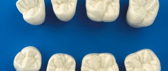
Location and anatomical features of molars
Since man is an omnivore, for a healthy metabolism he needs different foods, including rough, animal, and plant foods. In order for the body to absorb it, you need to chew the food well, which is what molars do. Their chewing area reaches 1 cm2. Each such organ can withstand pressure of up to 70-75 kilograms. In total, there are up to 12 such units in the oral cavity of an adult. Six of them are located on the lower jaw, six on the upper jaw, three on each side.
The last elements in the series are the third molars. They are also called wisdom teeth. They cut through in adults, and often may not cut through at all. Their roots can have two, three or more branches. The size of the crowns of molar units can reach from 7 millimeters, in some organs up to nine. Their chewing surface is diamond-shaped with rounded corners. It has four tubercles, which are separated by 3 grooves located transversely. Classification of roots:
- palatal;
- buccal-distal;
- buccal-mesial;
The last type of roots is the largest. The palatine root belongs to the middle ones. The shortest in size is the buccal-distal. Each subsequent molar unit is endowed with smaller roots and a crown portion than the previous one. The first molar organs are large, their coronal surface is significant in area, and the size of the roots is the largest. The second units are already smaller in size. The extreme units do not have milk precursors. Their root length and crown size vary significantly. Usually, there are 3 tubercles on the crown, one of which is lingual and 2 buccal. As a rule, their roots are not too long, but they are often displaced and deviated to the side. Wise teeth can come into contact with 2 antagonists at the same time. This is considered normal.
On the upper jaw
The top row has its own characteristics and differences. Their main aspect is location. They form a straight line that is only slightly curved and diverges to the sides. The slope of the units increases from the first to the third segment. In addition, in the upper teeth:
- Larger in size than the organs located in the lower jaw;
- The root system is more powerful, it has three roots;
- The tooth surface area is extensive;
- Good ability to chop and chew food;
- The outer side is covered with sharp tubercles with protruding forms, there are 3 or more of them;
The second molars of the upper row in the coronal part are similar to the first molar units, although they are more massive and rectangular. They are easier to dissect. The roots are usually grouped, sometimes merging with each other. The canals, which are parallel, often overlap each other when X-rays are taken. The roots are shorter compared to previous teeth. The three mouths often form a flat triangle, less often a straight line. The convex bottom of the chamber gives the mouths a funnel shape. It happens that the channels to the bottom of the chamber bend at a very acute angle.
Rarely do the upper posterior units have four roots. The first molar large organ of the upper jaw is equipped with more powerful roots than its lower antagonist. The peculiarity of the second molar teeth is that their crown can have any shape, while the lower ones can only have a certain shape. Properties of the shape of the upper units: cubic, regular, with a clear cross-shaped groove that divides the surface into four parts. The eights that are located on top are smaller in size than the bottom ones. In addition, they can develop from 1 to 4 or more roots.
On the lower jaw
Molars on the chewing side have tubercles, as well as fissures, that is, grooves. They can be shaped like cones, drops, or grooves. Reliefs are formed during the formation of organ rudiments. Since they grind food, there is a high risk of caries. To avoid such a pathology, it is necessary to seal the fissures. The procedure prevents food particles from getting into the grooves. In addition, sealants nourish the enamel layer with fluoride, helping it become healthier. To achieve maximum effectiveness, the session is carried out immediately when the units erupt, or within 12 months.
The molar organs of the lower row differ from the segments of the upper jaw. Their features:
- Such units have a different anatomical structure.
- They most often have 3 canals and 2 roots.
- They are smaller in size than their antagonists.
- The number of tubercles varies from three to six.
- The distal and medial roots are parallel.
- Root fusion is often diagnosed.
- The crown of the first unit has the shape of a cube, it is slightly elongated in the direction of the dentition.
- The most posterior wise crowns are always large in size, they are larger than the upper ones.
- As a rule, they have two roots that can grow together into one.
- The fissure tubercles are lower, blunt, and have a rounded shape.
The bottom row is located almost in a straight line. The crowns are inclined in the opposite direction than on the upper segment. Because of this, when the jaw bones are closed, the upper row is superimposed on the units of the lower jaw with a slight shift outward. The crowns of the 2nd molar tooth are slightly smaller than the coronal part of the first unit. The approximal surfaces are located almost parallel. They taper slightly at the neck. The vestibular surface is divided into 2 parts by a rather deep notch.
Notes
- Rozkovcová, E; Markova, M; Dolacey, J (1999). "Studies of agenesis of third molars among populations of different origins." Collection Lekarsky
.
100
(2):71–84. PMID 11220165. - ^ a b
Zhao, Weiss & Stock 2000, Acquisition of multicusped cheek teeth in mammals, p. 154 - Myers et al. 2013b
- Stokstad 2001
- Luo, Cifelli and Kielan-Jaworowska 2001
- Luo, Ji and Yuan 2007
- ^ a b c d f g gram h i j k l
Myers et al. 2013a - ^ a b c d
Lawlor 1979, pp. 13–4 - Flynn, Wyss and Charrier 2007
- ^ a b
Kwan, Paul W. L. (2007). "Digestive System I" (PDF). Tufts University. Archived from the original (PDF) on 2012-09-13. Retrieved 2013-05-18.
When do the first primary molars appear?
Molar teeth appear early in children. After a tubercle grows on the gum, little white spots appear. The milk molar organ appears between 1 and 2.5 years. They erupt painfully. By this time, the children’s chewing reflex has already developed. Children immediately use these chewing organs, because the consumption of solid foods without them was problematic. Tough food speeds up the process of jaw formation.
The deciduous bite contains eight molar units. Although the organs are located behind the fangs, they appear earlier from them. They come out in pairs. The chewing organ in the bottom row is shown first. Behind it appears a unit on the upper jaw. Second primary molars appear after 2 years of age. Sometimes it happens a little later or earlier. Dentists consider this normal. In order not to miss pathologies, if teething occurs later or earlier, it is worth showing the baby to a pediatrician and dentist. Deviation from the generally accepted pattern may be due to heredity or genetic predisposition.
The activity of the appearance of units increases by 24 months, that is, during the period of emergence of the second temporary chewing organs. The whole set appears by 30 months. Teeth replacement usually begins at age five and continues until age twelve. Molar units change the fastest. The order of replacement is the opposite of the pattern of appearance. Bone elements can grow in the free space that appears due to the fact that the jaw has grown. The first signs of loss of a time unit are swaying. You should wait until the rudiment falls out on its own. If it is removed, the permanent unit may appear in the wrong place, causing displacement of the masticatory organs.
Symptoms of appearance
Since the first molars are the largest rudiments, their chewing surface is extensive and their roots are large. Therefore, all mothers and fathers remember their birth, because they come out painfully. Only some babies do not experience discomfort. But this is a rare case. In other situations, many problems arise. Main symptoms:
- Rapid swelling of the gums.
- Increased salivation.
- The little ones become very restless.
- Sleep gets worse.
- Kids refuse to eat.
- Body temperature rises.
- If the body's defenses are reduced, a runny nose may appear.
- Sometimes babies suffer from diarrhea.
When tubercles with sharp edges sharply break through the gum tissue, babies suffer greatly from this. It happens that at the same time the growth of fangs begins. And these are the most painful sensations. The main symptom of the appearance of permanent units is the growth of the jaw, which entails the appearance of free spaces behind the temporary rudiments. At this point, permanent chewing organs will emerge. Gaps also appear between teeth. They ensure the even position of the indigenous bone organs.
To alleviate the condition of the little ones, pediatric dentists prescribe painkillers that will not cause harm to the body, and special rings for massaging the gums. A tasty syrup for toddlers is often recommended. When temporary rudiments change to permanent ones, the child needs to rinse his mouth. If the wound is bleeding, a soda solution will help. When severe swelling appears, the baby should be shown to the dentist. Dangerous complications can be avoided if you regularly visit the dentist and take good care of your oral cavity.
Children's adult dental problems
Permanent teeth that are barely emerging may already have developmental deviations, and parents should be prepared for this.
- Lack of permanent teeth. If all the normal deadlines have passed, but they still haven’t appeared, the dentist examines an x-ray, on which you can see the jaw with new teeth. The reasons may be heredity (this is noticeable in the picture) or adentia - the absence of the formation of rudiments in the womb. Sometimes newborn teeth die due to inflammation. In such cases, children are given prosthetics.
- Molar tooth pain. The new tooth does not yet have a normal layer of minerals. Due to weak mineralization, it is easy for a child to catch caries, and with deep destruction, pulpitis with periodontitis. In such cases, toothache will be accompanied by fever and weakness. Postponing a visit to the dentist risks losing an adult tooth. In case of weak enamel and milk caries, fissure sealing is sometimes recommended - closing the recesses on permanent teeth with a composite material.
- Uneven growth of permanent teeth. If the growth of an adult tooth outpaces the loss of a temporary one, the bite is disturbed. Orthodontic therapy is required, in which the temporary tooth is removed. There is no need to loosen it or remove it at home.
- Loss of adult teeth. It happens both with inflammation of the gums, pulpitis, caries, and with general diseases (diabetes mellitus, systemic pathologies of connective tissues). The loss of anterior teeth is a serious problem: in order for the maxillofacial apparatus to form normally, the baby needs temporary prosthetics. When the jaw is fully formed, temporary dentures are replaced with permanent ones.
- Injury to molars. Most modern children are hyperactive, so there is always a risk of mechanical damage to the teeth, especially since they fully mature only a few years after their appearance. For minor fractures and cracks, the volume is increased with composite material.
When do primary molars become molars?
To keep the chewing organs strong, fluoridation of the teeth is often required. This technology ensures the prevention of carious lesions due to additional mineralization of the enamel layer. Fluoride ions create an antibacterial environment in the oral cavity, prevent acid damage to crowns, reduce the vulnerability of units, and reduce the formation of soft plaque and stones. After the procedure, patients develop gum disease much less often.
When the milk organ is replaced by the root organ, children experience slight discomfort. The first permanent posterior tooth appears after six to seven years. New units grow under the temporary ones, appearing above the gum after they fall out. You should not speed up the process of hair loss, as this can cause:
- Incorrect growth of permanent units;
- Malocclusion;
- Disproportional growth of the jaw bone;
You should visit the dentist regularly during this period. If the units grow crookedly, the doctor will immediately respond to the problem, correcting the child’s bite in a timely manner. Scheme for cutting through constant molar units:
- In the period of 6-8 years, permanent sixes appear;
- When the child turns 12-13 years old, sevens, both upper and lower, are cut through;
- After 17 years, only a few wise people make it through. Their growth can last up to 30 years or more;
Eights may not erupt at all, remaining under the gum tissue or in the jawbone. It is important to visit the dentist regularly to prevent serious complications.
Recommendations
- Flynn, John J.; Wyss, André R.; Charrier, Reynaldo (May 2007). "The Lost Mammals of South America". Scientific American
.
296
(5):68–75. doi:10.1038/scientificamerican0507-68. OCLC 17500416. PMID 17500416. Retrieved 2013-05-11.CS1 maint: ref=harv (communications) - Lawlor, T. (1979). "Mammal Skeleton" (PDF). Handbook of the Orders and Families of Living Mammals
. Mad River Press. ISBN 978-0-916422-16-5. OCLC 5763193. Archived from the original (PDF) on 2010-07-01. Retrieved 2013-05-12.CS1 maint: ref=harv (communication) - Luo, Zhe-Xi; Cifelli, Richard L.; Kelan-Jaworowska, Zofia (4 January 2001). "Dual origins of tribosphenean mammals". Nature
.
409
(6816):53–7. Doi:10.1038/35051023. PMID 11343108. S2CID 4342585.CS1 maint: ref = harv (communication) - Luo, Z.-X.; Ji, Q.; Yuan, K.-H. (November 2007). "Convergent dental adaptations in pseudotribosphenic and tribosphenic mammals". Nature
.
450
(7166): 93–97. Doi:10.1038/nature06221. PMID 17972884. S2CID 609206.CS1 maint: ref = harv (communication) - Myers, P.; Espinosa, R.; Parr, C. S.; Jones, T.; Hammond, G.S.; Dewey, T.A. (2013a). "Basic structure of cheek teeth". Animal Diversity Network, University of Michigan. Retrieved 2013-05-12.CS1 maint: ref=harv (communication)
- Myers, P.; Espinosa, R.; Parr, C. S.; Jones, T.; Hammond, G.S.; Dewey, T.A. (2013b). "Diversity of cheek teeth". Animal Diversity Network, University of Michigan. Archived from the original on 2013-04-05. Retrieved 2013-05-12.CS1 maint: ref=harv (communication)
- Stokstad, E. (January 2001). "Dental theory reconsiders the history of mammals." The science
.
291
(5501): 26. doi:10.1126/science.10.1126/science.291.5501.26. PMID 11191993. S2CID 6297739.CS1 maint: ref = harv (communication) - Zhao, Z.; Weiss, K. M.; Stock, D. W. (2000). "Development and evolution of dentition and their genetic basis." In Teaford, Mark F; Smith, My Meredith; Ferguson, Mark W. J. (ed.). Development, function and evolution of teeth
. Cambridge University Press. pp.152–72. ISBN 978-0-511-06568-2 .CS1 maint: ref=harv (communication)