27.08.2020
At Unit dental clinics, we educate our patients about oral hygiene. There is an opinion that if there are no bacteria and microbes in the oral cavity, then caries and other diseases cannot occur. Is it so?
Our body, including the oral cavity, is inhabited by millions of bacteria. They form a harmonious microflora. When the balance of healthy and pathogenic bacteria tilts toward the latter, then a person begins to get sick.
What is the normal and pathological state of the oral microflora?
What bacteria inhabit the oral cavity?
The permanent microflora of the mouth consists of 30 strains of microorganisms that are responsible for the most important functions. These strains include: streptococci, veillonella and diphtheroids.
Almost 300 species of microorganisms constantly reside in the oral cavity. Among them there are pathogenic - harmful. Suitable habitats for parasites are braces and plates, carious cavities, crowns, gum pockets, and the back of the tongue. Some teeth have a soft but thick layer of yellow plaque, which over time turns into dark tartar - this is the focus of harmful microorganisms.
Bacteria and microbes form a unique environment that is maintained in the normal state of the body and is capable of self-regulation. In the normal state of the oral microflora, beneficial bacteria outperform pathogenic bacteria.
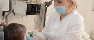
Systematic hygienic teeth cleaning at the dentist is an essential attribute of a healthy and beautiful smile.
Every time we accept another patient for caries treatment, we are convinced that the price of professional hygiene is disproportionate to the consequences and costs that will have to be borne if it is not done.
It has been proven that even correct, scrupulous and constant home care is ineffective. 90% of adults brush their teeth ineffectively.
Teeth cleaning at Unit using modern technologies: Air Flow and ultrasound gives the maximum effect in maintaining a healthy balance of microflora and local immunity.
Sign up for professional cleaning
Oral microflora: what you need to know
When patients ask what causes tooth decay, do you usually tell them “sugar” or “germs in the mouth”? We know that oral microflora plays an important role in maintaining oral health. Understanding the relationship between the body and the bacteria that inhabit it can help you teach your patients how to prevent dental disease.
What is oral microflora?
The microflora of the oral cavity is the totality of microorganisms found in it; As noted in a study published in the Journal of Bacteriology, more than 700 species and strains of bacteria have now been found in the oral cavity. Bacterial microflora in the mouth forms a structure that covers the teeth, dentists call this plaque or dental biofilm.
Many oral bacteria are associated with dental diseases that result from dysbiosis. An imbalance of different types of bacteria creates conditions for the excessive proliferation of potentially pathogenic microorganisms, playing a leading role in the development of a number of diseases. The current hypothesis is that the entire biofilm may be involved in this process. This is supported by recent research and the theory of a key pathogenic factor.
The main causes of dental and gum disease are two types of bacteria: Streptococcus mutans (S. mutans) and Porphyromonas gingivalis (P. gingivalis); In addition, researchers note a connection between lactobacilli and the occurrence of tooth root caries. Publications in the journal PloS One note that S. mutans consumes easily fermentable sugars and, as a result of metabolism, produces acid, which leads to demineralization of dental hard tissues, which causes caries. S. mutans also produces extracellular polysaccharides early in the biofilm formation process, essentially providing the “glue” for its formation and attachment to dental and oral soft tissue surfaces. According to the authors of Frontiers in Microbiology, P. gingivalis is the main microorganism involved in the development of chronic periodontitis.
Causes of changes in oral bacterial flora
The microflora of the oral cavity of each person changes throughout life. As children grow, bacteria take up residence in their mouths. S. salivarius colonizes soft tissue shortly after birth. As teeth erupt and gingival sulci form, microorganisms such as S. sanguinis and S. mutans colonize tooth and soft tissue surfaces. As a person grows and ages, the composition of the intraoral environment changes and the number of strains present increases.
Imbalance of the oral microflora can occur for many reasons, for example due to a diet high in fermentable carbohydrates, which changes the pH, or due to the formation of a dense biofilm, which is particularly protective of microorganisms located in its thickness (that is, it prevents the penetration of antimicrobial agents). Finally, opportunistic microorganisms such as Helicobacter pylori can penetrate the oral cavity. This microorganism is present in patients with gastroesophageal reflux disease (among other stomach diseases) and is involved in the development of periodontitis, according to publications in the International Journal of Clinical and Experimental Medicine.
Helping patients control oral microflora
How can you help patients balance their oral flora? The main thing is not to allow the balance to shift in any direction favorable to the development of diseases. Provide patients with several practical steps to help avoid creating unfavorable conditions and developing oral dysbiosis.
- Reduce your sugar intake. This will reduce the risk of creating a cariogenic environment by bacteria that metabolize carbohydrates. Instead, encourage patients to drink unsweetened and non-acidic beverages, as well as milk, and consume dairy products as part of a healthy diet that includes adequate calcium.
- Brush your teeth twice daily with an optimal fluoride toothpaste to break down biofilm and protect tooth enamel.
- Floss daily or use other interdental cleaners, such as interdental brushes, to remove plaque buildup between teeth and in the sulcus. Simply brushing your teeth will not eliminate bacteria from all of these hard-to-reach areas. Finish with an antibacterial mouth rinse. This can help remove food debris from your mouth and also reduce bacteria.
- Probiotics have been found to be beneficial for digestive disorders in healthy patients, and preliminary evidence from several studies suggests that probiotics may also help protect teeth and gums.
- Encourage patients to visit your office regularly for examinations, with the frequency of visits based on your and their own assessment of the risk of developing tooth decay and other oral diseases. Despite proper oral care at home, some people are at very high risk for dental disease and will need more frequent visits.
Understanding the role of the intraoral microflora is the first step in teaching patients how to control its balance to maintain oral health. Educating patients about the role of bacteria in dental disease can help them achieve overall oral health.
Why do bacteria and germs cause oral diseases?
Even stable microflora of the oral cavity cannot be called completely safe. Thus, any damage to the mucous membrane or tooth enamel and subsequent inflammation contribute to the development of pathogenic processes. Even normal microflora can become unfriendly with weakened immunity.
The most dangerous are “alien bacteria”. They reproduce as a result of disruption of the normal functioning of the ecosystem. This phenomenon is common in periodontitis, caries, stomatitis, gingivitis and other diseases. The uncontrolled spread of bacteria, fungi and microbes leads to the release of toxins and the development of the inflammatory process. Often such microorganisms demonstrate a tendency to constant relapses, especially if therapy is not accompanied by restoration of the microflora.
Oral candidiasis - symptoms and treatment
If treatment is untimely or incorrect, acute candidiasis can become complicated and become chronic or invasive, which is difficult to treat. In addition, complications such as fungal esophagitis, tracheitis, gastrointestinal candidiasis, and candidal sepsis may occur. There is a high probability of inflammation of the genital organs in women and men.
Candidal esophagitis (inflammation of the esophagus). Symptoms: signs of intoxication of the body, bloating, heartburn, sore throat, dysphagia (difficulty swallowing solid food), bitterness in the mouth or sour taste. Can lead to esophageal ulcers, rupture of the esophageal tube, internal bleeding, stricture (narrowing of the lumen) of the esophagus.
Candidal tracheitis (inflammation of the trachea). Symptoms: increased body temperature up to 37 °C, severe pain in the trachea, chest, shortness of breath and a feeling of suffocation when coughing, itching, burning, pain behind the sternum or between the shoulder blades, the appearance of ulcers on the skin. Through abscesses on the walls of the trachea, the fungus can penetrate into the blood, which will subsequently lead to sepsis.
Candidiasis of the gastrointestinal tract . Candida penetrates the intestines and injures the mucous membrane, causing a severe form of dysbiosis. Symptoms: nausea, vomiting with blood and whitish films, increased body temperature to 37-38 °C, bloating and pain in the upper abdomen, diarrhea mixed with white flakes. If the stomach wall is perforated, serious consequences can occur, such as peritonitis (inflammation of the peritoneum) or internal bleeding.
Also a severe complication of oral candidiasis is candidal meningitis , which can be fatal. Most often, the complication occurs in patients with severe concomitant diseases (immunodeficiency, decompensated diabetes mellitus) and in people with reduced immunity. Candidal meningitis is manifested by the following symptoms: increased body temperature to 37.2-37.9 °C, cephalalgia (constant headaches), drowsiness, hemorrhagic rash throughout the body, vomiting, which does not bring relief to the patient, often without preceding nausea. Neurological symptoms may appear: disturbances of consciousness, higher nervous activity, movement disorders, sensitivity, vision.
The danger of the disease lies in a sharp decrease in the body's immunity and the rapid spread of infection to the spinal cord and brain. There are also difficulties in diagnosing the disease due to the erased symptoms and the absence of pronounced meningeal syndrome (signs of irritation of the meninges).
Attachment of a secondary infection. Inflamed organs and tissues are very sensitive to other infections. They penetrate most quickly through ulcers and cracks with bleeding. When a secondary infection is added, ulcerative necrotizing stomatitis or Vincent's tonsillitis, syphilitic seizure, syphilitic dysphonia (impaired vocal function), etc. may occur.
Candidal sepsis is the introduction of a fungal infection into the general bloodstream. A serious condition in which candida penetrates into all organs and tissues. Signs of intoxication of the body: nausea, vomiting, abdominal pain, tachycardia and body hyperthermia develop (increase in temperature above 38 ° C), multiple organ failure occurs (disruption of several functional systems of the body), and the activity of the central nervous system (CNS) is disrupted. In this condition, treatment is carried out only in intensive care units, since if assistance and special therapy are not provided in a timely manner, the outcome can be fatal [2].
ODONTOGENIC AND PERIODONTAL INFECTION
PULPITIS
Main pathogens
Viridans streptococci ( S. milleri
), non-spore-forming anaerobes:
Peptococcus
spp.,
Peptostreptococcus
spp.,
Actinomyces
spp.
Choice of antimicrobials
Antimicrobial therapy is indicated only in case of insufficient effectiveness of dental procedures or spread of infection into surrounding tissues (periodontal, periosteal, etc.).
Drugs of choice
: phenoxymethylpenicillin or penicillin (depending on the severity of the disease).
Alternative drugs
: aminopenicillins (amoxicillin, ampicillin), inhibitor-protected penicillins, cefaclor, clindamycin, erythromycin + metronidazole.
Duration of use
: depending on the severity of the current (at least 5 days).
PERIODONTITIS
Main pathogens
Microflora is rarely detected in the periodontal structure and is usually S.sanguis, S.oralis, Actinomyces
spp.
In periodontitis in adults, gram-negative anaerobes and spirochetes predominate. P.gingivalis, B.forsythus, A.actinomycetemcomitans and T.denticola
are highlighted most often.
In juveniles, there is rapid involvement of bone tissue in the process, with A. actinomycetemcomitans
and
Capnocytophaga
spp.
P.gingivalis
is rarely isolated.
In patients with leukemia and neutropenia after chemotherapy along with A. actinomycetemcomitans
C.micros
is isolated , and in prepubertal age -
Fusobacterium
spp.
Choice of antimicrobials
Drugs of choice
: doxycycline; amoxicillin/clavulanate.
Alternative drugs
: spiramycin + metronidazole, cefuroxime axetil, cefaclor + metronidazole.
Duration of therapy
: 5-7 days.
For patients with leukemia or neutropenia after chemotherapy, cefoperazone/sulbactam + aminoglycosides are used; piperacillin/tazobactam or ticarcillin/clavulanate + aminoglycosides; imipenem, meropenem.
Duration of therapy
: depending on the severity of the course, but not less than 10-14 days.
PERIOSTITIS AND OSTEOMYELITIS OF THE JAWS
Main pathogens
With the development of odontogenic periostitis and osteomyelitis, S. aureus
, as well as
Streptococcus
spp., and, as a rule, anaerobic flora prevails:
P.niger, Peptostreptococcus
spp.,
Bacteroides
spp.
Specific pathogens are less commonly identified: A.israelii, T.pallidum
.
Traumatic osteomyelitis is often caused by the presence of S.aureus
, as well as
Enterobacteriaceae
spp.,
P.aeruginosa
.
Choice of antimicrobials
Drugs of choice
: oxacillin, cefazolin, inhibitor-protected penicillins.
Alternative drugs
: lincosamides, cefuroxime.
When P.aeruginosa
, antipseudomonas drugs (ceftazidime, fluoroquinolones) are used.
Duration of therapy
: at least 4 weeks.
ODONTOGENIC MAXILLARY SINUSITIS
Main pathogens
The causative agents of odontogenic maxillary sinusitis are: non-spore-forming anaerobes - Peptostreptococcus
spp.,
Bacteroides
spp., as well as
H.influenzae, S.pneumoniae
, less often
S.intermedius, M.catarrhalis, S.pyogenes
.
Isolation of S.aureus
from the sinus is characteristic of nosocomial sinusitis.
Choice of antimicrobials
Drugs of choice
: amoxicillin/clavulanate. For nosocomial infection - vancomycin.
Alternative drugs
: cefuroxime axetil, co-trimoxazole, ciprofloxacin, chloramphenicol.
Duration of therapy
: 10 days.