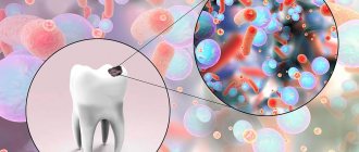
Surface formations on teeth
On the surface of the tooth there is a cuticle, pellicle, as well as plaque and tartar (with poor oral hygiene).
The cuticle, or reduced epithelium of the enamel organ, is lost soon after eruption, and therefore does not play a significant role in the physiology of the tooth. This formation, found mainly in the subsurface layer of enamel, sometimes comes to the surface in the form of a microscopic film.
In some places, the cuticle in the form of a tube reaches the enamel-dentin junction.
Pellicle (acquired cuticle) is formed from salivary glycoproteins on the surface of the tooth after its eruption. If the tooth comes into contact with saliva, then when the pellicle is removed with an abrasive, it is quickly restored. The pellicle is a structureless formation, tightly fixed on the surface of the tooth, and plays an important role in the selective attachment of bacteria.
The processes of diffusion and permeability in the surface layer of enamel depend on the state of the pellicle. To a certain extent, this shell protects the integrity of the enamel structure, but a large number of pellicles is not an indicator of enamel resistance.
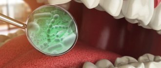
Above the pellicle you can find dental plaque - a dense formation consisting of bacteria located inside a matrix, which is formed by proteins, polysaccharides, lipids and some inorganic substances (calcium, phosphates, magnesium, potassium, sodium, etc.)
Plaque is attached to the tooth surface less tightly than pellicle, and at the same time, unlike food debris, it cannot be removed by simple rinsing. Plaque begins to accumulate soon after brushing your teeth; it is formed by the adsorption of microorganisms on the surface of the enamel and grows due to the constant layering of new bacteria, and in a certain sequence: first, coccal flora, and then rod-shaped and filamentous bacteria. As plaque grows and its thickness increases, anaerobic forms of bacteria begin to predominate.
Plaque has a porous structure, which allows carbohydrates to freely penetrate into its deep layers. When eating soft foods and consuming a significant amount of easily fermentable carbohydrates, its significant and rapid growth occurs.
Most often, dental plaque is located above the gum, in the cervical region, in fissures, and more microorganisms accumulate at the entrance to the fissures than in the depths.
80-85% of dental plaque consists of water. As for mineral components, calcium, general and inorganic phosphates, and fluorides predominate. Calcium in plaque may be associated with bacteria, extracellular proteins, or phosphates, which in turn may exist as inorganic orthophosphate or organic compounds. Fluoride is present in low concentrations in plaque fluid and in high concentrations in solid plaque. Although the mechanism of fluoride binding in plaque has not been fully elucidated, there are suggestions that the ion accumulates inside bacteria and forms extracellular complexes with calcium. The aqueous phase (plaque fluid), constituting 25-35% of the total volume, is located extracellularly and is an “incubation medium” for bacteria.
Plaque can be white, green or brown.
Soft white plaque, visible without staining with special solutions, accumulates mainly during the period of rest of the speech and chewing apparatus and in the absence of proper oral hygiene. This type of plaque can cause bad breath, distort the sense of taste, and also serve as a center of mineralization during the formation of tartar.
Green plaque, more often observed in children and young patients, is located in a thin layer on the labial surfaces, mainly of the front teeth. The appearance of this plaque is associated with the vital activity of chromogenic microorganisms containing chlorophyll.
Brown plaque is more common in smokers, and its color depends on the nicotine and intensity of smoking. It is difficult to clean with toothbrushes and toothpastes, but dental plaque can be white, green and brown. To remove it, teeth should be treated with hard brushes and special fine pastes.
Brown plaque can also occur in non-smokers due to a large number of copper amalgam fillings, as well as in people who work on the manufacture of copper, brass and bronze products. In children, plaque of this color often forms on baby teeth when a large amount of unreduced iron is secreted with saliva, which, combining in the oral cavity with sulfur from decaying protein substances, causes staining.
Calcification of dental plaque leads to the formation of tartar, hard deposits of varying consistency and color. Calcium phosphate crystals that are deposited within plaque may be closely associated with the enamel surface. Sometimes, especially in the presence of demineralization, it is difficult to determine where the enamel ends and the stone begins. For the formation of supragingival calculus, minerals coming from saliva are used mainly, while subgingival calculus is used from gingival fluid. The organic part of the stone is a protein-polysaccharide complex, including epithelial cells, leukocytes, microorganisms, and food debris.
Stone deposition, sometimes of considerable thickness, occurs in both the subgingival and supragingival areas. Calcification begins in plaque, which is present on the teeth for at least a few days.
Supragingival tartar is most often localized in the area of the lower frontal teeth and the buccal surfaces of the upper molars, where the ducts of the salivary glands open. In the absence of hygienic care, stone formation occurs on teeth that are not involved in the act of chewing. The color of the stone (white, yellow, brown) depends on the effects of food, nicotine, as well as oxides of iron, copper and other substances.
Subgingival tartar is revealed only by probing. It is usually dark brown in color with a greenish tint, and is formed on the neck of the tooth within the gingival groove, on the root cement, in the periodontal pocket. The stone surrounds the neck of the tooth, often forming projections, and is firmly attached to the underlying surface.
If a patient develops a significant amount of tartar, this may be due to a decrease in the concentration of pyrophosphate, a tartar inhibitor, or the absence of a specific salivary protein that prevents calcium phosphate precipitation and crystal growth.
Dental plaque: impact on dental and oral health
The article reflects the multicomponent composition, pathogenesis, chemical composition and complexity of formation, the role of deposits on teeth on the mechanism of development of dental diseases, their effect on the tissues of teeth and gums. The author strives to highlight pressing theoretical and practical issues regarding dental plaque, its harm, removal and prevention.
Key words : dental plaque, dental calculus, dental plaque, mineralization
The article reflects the multicomponent composition, pathogenesis, chemical composition and complexity of formation, the role of deposits on the teeth on the mechanism of development of the dental cavity, their effect on the tissues of the teeth and gums. The author strengthens the pressing theoretical and practical issues related to dental deposits, their harm, removal and prevention.
Key words: dental plaque, dental calculus, dental plaque, mineralization.
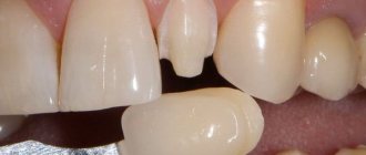
Dental deposits are, consisting of organic and inorganic substances, soft (dental plaque, dental plaque) and hard (tartar) formations on the surface of teeth, crowns and roots. [2] In contact with the hard tissues of the tooth, the mucous membrane of the gums surrounding the tooth, as well as other areas, they contribute to the development of caries, various periodontal diseases and the oral mucosa; under certain conditions, they have an adverse effect on the human body as a whole, and also create a significant aesthetic defect, changing the color of the teeth from yellow to dark brown.
The primary form of dental deposits is dental plaque - an organic base that, during the process of mineralization, turns into dental calculus [1].
The terms “dental plaque” and “tartar” are etiologically correct. Indeed, in reality, “dental plaque” and “tartar” are not of dental origin. The formation of tartar and plaque occurs on artificial crowns, phantoms, and removable dentures. They are formed by the environment of the oral cavity, which is largely associated with the function of many systems of the body and its organs. Based on this, we should assume that the correct terms are “dental deposits”, “dental plaque” and “tartar”, and not “dental deposits”, “dental plaque” or “tartar”. Both types of dental deposits play a significant role in the pathogenesis of periodontal and dental diseases [2].
There are mineralized and non-mineralized dental deposits. The first include: cuticle (remnants of the membrane after tooth eruption), pellicle (or acquired cuticle), soft dental plaque (sticky layers of bacteria and microbes, their waste products, saliva, food debris and destroyed cells) and dental plaque (dense formation that creates acidic environment that destroys enamel).
Dental plaque forms on the surface of the tooth, mainly in places that are poorly accessible for mechanical cleaning: the cervical area, especially the interdental spaces (from the gum pocket to the equator of the tooth crown), pits and fissures, proximal surfaces and cervical parts of the teeth, areas from the gums to contact points [2].
The rate of plaque formation during the first day (after thorough cleaning of the teeth) is not the same. The maximum speed is in the first 4 hours, the next 4 hours it is reduced, then it gradually increases again, after which it returns to the initial level by the end of the day. This is of practical importance for establishing an oral hygiene regimen. Dental plaque matures by the end of the 9th day at an uneven rate: the highest on the 1st day, then the rate of formation is reduced, and from the 5th to the 9th day it is maintained at a minimum level
The formation of soft dental plaque and dental plaque is influenced simultaneously by several factors:
- lack, stagnation and increased acidity of saliva, especially during sleep;
— diversity and quantity of oral microflora;
- level of carbohydrate consumption;
- insufficient and irregular oral hygiene.
In turn, mineralized deposits are supragingival and subgingival calculus (hardened plaque):
Tartar is a hardened dental plaque; the lighter it is, the denser and faster it forms. It is located mainly in the area where the ducts of the major salivary glands open: on the lingual surface of the lower frontal teeth, in the sublingual area where the ducts of the sublingual and submandibular salivary glands are located, on the buccal surface of the upper molars.
Supragingival tartar is located on the surface of the teeth at the supragingival margin and is visible to the naked eye. It has a white or yellowish color and a clay-like or hard consistency. When exposed to a special tool, it is easily separated from the tooth surface. The subgingival is located on the surface of the tooth root below the gum level. It is usually hard and dense and can only be detected by a dentist using special instruments (such as a probe). It has a greenish-black or dark brown color and fits tightly to the surface of the tooth root. This type of stone is found in patients with various periodontal diseases (the tissues surrounding and supporting the tooth) [5].
The chemical composition of dental deposits varies significantly in different parts of the oral cavity and depends on age, the composition and quality of food consumed, and the condition of periodontal tissues.
Studies of the chemical composition of dental plaque were carried out mainly to identify the quantitative content of Ca, P, K and Na. Water makes up 78.7–80.3% of its mass. The dry residue of dental plaque contains 1.65% total P, 0.43% inorganic P, 0.45% Ca, 0.44% Na. In dental plaque, 14–45% of P coming from the enamel was detected.
Ca, P, Mg, Fe and Cu were detected in soft dental plaque and saliva using chemical and spectral analyses. The content of these elements is highly variable. According to the level of concentration in the substrate, the ions are located in the following increasing order: in dental plaque - Cu, Fe, Ca, Na, P; in saliva - Cu, Fe, Na, Mg, Ca, P. Most samples of dental plaque contain fewer ions than saliva. Some ions are detected only in saliva or only in dental plaque. This made it possible to consider it as an autonomous ecological system, having its own metabolism and at the same time exposed to numerous factors of the oral cavity [2]
The structure of dental calculus was studied in detail using chemical structural analysis, which showed that dental calculus is 80% composed of inorganic substances, the main of which are calcium phosphates. In particular, octacalcium phosphate (brushite) makes up almost 50% of dental calculus. Depending on the amount of mineral substances, the consistency of tartar changes: with 50–60% of mineral compounds it is soft, 70–80% is medium and more than 80% is hard. A study of dental calculus conducted by A. A. Kolesov (1957) showed that the crystalline properties of this formation vary depending on the ratio between crystalline and amorphous substances [1].
Dental plaque has a large number of consequences. If plaque is not removed, it turns into tartar and leads to inflammation of the mucous membrane of the mouth and gums. Swelling of the gingival margin contributes to the formation of false pockets and at the same time increases the secretion of gingival fluid. This creates favorable conditions for the development of bacterial microflora, which is a key factor in severe complications: caries, gingivitis, periodontitis, stomatitis, halitosis, infective endocarditis.
The very first and main complication caused by dental plaque is caries - softening of the hard tissues of the tooth by acids secreted by plaque bacteria. The main reason is frequent consumption of sugars, high acidity in the mouth, decreased salivation (during sleep and thirst).
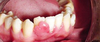
Gingivitis, an inflammation of the gums that is accompanied by redness, swelling, bleeding and bad breath, is not so rare. Irregular hygiene allows microbial plaque to accumulate between the gums and teeth, leading to inflammation. At the same time, the grooves between the teeth and gums increase and gum pockets appear. They contain bacteria that, in addition to gingivitis, can cause tooth root caries and periodontitis. The most common cause of gingivitis is dental plaque. Minor factors include: malocclusion, tartar, poor dental restoration, dry mouth, hormonal changes, systemic disorders (diabetes mellitus, AIDS, vitamin deficiency, leukemia, leukopenia), taking medications..
Periodontitis is another type of inflammatory gum disease, in which the tissue, including bone, that holds the tooth in the socket atrophies. The leading role in the development of the disease is played by pathogenic microorganisms of dental plaque. Bacterial waste products increase the permeability of blood vessels, which leads to the body's immune response and chronic inflammation. With periodontitis, there is bleeding, redness and swelling of the gums, its separation from the roots of the teeth, and in severe cases, purulent discharge from periodontal pockets. Also, the mobility of teeth when chewing increases, and food debris begins to get stuck between the teeth and gums. Periodontitis is an irreversible process, as it is already accompanied by tissue destruction.
Stomatitis is an inflammatory disease of the oral cavity with the formation of ulcers and erosion. Pathology is caused by bacteria that affect areas with microtraumas. In severe cases, blisters and areas of cell death (necrosis) appear on the mucous membrane.
Infectious endocarditis is an inflammation of the inner lining of the heart, which is accompanied by damage to the valve apparatus and vascular cells by opportunistic pathogens (in most cases streptococci and staphylococci). Bacteria from the gum pockets enter the bloodstream, then adhere to the cells of the heart or vessel (usually already damaged) and cause inflammation.
Having considered all the possible complications caused by dental plaque, it is worth assessing the importance of dental hygiene and prevention of possible consequences.
Deposits of dental plaque and tartar are a certain pathological phenomenon, for the elimination of which all provisions of primary prevention are fully applicable - elimination of the causes and conditions for the formation of dental plaque (social - health education, hygienic - training and education of the need for oral hygiene, medical measures - medical examination and elimination factors in the formation of dental plaque, increasing the body’s resistance to environmental factors that provoke their formation).
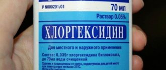
If you visit the dentist regularly, dental plaque is detected at the initial stage and is easily removed. It is recommended to carry out hygienic teeth cleaning once every six months. If the plaque is not removed, it turns into a dense formation and caries develops. With timely consultation with a doctor and proper treatment, the prognosis will be favorable. In case of acute caries, you should visit the dentist 2–4 times a year, treat the oral cavity with chlorhexidine, eat less carbohydrates and monitor the microbiological parameters of saliva every 2–3 years.
Preventive examinations, timely treatment, and removal of plaque significantly improve the health of the oral cavity and prevent the development of diseases of the gums and teeth, but without satisfactory self-hygiene it is impossible to achieve favorable long-term results.
Prevention of dental plaque is equally the responsibility of the doctor and each person. Its effectiveness depends on the validity of medical prescriptions, actions and manipulations and the scrupulous implementation of recommendations, hygiene regimens prescribed by the doctor and carried out by the patient himself. The widespread prevalence of caries and periodontal diseases deprives practical dentistry of the opportunity to carry out effective prevention of these diseases by doctors alone. In the best case, medical care can be provided to everyone who consults a doctor. But this is no longer prevention - as a rule, people go to the doctor in a state of illness identified by the patient himself, and it is very rare for people to go for individual medical examinations.
The basic rules of hygiene and prevention include:
Brushing your teeth twice: depending on the intensity of plaque formation, with tooth powder or toothpaste (as recommended by your doctor).
Intensive rinsing (after each meal) with water or elixir solutions, or decoctions of medicinal plants after removing food debris from the interdental spaces with a toothpick.
Alkaline rinses and soda paste with hydrogen peroxide to dissolve and eliminate heavy plaque and thick saliva.
Hydrotherapy at home or in a physiotherapy room, rinsing with alkaline mineral waters.
Oral care eliminates or reduces factors that damage periodontium. Hygiene products and the correct use of them reduce the pathogenicity of oral microbes, thereby eliminating the factors of destruction and sensitization of the gums. They enhance the therapeutic effect of medications, prevent inflammation and tissue destruction.
A decrease in the body's reactivity causes a decrease in the resistance of periodontal tissues, which creates favorable conditions for the implementation of local aggressive microbial factors and the occurrence of gingivitis and periodontitis. Compliance with the rules of oral hygiene breaks the vicious circle of factors leading to gum inflammation. Optimal hygienic conditions slow down and reduce the intensity of inflammation and periodontal destruction.
Mechanical (often in combination with chemical) cleaning is performed by chewing gum. Modern industry produces chewing gum, which can be recommended after brushing your teeth in the morning and evening for 15–20 minutes (as long as it retains its aroma and taste). However, chewing gum cannot replace mechanical cleaning of teeth with a brush.
There are also medicinal (chemical) means for the prevention and removal of dental plaque, which can be divided into 5 groups:
- means that prevent the adsorption of organic matrix components on the tooth surface - desorbents, hydrophobic film coatings;
- agents that suppress the formation and growth of the organic matrix and reduce the virulence of its constituent microorganisms - antibiotics, antiseptics;
- agents that cause desorption of the organic matrix - enzymes, surfactants;
- agents that prevent the mineralization of the organic matrix - deflocculants, crystallization inhibitors, competitive inhibitors of cations and anions;
- agents that destroy mineralized dental deposits - chelates, acids [2].
The instrumental method of removing tartar is the most radical, but at the same time the most common, accessible and proven in the practical work of a dentist.
Instruments for removing tartar can be classified into 5 groups according to their purpose: for removing tartar from the surfaces of the labial and buccal, lingual, proximal, subgingival parts of the teeth, for polishing the surfaces of teeth freed from tartar.
Conclusion. Considering the importance of the factor of the appearance of dental plaque when making a dental diagnosis, as well as based on existing research and observations, it has been established that, using the described methods for removing dental plaque, treating patients with periodontal diseases, preventing the formation of dental plaque, following recommendations for oral care , you can reduce the frequency of formation of dental plaque, and therefore the frequency of diseases of the teeth, oral cavity and the body as a whole.
Literature:
- Dental plaque / Levitsky A.P., Mizina I.K.—2nd ed., revised. and additional - K.: Zdorovya, 1987. - 80 p. - (Book of a practical doctor). —
- Dental deposits: their effect on teeth, periodontal tissues and the body / A. P. Grokholsky, N. A. Kodola, T. D. Tsentilo. - K.: Health, 2000. - 160 p.
- Boychenko O. N., Kotelevskaya N. V., Nikolishin A. K., Zaitsev A. V. Analysis of ideas about dental deposits // Bulletin of problems of biology and medicine, vol. 3, volume 1. – 2022. –. - With. 19–24
- New generation of therapeutic and prophylactic toothpastes and elixirs based on biologically active substances / V. A. Drozhzhina [et al.] // Proc. report region scientific - practical conf. dentists. - Rostov n/d., 1997. - P. 19.
- Features of the spread and manifestation of periodontal diseases and oral mucosa among schoolchildren in St. Petersburg E. V. Leonova [et al.] // Periodontology. –1998. — No. 4(10). — P.49–50.
- Pakhomov G.N. Primary prevention in dentistry / G.N. Pakhomov. - M.: Medicine, 1982. - 238 p.
- Pashaev Ch. A. New approach to the prevention of dental caries / Ch. A. Pashaev, L. K. Ibragimova, B. M. Gamzaev // New in dentistry. - 2004. - No. 7. - P.24–25.
- Persin L. S. Remineralizing therapy of early carious lesions of teeth - complications of orthodontic treatment / L. S. Persin, G. M. Barer, O. A. Varavina // Materials of All-Russia. conf. - M., 2003. - P.23
Features of ciliate pellicles
Ciliates have a very diverse shape. They can be:
- oval;
- oblong.
The sizes of ciliates vary from thirty to forty micrometers. These are the most complex protozoa, which have very original integuments. Their cytoplasm always has two layers:
- external – ectoplasm or elastic pellicle;
- internal – endoplasm.
The outside of the pellicle often has a sculptured structure with regularly spaced thickenings. The thickenings of the ciliate slipper in the pellicle area have the shape of regular hexagons, reminiscent of a honeycomb. For a pellicle, such a structure is very justified, since it significantly increases the strength of this formation.
Are you an expert in this subject area? We invite you to become the author of the Directory Working Conditions
Externally, the body of the ciliate is covered with cilia, which begin from the pellicle and its basal bodies. The number of cilia is very large and can be up to 10 thousand. According to the internal structure, cilia consist of two fibrils and nine fibrils reaching the periphery.
They continue in the kinetosome and acquire a triple structure. If the cilia are not arranged in rows, but evenly, then this is a primitive sign of ciliates. The locomotor apparatus of ciliates specializes in two directions:
- cilia concentrate on certain areas of the body;
- the cilia stick together and retain their own individuality as part of large working complexes.
When the cilia are connected in one or more rows, a ciliated membrane is formed, which is located on the pellicle. All these structures are called membranellas. If the cilia located on the pellicle in the form of a brush connect, a cirri is formed.
The most complex apparatus of cilia is found in ciliates in the area of the mouth. There are ciliates that have contractile fibers or myonemes; they are capable of sharp contraction. This significantly increases the possibility of movement of ciliates, despite the density of the pellicle.
Finished works on a similar topic
Coursework Pellicle of ciliates 420 ₽ Abstract Pellicle of ciliates 250 ₽ Examination Pellicle of ciliates 230 ₽
Receive completed work or specialist advice on your educational project Find out the cost
The pellicle of ciliates contains specialized formations called trichocysts.
Definition 2
Trichocysts are small protective spindle-shaped formations that are located in the cytoplasm of the ciliate perpendicular to the surface of its body. When exposed to any external irritants, they are thrown out and take the form of long threads with sharp points at the ends.
In addition, in the pellicle of ciliates there are peculiar structures in the form of plaques and hexagons, similar to trichocysts. Their threads are also capable of twisting and unwinding. In some ciliates, the pellicle may have a glassy structure, and it may be arranged in folds.
Other ciliates have a delicate, armor-like pellicle. The pinnate is in the form of a narrow strip from the anterior pole to the middle of the body. The edge of the peristome (the primary part of the cell mouth) is surrounded by two membranes.