To the reference book Palpation of the larynx and neck is one of the diagnostic methods that are used in otolaryngology in the treatment of diseases of the ENT organs.
Author:
- Oganesyan Tigran Sergeevich
ENT pathology expert
3.75 (Votes: 4)
Palpation of the larynx and neck is a visual diagnosis of diseases of the pharynx and larynx, which allows you to assess the condition of the skin, the severity of the local vascular network, the shape and position of the organ.
Much becomes clear already during a conversation with the patient, for example, by the sound of his voice (the presence of nasality, rattling, shortness of breath and other characteristic symptoms), the complaints presented (their dynamics, concomitant diseases in the anamnesis). After examining and questioning the patient, the doctor proceeds to palpation of the larynx and neck. Depending on the indications, it can be superficial or deep.
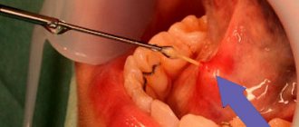
Throat bone (fish bone)
Among cases of foreign bodies of the upper respiratory tract in the practice of an otorhinolaryngologist, fish bones are the most common. The peak demand for removal of fish bones occurs in the summer months, when the diet contains a lot of freshly caught river fish. Samara is no exception, as it is located on the Volga River. Removing and pushing through fish bones is done at home with a crust of bread. Most often, small, thin bones—ribs—get stuck. The bone becomes lodged in the upper respiratory and digestive tracts at the time of ingestion. The most favorite places for bone fixation in the pharynx are the palatine tonsils, lingual tonsil, lateral ridges, posterior palatine arches, and pyriform sinuses. The palatine tonsils become a target for fish bones, since they actively accompany the bolus of food at the moment of swallowing. The lingual tonsil suffers for the same reasons. The tissue of the palatine and lingual tonsils is represented by lymphadenoid tissue, which is very loose and easily threaded onto a thin fish bone. Concomitant pathology in the form of chronic tonsillitis with hypertrophy of the tonsils increases the risk of bone entering the tissue. In the case where the bone is stuck in the upper parts of the pharynx and is in the line of sight, removing the fish bone in such a situation is not difficult. The situation with bone fixation in the lower parts of the pharynx requires the participation of a specialist. It is extremely difficult to remove such a bone without the help of an otolaryngologist. Complications of pharyngeal trauma caused by fish bones are rare. This form of sore throat is classified as traumatic; if the bone remains in the tonsil tissue for a long time, paratonsillitis can develop, which will end in a peritonsillar abscess. Acute pharyngitis, lateropharyngeal abscess, mediastinitis, phlegmon of the pharynx, neck, sepsis, laryngeal stenosis as a complication are quite rare. Removal of fish bones in Samara is performed by ENT doctors at Outpatient Center No. 1. First medical aid. During pharyngoscopy, you should carefully examine the palatine tonsils, moving away the palatine arches; with indirect laryngoscopy, you should carefully examine the root of the tongue, the vallecula of the tongue, and pear-shaped pouches. Finger examination is allowed. The foreign body is removed with a forceps or tweezers under visual control, after which it is recommended to rinse the oropharynx with an antiseptic solution and adhere to a gentle diet. If the foreign bodies are located in a different location in the pharynx, the patient should be urgently hospitalized in the otorhinolaryngology department. Specialized assistance. Foreign bodies of the lingual tonsil, vallecula of the tongue root and pyriform pouches are removed during indirect laryngoscopy in adults and direct hypopharyngoscopy in children using a laryngeal forceps or forceps. Anti-inflammatory therapy is prescribed. If a foreign body is not detected in the pharynx, and the pain syndrome persists, it is necessary to exclude a foreign body of the esophagus. For this purpose, fibrohypopharyngoscopy and esophagoscopy are performed.
Snoring and obstructive sleep apnea syndrome
Otorhinolaryngologist, Scientific and Practical Center MSR named after. L.I. Shvetsova
Kuznetsova Elena Nikolaevna
What is snoring and why is it dangerous?
Snoring (ronchopathy, benign nocturnal snoring) is a sound phenomenon that occurs when the soft structures of the pharynx beat against each other against the backdrop of a stream of air passing through the narrowed airways. This is a specific process that accompanies a person’s breathing during sleep, expressed by a distinct low-frequency rattling sound.
*from 30 to 50% of the adult population snores in their sleep
*30% of snorers develop obstructive sleep apnea syndrome.
Obstructive sleep apnea syndrome (OSA) is a disease characterized by snoring, periodic collapse of the upper airways at the level of the pharynx and cessation of pulmonary ventilation, decreased blood oxygen levels, severe sleep fragmentation and excessive daytime sleepiness (Guilleminault C., 1978).
Often witnesses to this disease are waking loved ones who watch with alarm as the snoring suddenly stops and a frightening cessation of breathing occurs, then the sleeper snores loudly and begins to breathe again. Sometimes there can be up to 500 respiratory arrests per night for a total duration of up to 4 hours, which leads to both acute and chronic lack of oxygen and significantly increases the risk of developing arterial hypertension, cardiac arrhythmias, myocardial infarction, stroke and sudden death in sleep.
Patients with breathing disorders during sleep experience restless, shallow, unrefreshing sleep, sweating and frequent urination at night, fatigue and headaches in the morning, severe daytime sleepiness, irritability, decreased memory and attention, and impotence.
The causes of OSA are the same as those of snoring. The difference lies only in the degree of their expression. If during snoring the walls of the respiratory tract only vibrate when a stream of air passes, then during illness they periodically collapse completely, stopping the access of air to the lungs.
The causes of snoring are:
1.Anatomical disorders leading to narrowing of the airways:
— Impaired nasal breathing
- Deviated nasal septum
- Chronic rhinitis
- Congenital narrowness of the nasal passages and/or pharynx
- Nasal polyps
— Pathology of the pharynx
- Elongated uvula
- Disturbance of the innervation of the muscles of the soft palate due to various degenerative neuromuscular processes or in connection with the use of substances with a muscle relaxant effect
(sleeping pills, tranquilizers, sedatives, alcohol) - Small, posteriorly displaced lower jaw (with malocclusion)
- Enlarged palatine tonsils
— Obesity
The risk of snoring in obese patients is 8-12 times higher than in patients with a normal body mass index. In patients with grade 3 obesity, a severe form of obstructive apnea syndrome (OSA) develops in 60% of cases. The risk of developing OSA increases significantly when the neck circumference in men is more than 42 cm and in women more than 40 cm.
Fat deposits in the area of the lateral walls of the pharynx lead to a narrowing of the lumen of the pharynx, increasing the likelihood of its collapse at the level from the uvula to the epiglottis.
2.Functional factors and diseases that reduce the tone of the pharyngeal muscles:
- Actually, sleep (decreased muscle tone)
- Sleep deficiency and fatigue
- Alcohol intake
- Taking sleeping pills
- Smoking
- Decreased thyroid function (hypothyroidism and acromegaly)
- Menopause in women
- Aging
Snoring, as a sound, is more of a social problem for others than for the person himself. However, snoring can be a harbinger and one of the main symptoms of a serious disease - obstructive sleep apnea syndrome.
OPTIMAL DIAGNOSTIC TACTICS
The diagnosis of snoring itself, as a rule, does not cause any particular difficulties. It should be noted, however, that the person himself usually underestimates his snoring. People around you overestimate. For example, a person may snore softly 10 times during the night (total duration is 10-20 minutes), but this will wake up his roommate 10 times and he will have the impression that the whole night was ruined by loud snoring.
The situation with the diagnosis of obstructive sleep apnea syndrome is much more complicated. For preliminary self-diagnosis, you can use a screening rule or a daytime sleepiness test, which allows you to suspect the disease.
An accurate diagnosis can only be established by a doctor based on consultation and polysomnogaphy.
Polysomnography and cardiorespiratory monitoring
Polysomnography is a method of long-term recording of various functions of the human body during night sleep.
The following parameters are registered:
- Snore.
- Breath.
- Blood oxygen saturation.
- Respiratory movements of the chest and abdominal wall.
- Body position.
- Electroencephalogram (brain activity).
- Electrooculogram (eye movements).
- Electromyogram (tonus of the mental muscles).
- Movements of the lower extremities.
Continuous video recording of sleep can also be carried out. Polysomnography is performed in sleep laboratories that have appropriate diagnostic equipment.
Cardiorespiratory monitoring
The same parameters are recorded as with polysomnography, with the exception of paragraphs. 6,7,8. The main difference between cardiorespiratory monitoring (CRM) and polysomnography is that the sleep structure itself is not analyzed. To a certain extent, this reduces the diagnostic accuracy of the method, however, for the diagnosis of breathing disorders during sleep (snoring and sleep apnea), CRM is often sufficient.
If it is necessary to diagnose sleep disorders themselves (insomnia, narcolepsy, etc.) or movement disorders during sleep (periodic limb movement syndrome during sleep), polysomnography is necessary. Polysomnography should also be performed in cases of suspected combination of sleep-disordered breathing with sleep disorders or movement disorders during sleep.
OPTIMAL TREATMENT TACTICS
There is no single and 100% effective treatment for snoring and obstructive sleep apnea. Accordingly, one should be treated with a dose of healthy skepticism towards announcements of miraculous interventions that eliminate snoring in all patients in one session.
The diversity of snoring and the varying severity of the condition require an individualized treatment approach. Sometimes it is enough to reduce your weight by 5-7 kg to stop snoring. An accurate assessment of difficulty in nasal breathing during the occurrence of snoring is of great importance. In some cases, eliminating a deviated nasal septum eliminates snoring. Laser plastic surgery and cryoplasty of the palate are effective only for uncomplicated snoring or mild forms of obstructive sleep apnea syndrome. With severe hypertrophy of the tonsils, it is necessary to raise the question of tonsillectomy, especially in the case of severe breathing disorders during sleep. The use of intraoral snoring devices is most effective in patients with retro- and micrognathia.
In severe cases of obstructive sleep apnea syndrome and the absence of obvious anatomical defects that could be corrected surgically, the only effective treatment is the creation of continuous positive airway pressure (CPAP therapy)
CPAP (English CPAP ) - therapy (an abbreviation for the English words C ontinuous P ositive A irway Pressure , which reflect the meaning of the method). The mechanism of action of CPAP therapy is quite simple. If excessive positive pressure is created in the airways during sleep, this will prevent them from collapsing and eliminate the main mechanism for the development of the disease, which is the cyclic closure of the airways at the level of the pharynx.
Thus, specific treatment approaches should be determined by a qualified professional depending on the cause and severity of the condition. Below is a brief description of all the possible treatments for snoring and obstructive sleep apnea.
GENERAL PREVENTIVE MEASURES AND SELF-MEDICATION
Do not increase or decrease body weight
If you have certain problems with breathing disorders during sleep, an increase in body weight by 10% of the initial one can worsen breathing parameters by 50%. This is usually accompanied by a transition to the next most severe stage of OSA. Patients were observed who gained 15-20% of their body weight over a period of one and a half to two years. These patients with a mild form of OSA, after gaining weight, received a severe form with all the ensuing consequences. This includes a significant deterioration in the quality of sleep, increased frequency of urination at night, crisis morning hypertension, morning headache, severe daytime sleepiness, irritability and a number of other symptoms. A significant reduction in body weight can, accordingly, significantly improve the situation.
Quit or limit smoking
Smoking often causes chronic inflammation of the pharynx and trachea, accompanied by swelling of their walls and decreased tone of the pharyngeal muscles. This, in turn, causes narrowing of the airways and increases snoring and OSA. However, one should be extremely careful when approaching the issue of smoking cessation in patients with OSA and obesity. This can lead to further weight gain and disease progression (see above), negating the benefits of quitting smoking. In this situation, you must first attempt significant weight loss, and only then resolve the issue of smoking. If you continue to smoke, it is advisable to at least abstain from it two hours before bedtime.
Avoid taking tranquilizers and sleeping pills
Tranquilizers and sleeping pills tend to reduce muscle tone and promote relaxation of the pharyngeal muscles, which in turn can worsen snoring and OSA.
Don't drink alcohol before bed
Alcohol also relaxes the pharyngeal muscles, causing snoring and OSA. The human liver processes about 10-15 ml of pure alcohol per hour. For example, 100 ml of vodka contains 40 ml of pure alcohol, respectively, the negative effect of this dose will last about 2.5-4 hours. You should especially refrain from taking alcohol and sleeping pills at the same time.
Treatments that do not require medical intervention
Positional treatment
- Mild forms of snoring and OSA are often position-dependent and occur only on the back. This is due to the retraction of the tongue. There is a simple and effective way to wean a person from sleeping on their back. On night pajamas or a special vest, a pocket is sewn between the shoulder blades in which a tennis ball is placed. In this case, every attempt to lie on your back will end in waking up and turning on your side. At the beginning of using this method, the quality of sleep may deteriorate, especially in people who are accustomed to sleeping on their backs, but within 3-4 weeks a strong conditioned reflex is developed not to sleep on their backs.
- It is necessary to ensure an elevated position of the headboard. An elevated position of the torso reduces tongue retraction even in a supine position. Moreover, the fluid in the body shifts downward, which leads to a decrease in swelling of the mucous membrane at the level of the nose and pharynx, an increase in their lumen, and, accordingly, a weakening of snoring.
- You should not use tightly stuffed large pillows to ensure an elevated position of the head, since this tends to cause the torso to slide off the pillow and the head to be tilted too much, which can even increase snoring.
- The head should be positioned as parallel to the body as possible. To achieve this, it is advisable to use small flat pillows, orthopedic or special contour pillows. The listed positional treatment methods help not only with snoring, but also with belching of gastric contents, which is often observed in overweight people who snore. Our pharmacies offer a number of drugs for the treatment of snoring. Drops for gargling “Good night” These drops are a mixture of 10 different essential oils, used diluted for gargling. According to manufacturers, drops have the following effects:
- They have a tonic effect on the pharyngeal muscles, which reduces snoring.
- Reduce or eliminate dry mouth during sleep, sore throat and other unpleasant sensations in the throat in the morning.
They lubricate the tissues of the pharynx, which reduces their injury and swelling during snoring.
Unfortunately, the initial enthusiasm for these drops gave way to moderate skepticism. Based on the results of controlled studies reported at the V World Congress on Sleep Apnea (Marburg, Germany, 1996), it was concluded that there was no clear clinical effect of the drops on OSA.
Despite this, it is quite possible to recommend this drug, since, firstly, it clearly reduces the subjective discomfort in the throat associated with snoring (dryness, soreness, pain), and secondly, it may have a preventive effect regarding the progression of the disease by reducing chronic trauma to muscle tissue at the level of the pharynx (works as a lubricant). The latter position has not yet been confirmed, but has not been refuted either, although from a pathophysiological point of view it has a right to exist.
Drug SnorStop (SnoreStop)
Snorstop is a complex homeopathic medicine
Dosage form: Tablets for sublingual administration (under the tongue).
Indications for use: Snorstop is used as a symptomatic remedy to reduce the intensity of snoring during sleep.
Contraindications: There are no absolute contraindications. The drug should be used with caution in cases of obstructive sleep apnea (sleep apnea) and pregnancy.
Take Snorstop once immediately before bed (put the tablet under the tongue until the drug is completely dissolved in the oral cavity). Weight up to 72 kg – 1 tablet before bedtime. If you weigh more than 72 kg – 2 tablets before bedtime. If the severity of snoring decreases, take Snorstop every other day; if the dynamics are positive, gradually reduce the frequency of use until you stop taking it.
The drug Snorstop has been used in Russia since 2000. Snorstop is intended for situational relief of uncomplicated snoring. It does not have a healing effect. After stopping the course of treatment, snoring appears or intensifies again after some time. In addition, Snorstop is practically useless for obstructive sleep apnea syndrome, when, in addition to snoring, there are pauses in breathing followed by loud snoring. It is usually recommended to use Snorstop for situational elimination or relief of snoring, for example, when traveling on a business trip, being in a hospital, etc. The effect of treatment is usually noticeable on the first night, but usually increases and reaches a maximum within 3-4 days. Thus, it is advisable to start taking the drug early. Snorstop is also recommended for situational use in patients whose snoring appears or worsens after drinking alcohol. The drug is also indicated for the treatment of snoring that occurs after a sore throat or pharyngitis.
It is not recommended to take Snorstop for more than 40 days. After this, it is necessary to take a break for approximately the same period during which the drug was used, since any long-term use of pharmacological agents carries the risk of potential complications
In the case of chronic snoring and, especially, obstructive sleep apnea syndrome, it is better to contact a specialized sleep center to clarify the causes and severity of the disease and choose the most effective treatment.
It is also worth mentioning various sprays to relieve snoring, “Hrapex”, “Slipex”. The mechanism of action of these drugs also lies in toning the muscles of the pharynx.
Ensuring maximum free nasal breathing
Difficulty in nasal breathing can significantly increase snoring and OSA. Let us dwell on therapeutic methods for improving nasal breathing, especially at night. The best effect is achieved by using special stickers on the nose (see picture). These stickers are a springy strip that sticks to the wings of the nose and pushes them apart, which makes nasal breathing much easier. You can predict the effectiveness of the strips quite simply by asking the patient to grab the wings of the nose with his fingertips, spread them apart and take a few breaths through the nose. These stickers are installed throughout the night. They can be used constantly or situationally, when you need to alleviate snoring as much as possible or there are factors that provoke snoring (alcohol consumption, ARVI).
For transient nighttime nasal congestion, it is necessary to consider the possibility of an allergic reaction to bedroom objects (dust, feathers, house mites), as well as a reaction of the nasal mucosa to dry air. In the latter case, humidification of the air during the night has a good effect. Constant use of drugs such as glazolin or sanorin is not advisable due to the rapid decrease in their effect and the development of dependence. In case of chronic nasal obstruction, consultation with an otolaryngologist is necessary.
Exercises to reduce snoring
There is a set of exercises for training the muscles of the tongue, lower jaw and pharynx, aimed at reducing snoring:
USING INTRAORAL DEVICES FOR SNORING
There is a domestic intraoral device for the treatment of snoring prevention and treatment “EXTRA-ENT”. In appearance, it vaguely resembles a regular baby pacifier and consists of a cup-shaped petal that touches the tongue, and fixing rims that protect it from swallowing or falling out of the mouth. The mechanism of action is to fix the tongue in a certain position, which prevents snoring. However, this mechanism does not give 100% results. The cup-shaped petal cannot permanently fix the tongue.
The therapeutic effect of the device is more logically explained by its reflex action on the muscles of the tongue and pharynx. If there is a foreign body in the oral cavity, the tongue constantly tries to touch it, sometimes even against our desire. The presence of a cup-shaped lobe in the anterior cavity of the mouth reflexively causes the tongue to move forward and tension its muscles, as well as the muscles of the pharynx, which prevent vibration and collapse of its walls.
This device has the greatest effect in individuals without severe obesity, with uncomplicated snoring and normal nasal breathing. The main obstacle to using the device is the possibility of deterioration in sleep quality due to the presence of a foreign body in the mouth.
There are also intraoral devices that push the lower jaw forward, which increases the anteroposterior size of the airway and, accordingly, reduces snoring and the severity of OSA. These can be blanks made of thermolabile material, which, when heated, are installed on the upper jaw (like a boxer’s mouthpiece), after which the patient closes his mouth with the lower jaw extended forward. The blank hardens and, when used again, allows the lower jaw to move forward (picture on the right). Intraoral devices provide the best effect for uncomplicated snoring, mild and moderate forms of OSA in patients with micro- and retrognathia. The main disadvantages of simulated devices are their high cost and the need for the participation of a doctor in their installation.
LASER PLASTY AND CRYOPLASTY OF THE PALATE
The therapeutic effect of these methods is based on causing a thermal or cold burn of the mucous membrane of the soft palate. A laser or cryoapplicator causes linear or pinpoint burns in the area of the soft palate and uvula, which causes inflammation. As the tissue heals, there is a decrease in volume and thickening of the palate, which reduces its vibration and the sound phenomenon of snoring. Technically, the procedure is quite simple and is performed on an outpatient basis. However, the patient experiences significant pain, reminiscent of a severe sore throat. The procedure can be repeated 1-3 times at intervals of 2-3 weeks until the desired effect is achieved.
Plastics are effective for uncomplicated snoring and mild OSA in patients with a low-lying soft palate and an elongated uvula. In moderate and severe forms of obstructive sleep apnea syndrome, especially in obese patients, the effectiveness of these techniques is low.
SURGICAL REMOVAL OF ANATOMICAL DEFECTS AT THE LEVEL OF THE NOSE AND PHYNAX
Difficulty in nasal breathing is one of the causes of snoring and obstructive sleep apnea syndrome (OSA). Moderate and severe forms of OSA should be considered an absolute indication for the elimination of chronic nasal obstruction caused by a deviated nasal septum, polyps or thickening of the nasal mucosa. For mild forms and uncomplicated snoring, especially in older age groups, it is necessary to carefully weigh the possible benefits and risks. In this case, it is necessary to take into account the social significance of snoring for the patient and his desire to eliminate this sound phenomenon.
The main surgical intervention at the pharynx level is uvulopalatopharyngoplasty. This operation involves excision of the uvula, part of the soft palate and palatine arches, as well as removal of the tonsils. This operation is very traumatic and must be used according to strict indications, since a number of complications may develop (postoperative respiratory arrest or bleeding; in the long term - a nasal voice and food getting into the respiratory tract). It should also be noted that in severe forms of OSA in obese patients, the positive effect of this surgical intervention is achieved only in 20-30% of those operated on. This is due to the persistence of narrowing of the underlying parts of the pharynx at the level of the root of the tongue and the epiglottis.
In any case, the decision on surgical treatment should be made by an ENT surgeon, taking into account clinical examination data, polysomnography results and other additional examination methods.
TREATMENT WITH CONTINUOUS POSITIVE AIRWAY PRESSURE
CPAP (CPAP therapy)
This type of treatment has already been mentioned above. This method is non-invasive and easy to use, but requires special, expensive equipment.
Treatment is carried out using a small compressor that delivers a constant stream of air at a certain pressure into the airways through a flexible tube and a sealed nasal mask. If in a healthy person the presence of a tube and a mask on the face can cause a deterioration in the quality of sleep, then in a patient with a severe form of OSA, accompanied by hundreds of pauses in breathing per night, and, accordingly, hundreds of micro-awakenings of the brain, sleep with the device is perceived as a much lesser evil. Moreover, daytime sleepiness is eliminated almost immediately and the overall quality of life improves.
The procedure for selecting therapeutic pressure is carried out in sleep clinics. Subsequently, the patient independently uses the device at home.
In severe cases, the device must be used every night. With a milder course of the disease, periodic use of the device is possible (4-5 times a week). The treatment has virtually no side effects. Cancellation of hardware treatment (even if it has been carried out for several years) does not entail any complications, except for the gradual return of the original symptoms. The use of the device does not cure a person, but provides normal sleep, improves the quality of life and prevents serious complications. Even very seriously ill patients can feel a significant improvement in their well-being within a few days.
CONCLUSION
In today's lecture we tried to find out what snoring is and what are the reasons for its occurrence.
-Treatment of snoring is not an easy task, requiring increased attention to the diagnosis of not only ENT organs, but also the general somatic condition of the patient.
-In most clinical cases, snoring treatment is a step-by-step process.
-None of the existing methods can give a 100% guarantee of getting rid of snoring. The average effectiveness of modern methods is 70%.
-For uncomplicated snoring, you can try conservative treatment methods yourself (gymnastics, sprays, positional treatment). But with obstructive apnea, this is a waste of precious time.
Thank you for your attention. Be healthy!
Foreign bodies of the pharynx
Causes.
They are usually localized in the oropharynx and laryngopharynx, where they enter with food, sometimes during manipulations in the mouth (pin, needle, toothpick). The most common foreign body in the pharynx is a fish bone, which penetrates into the loose tissue of the palatine, lingual tonsils, and into the vallecula of the root of the tongue. Less often, foreign bodies (coin, meat bone) are fixed in pear-shaped pockets. Foreign bodies enter the nasopharynx from the nasal cavity (needle), or from the lower parts of the pharynx during vomiting. This occurs more often in children and the elderly.
Symptoms.
Pain in the throat when swallowing with irradiation into the ear (stabbing with a fish bone), discomfort in the projection of a foreign body, sometimes hypersalivation, vomiting, difficulty swallowing.
Complications.
Bleeding, acute pharyngitis, lateropharyngeal abscess, mediastinitis, phlegmon of the pharynx, neck, sepsis, laryngeal stenosis.
First medical aid.
During pharyngoscopy, you should carefully examine the palatine tonsils, moving away the palatine arches; with indirect laryngoscopy, you should carefully examine the root of the tongue, the vallecula of the tongue, and pear-shaped pouches. Finger examination is allowed.
The foreign body is removed with a forceps or tweezers under visual control, after which it is recommended to rinse the oropharynx with an antiseptic solution and adhere to a gentle diet. If the foreign bodies are located in a different location in the pharynx, the patient should be urgently hospitalized in the otorhinolaryngology department.
Specialized assistance.
Foreign bodies of the lingual tonsil, vallecula of the tongue root and pyriform pouches are removed during indirect laryngoscopy in adults and direct hypopharyngoscopy in children using a laryngeal forceps or forceps. Anti-inflammatory therapy is prescribed. If a foreign body is not detected in the pharynx, and the pain syndrome persists, it is necessary to exclude a foreign body of the esophagus. For this purpose, fibrohypopharyngoscopy and esophagoscopy are performed.
Pharynx
Pharynx
(lat.
pharynx
) - the upper part of the human digestive tract, shaped like a tube, with one end opening into the oral cavity and the other into the esophagus. The pharynx extends from the base of the skull to the 6th-7th cervical vertebrae. The internal space of the pharynx makes up the pharyngeal cavity. The pharynx is located behind the nasal and oral cavities and larynx, in front of the main part of the occipital bone and the upper six cervical vertebrae.
The pharyngeal cavity is separated from the esophagus by the upper esophageal sphincter.
Compression of the pharynx is carried out by muscles - constrictors (squeezers) of the pharynx: upper, middle and lower. The opposite function to them is performed by the muscles - the levators of the pharynx: stylopharyngeus and tubopharyngeus
The length of the pharynx is 12-14 cm, it is divided into 3 parts:
a) upper - nasal part (nasopharynx); b) middle - oral part (oropharynx); c) lower - laryngeal part (larynx).
The opening connecting the oral cavity with the pharynx is called the pharynx.
Among the anatomical features, it is necessary to pay attention to the abundance of lymphoid tissue in the pharynx, which is of great clinical importance. As already mentioned, between the arches of the soft palate on the left and right there is an tonsil. At the border between the upper and posterior walls of the pharynx in the midline there is a focus of accumulation of lymphoid tissue - the pharyngeal tonsil (third tonsil). The fourth tonsil is located on the anterior wall of the lower part of the pharynx at the root of the tongue. The entire complex of lymphoid tissue located on all sides of the pharynx is called the lymphoid pharyngeal ring. On the back wall of the pharynx in the mucous membrane there are many follicles of accumulation of lymphoid tissue (Ionov A.Yu. et al.).
Acidity of pharyngeal secretions in healthy people and patients with chronic laryngitis and pharyngolaryngeal reflux
The acidity of pharyngeal secretions is different in healthy people and in patients with chronic laryngitis and pharyngolaryngeal reflux. In patients, when compared with a group of healthy individuals, the acidity of the pharyngeal secretion is higher, i.e. pH is lower (A.V. Lunev):
| Groups of surveyed | Average values of acidity of pharyngeal secretions, units pH |
| Healthy | 6,5 |
| Patients with chronic laryngitis without GERD | 5,6 |
| Patients with pharyngolaryngeal reflux | 4,9 |
| Patients with GERD without pharyngolaryngeal reflux | 5,2 |
On the GastroScan.ru website in the “Literature” section there is a subsection “ENT diseases associated with gastrointestinal diseases”, containing publications for healthcare professionals on this topic.
Patient Materials
The GastroScan.ru website also contains materials for patients on various aspects of gastroenterology:
- “Advice from doctors” in the “Patients” section of the site
- “Popular gastroenterology” in the “Literature” section
- “Popular gastroenterology” in the “Video” section
Back to section
Foreign bodies of the esophagus
Causes.
Hasty eating, missing teeth, inadequate dentures, decreased pharyngeal reflex, alcohol intoxication, cicatricial narrowing of the esophagus. Foreign bodies usually get stuck in the area of physiological narrowings, most often at the level of the first thoracic vertebra.
Symptoms.
The onset of the disease is sudden and associated with food intake. Characterized by pain in the throat or behind the sternum with irradiation to the back, interscapular area, dysphagia, aphagia, drooling, general weakness, malaise, pain on palpation of the neck (on the left), aggravated by tapping on the spine, possibly forced positioning of the head.
When a foreign body is localized in the area of the first physiological narrowing of the esophagus, the head is tilted forward, down, the patient holds it motionless, and turns the whole body. When a foreign body is localized in the thoracic esophagus, the patient is in a semi-bent position (“carrying posture”).
Indirect laryngoscopy reveals swelling, hyperemia of the mucous membrane in the area of the aryepiglottic folds, arytenoid cartilages, and accumulation of saliva in the pyriform pouch (usually the left one). Vomiting and coughing are possible. A large foreign body can cause difficulty breathing through the larynx.
Complications.
Perforation of the esophagus, periesophagitis, mediastinitis, bleeding from the great vessels.
First medical aid.
. Immediate evacuation to hospital. Attempts to push a foreign body by swallowing bread crusts or using bougies are prohibited.
Specialized assistance
provided by otorhinolaryngologists together with endoscopists. To do this, indirect laryngoscopy and x-ray examination of the cervical spine in two projections (according to G.M. Zemtsov) are performed, which makes it possible to detect the shadow of a foreign body, indirect signs of a non-contrast foreign body of the esophagus or damage to its walls.
These symptoms are:
- straightening of the cervical spine due to tension in the scalene muscles;
- expansion of the prevertebral space;
- the presence of a symptom of an air “arrow” - an accumulation of air released from the stomach below the level of a foreign body, the pointed end of the “arrow” indicating the location of the foreign body;
- striped clearings in the prevertebral space are a sign of air penetration into the retroesophageal tissue or the development of putrefactive inflammation with the formation of gas.
Fibroesophagoscopy is also performed for diagnostic and therapeutic purposes. If it is impossible to remove a strangulated foreign body of the esophagus during esophagoscopy, an esophagotomy is performed. Anti-inflammatory therapy is prescribed.
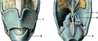
Types of palpation of the larynx and neck
Superficial palpation
This type of study is focused on obtaining objective data on the consistency and turgor of the skin in the neck area, which can be changed as a result of the development of a pathological process, for example, a malignant tumor. The patient is in an upright position.
The area is palpated in two positions - with the head in the normal position and slightly tilted back. The results make it possible to assess the nature of the relief of the skin covering the larynx and neighboring areas.
- hyoid bone;
- thyroid cartilage;
- cricoid cartilage;
- breastbone;
- prominence of the hyoid bone;
- the depression between the hyoid bone and the thyroid cartilage;
- Adam's apple;
- the cavity between the cricoid and thyroid cartilages;
- arch of cricoid cartilage;
- laryngeal prominence;
- depression above the breastbone.
Foreign bodies in the respiratory tract
Causes.
Aspiration of liquid or obstruction by particles of food or soil during a sudden deep breath, falling, crying, fright, talking, laughing. This is facilitated by the distraction of the victim’s attention while eating, the habit of holding foreign objects in the mouth, a decrease in the laryngeal-pharyngeal reflex, wearing removable dentures, alcohol intoxication, lack of consciousness due to traumatic brain injury, or poisoning.
Foreign bodies of the bronchi (88%) are more common, less common are the trachea (8.8%) and larynx (3.2%). The clinical picture depends on the nature, shape and level of presence of the foreign body in the respiratory tract.
Deep palpation
The doctor examines in detail the area of the hyoid bone and palpates the angles of the lower jaw. The movements of the doctor's fingers are directed from top to bottom, going down along the edges of the sternocleidomastoid muscle. After palpation of the supraclavicular fossa, it moves directly to the larynx itself. The otolaryngologist touches all its elements with the fingers of both hands, excluding/establishing pain, determines the shape and consistency of the structures.
Moving the larynx from side to side helps assess the degree of its mobility. In case of injuries, for example, in the case of a cartilage fracture, when the organ is displaced to the right or left, a crunching sound becomes audible.
By palpating the larynx, you can also identify the isthmus of the thyroid gland, which is located close to the cricoid cartilage. Feeling this area, the doctor asks the patient to swallow saliva. If during such actions a kind of push is felt, this may indicate the presence of an ectopic area in the tissues of the thyroid gland.
To make an appointment with an otolaryngologist, call one of the numbers listed or leave an online request on the website. All specialists are accepted by appointment. In emergencies and emergency conditions, you can use the service of calling a doctor at home.
Foreign bodies of the larynx
Symptoms.
When foreign bodies cover more than half of the glottis (live fish, a piece of food, adenoid tissue), fulminant stenosis develops: suffocation occurs, the voice disappears, and consciousness is lost.
Sharp and thin metal foreign bodies (pins, sewing needles, fish bones) initially do not cause severe breathing problems. A convulsive cough occurs, accompanied by sudden difficulty breathing, voice disorder, vomiting, and pain in the larynx are possible. During laryngoscopy, a foreign body can be detected that has stuck into the area of the arytenoid cartilage and aryepiglottic fold. The addition of mucosal edema causes an increase in inspiratory dyspnea.
Complications.
A foreign body obstructing the lumen of the larynx causes fulminant stenosis, and without proper assistance in the next few minutes leads to death. If the larynx is not completely obstructed by a foreign object, acute laryngeal stenosis develops in the coming hours.
First medical aid.
At the IV (terminal) stage of laryngeal stenosis, a conicotomy or cricoconicotomy is performed; at the III (decompensated) stage of stenosis, an urgent tracheostomy is performed. Dehydrating, diuretic, antihistamine, and corticosteroid drugs are administered. The victim is immediately evacuated to the ENT department.
Specialized assistance
consists of immediate removal of the foreign body during indirect (in children) or direct fibrolaryngoscopy with the participation of an endoscopist and an anesthesiologist, carrying out anti-edematous, anti-inflammatory, symptomatic therapy.
Tracheal foreign bodies
Symptoms.
Foreign bodies (nuts, beans, peas, watermelon seeds), carried away by the inhaled air, can pass through the glottis and become fixed on the mucous membrane of the trachea. This leads to paroxysmal convulsive dry cough, difficulty breathing, chest pain, vomiting during coughing attacks. Symptoms of “balloting” or “popping” of a foreign object in the trachea are detected. The addition of mucosal edema leads to inspiratory dyspnea.
Complications.
Foreign bodies capable of swelling (bean seeds), in combination with reactive edema of the tracheal mucosa, can lead to its stenosis, especially in young children, and the development of tracheitis.
First medical aid.
Prescribe sedatives, dehydrating, antihistamines, corticosteroids, antibiotics, oxygen inhalations. For decompensated stenosis, tracheostomy is performed.
Specialized assistance
consists of urgent removal of a foreign body during upper tracheoscopy under anesthesia using muscle relaxants. If it is impossible to remove a swollen foreign body through the glottis, a tracheostomy is performed on a bronchoscopic tube and removed through an incision in the trachea. Prescribe anti-inflammatory, decongestant, symptomatic therapy.