Find out more about nervous diseases starting with the letter “N”: Sleep disturbance; Narcolepsy; Hereditary cerebellar ataxia of Pierre-Marie; Spinal circulatory disorders; Trigeminal neuralgia; Neuralgia of the submandibular and sublingual nodes; Neuralgia of the glossopharyngeal node; Neuralgia of the ear ganglion; Neurasthenia; Neural amyotrophy of Charcot-Marie-Tooth; Acoustic neuroma; Neuroma; Optic neuritis; Pharyngeal neuritis; Neuritis of the facial nerve; Neuritis; Obsessive-compulsive neurosis; Pharyngeal neurosis; Neuroses; Neurosis-like stuttering; Femoral nerve neuropathy.
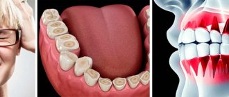
What is glossopharyngeal neuralgia?
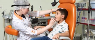
If the cranial nerve is affected by a quarter on one side, and paroxysms of pain appear in the tonsils, soft palate, pharynx and ear, then the patient may have neuralgia of the glossopharyngeal nerve. With this pathology, a violation of the taste perception of the posterior third of the tongue, on the affected side, is detected. Hypersalivation and decreased reflex of the palate and pharynx are also noted. To make a diagnosis, an examination by a neurologist or dentist is required. CT and MRI of the brain are indicated as hardware diagnostic methods. In most cases, treatment is conservative, including:
- Anticonvulsants;
- Analgesics;
- Sleeping pills and sedatives;
- Vitamin therapy;
- Physiotherapy;
- General strengthening procedures.
2. Reasons
The most common causes of glossopharyngeal neuralgia known today are trauma, acute respiratory infections and chronic infectious and inflammatory foci (for example, in the tonsils), inflammation of the meninges (meningitis, arachnoiditis), poisoning (in particular, lead compounds), oncological processes, vascular pathology (aneurysms, thrombosis), endocrine disorders.
In some cases, the cause remains unclear (idiopathic neuralgia).
Visit our Neurology page
Structure of the glossopharyngeal nerve
N. glossopharyngeus or glossopharyngeus nerve begins in the nuclei of the medulla oblongata. It is based on motor, sensory and autonomic parasympathetic fibers. Moreover, the sensory fibers begin in the sensory nucleus common to the vagus and glossopharyngeal nerves. They innervate the mucous membrane of the pharynx, soft palate, tongue, tonsils, and Eustachian tube. The sensation of taste in the anterior two-thirds of the tongue is provided by taste fibers emerging from the nucleus of the tractus solitarius. The taste fibers of the glossopharyngeal nerve are responsible for the taste sensations of the posterior third of the tongue and epiglottis.
The motor fibers originate in the nucleus ambiguus and innervate the stylopharyngeal muscle, which is necessary for raising the pharynx. Together with the vagus nerve, they form the reflex arcs of the pharyngeal and palatal reflexes.
Parasympathetic fibers originate in the salivary nucleus. Being part of the tympanic and lesser petrosal nuclei, they reach the autonomic ganglion, and with the branch of the trigeminal nerve they regulate salivation of the parotid gland.
Since the pathways and nuclei of the glossopharyngeal and vagus nerves are common, isolated pathology n. Glossopharyngeus. But more often symptoms of combined lesions are observed.
Pathogenesis
There are cases of idiopathic Sicard syndrome. At the same time, it is almost impossible to establish the etiology of the disease. Provoking factors may be:
- Acute or chronic intoxication;
- Otitis;
- Pharyngitis;
- Tonsillitis;
- Sinusitis;
- Atherosclerosis;
- Viral infections incl. flu.
Secondary neuralgia occurs due to:
- Arachnoiditis, encephalitis and other infectious lesions of the posterior cranial fossa.
- TBI.
- Hyperthyroidism.
- Diabetes mellitus.
- Meningiomas, gliomas, medulloblastomas and other intracerebral tumors of the cerebellopontine ganglion.
- Compression and irritation of any part of the glossopharyngeal nerve.
- Nasopharyngeal tumors.
- Intracerebral hematomas;
- Carotid artery aneurysm;
- Hypertrophy of the styloid process.
- Overgrowth of osteophytes of the jugular foramen.
- Ossification of the styloid ligament.
In some cases, pathology may be the first symptom of cancer of the larynx or pharynx.
Symptomatic picture
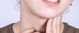
Clinical signs include painful paroxysms lasting from a few seconds to three minutes. A sharp acute pain originates at the root of the tongue and instantly spreads to the tonsils, soft palate, ear and pharynx. May radiate to the eye, neck and lower jaw. Pain can be triggered by coughing, chewing food, its temperature, yawning, swallowing and even talking. During paroxysm, dry mouth is noted, immediately after hypersalivation. But dryness is not a mandatory sign of diagnosis, because the secretory insufficiency of the parotid gland can be compensated by the remaining salivary glands.
Paresis of the levator pharyngeal muscle does not cause swallowing disorders. But difficulties in chewing and swallowing associated with impaired proprioceptive sensitivity, which is responsible for the position of the tongue, may be noted by patients.
The disease has a wave-like course, worsening in autumn and winter.
Diagnostic methods
The diagnosis is made by a neurologist, who, if necessary, can involve an otolaryngologist and dentist. During the examination, the doctor determines analgesia, that is, the absence of pain sensitivity at the base of the tongue, upper parts of the pharynx, and tonsils. Taste sensitivity is also studied, for which a taste solution is applied to symmetrical areas of the tongue. The diagnosis is confirmed by isolated unilateral taste disorder in the posterior third of the tongue. Bilateral disruption is typical for diseases of the oral mucosa, such as chronic stomatitis.
The pharyngeal reflex needs to be checked. To do this, touch the back wall of the throat with a paper tube, which provokes a swallowing movement and occasionally a cough. Normally, touching the soft palate should be accompanied by a lifting of the palate and uvula. Sicard syndrome is characterized by the absence of reflexes on one side. But a similar symptomatic picture can occur with damage to the vagus nerve. If the pharynx and pharynx are strewn with herpetic rashes, the doctor can diagnose ganglionitis of the ganglionitis of the glossopharyngeal nerve nodes, which differs in almost identical n. Glossopharyngeus clinical picture.
To establish the root cause of symptomatic neuritis, neuroimaging diagnostics is needed:
- MRI or CT scan of the brain;
- Electroencephalogram;
- Echo-EG;
- Ophthalmoscopy with consultation with an ophthalmologist.
It is necessary to distinguish between neuralgia of the glossopharyngeal nerve and diseases that cause painful paroxysms of the face and head. These include:
- Neuralgia of the ear ganglion;
- Trigeminal neuralgia;
- Oppenheimer's syndrome;
- Glossalgia;
- Ganglionitis of the pterygopalatine ganglion;
- Retropharyngeal abscess;
- Tumors of the pharynx.
The prevalence of orofacial pain in adults is up to 10%, men suffer twice as often as women, which differs from the general gender trends in the prevalence of pain syndromes, which are more common in women. An epidemiological analysis of patterns of facial pain (FP) in 7124 respondents showed that 7% had pain in the orofacial region during the last week, and at follow-up for 12 months this figure increased to 16%. Another epidemiological paradox of orofacial pain syndromes is a decrease in their prevalence with age, while the prevalence of pain syndromes of other localizations in the elderly increases [1, 2].
Orofacial pain syndromes are difficult to diagnose due to the diversity of the anatomical structure of individual formations and structures of the face and mouth, the morphological and functional characteristics of the peripheral and central parts of the nervous system, providing their afferent and efferent innervation, the influence of psychological factors on the clinical picture of L.B. Sensory innervation of the face is provided mainly by the trigeminal nerve, and in the oral cavity - also by the glossopharyngeal, vagus and facial nerves. A feature of the innervation of the skin and mucous membrane of the face is the high density of receptors, many of which can be activated by stimuli of various modalities (pain, temperature, chemical or mechanical). Most nociceptive information is transmitted by Aδ fibers. The temporomandibular joint (TMJ) and the masseter muscle are innervated by Aδ and C fibers. The cornea is also innervated by Aδ and C fibers, excited by mechanical, temperature and chemical stimuli at low receptor activation thresholds. The dental pulp is supplied with Aδ- and C-, Aβ- and sympathetic fibers. The lesion of each nerve is characterized by a set of clinical patterns, however, the lack of clear boundaries and the common innervation of orofacial anatomical structures can lead to difficulties in differential diagnosis. Chronicity of nociceptive pain syndromes in the head and face is caused by sensitization of the trigeminovascular and/or trigeminocervical systems and leads to the development of neuropathic and dysfunctional pain syndromes [3, 4].
Classification of orofacial pain syndromes
According to the beta version of the International Classification of Headache Disorders, 3rd edition (ICHD-3-beta) [5], PH is divided into those associated with pathology of the anatomical structures of the head and neck, cranial neuralgia and central pain (Table 1).
Table 1. Facial pain III part of ICHD-3-beta
The classification is intended for neurologists and is not always clear to doctors of other specialties. Some points of this scale, for example clause 13.9. (recurrent painful ophthalmoplegic neuropathy, in ICHD-2 was designated as “ophthalmoplegic” migraine) is still causing confusion even among neurologists. Psychogenic headaches are classified in Chapter 12, but psychogenic headaches are not mentioned in the beta version of ICHD-3; PILB (clause 13.11.) most corresponds to this nosology, but there is no direct indication of its psychogenic nature. It is more convenient to divide orofacial pain syndromes into dental and non-dental [6], which are also classified according to etiopathogenetic characteristics (Table 2).
Table 2. Clinical classification of facial pain
There are also central and peripheral pain syndromes in the orofacial region [6]. Peripheral lesions include local lesions of the teeth and adjacent tissues, as well as the ANS. Central pain syndromes develop as a result of intracranial pathology, for example, neurovascular compression, demyelinating and vascular diseases, and space-occupying neoplasms. There are three main mechanisms of pain development: nociceptive, neuropathic and psychogenic (dysfunctional).
Nociceptive pain
It occurs when nociceptors are irritated and, as a rule, occurs against the background of inflammatory changes, which in turn are divided into septic and aseptic. The pain is clearly localized, but if a nerve is involved in the process of inflammation, irradiation into the zone of its innervation is possible, i.e., a neuropathic component of pain is added. An important diagnostic criterion is the possibility of provoking pain with functional tests. Considering the diversity of tissues of the facial area, the causes of nociceptive pain are represented by a wide group of different nosologies, the treatment of which is carried out by different specialists. In case of nociceptive orofacial pain syndromes, it is important to timely clarify the source of pain and refer the patient to the appropriate specialist.
Toothache
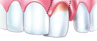
The most common causes of nociceptive facial pain are dental diseases, but epidemiological studies on this group are few [7, 8]. The typical social portrait of such patients is young patients or people who do not monitor their health, usually from the lower strata of society [9–11]. The cause of pain is diseases of the teeth and adjacent tissues (caries, tooth fractures, decreased dentin thickness). Pain may persist after a tooth fracture and after manipulations to restore it [12]. In diseases of periodontal tissues, pain is the least severe and occurs less frequently with gingivitis (6%), although it increases significantly as the disease progresses during the formation of periodontal pockets (25%) [13]. The pain syndrome is most pronounced when the pulp is involved in the pathological process, which is richly supplied with Aδ- and C-Aβ- and sympathetic fibers. Despite the wide range of stimuli, pulp receptors are predominantly monomodal, however, “sleeping” nociceptors are also found among them. The high pain sensitivity of intradental tissues also determines the closedness of the space; as a result, the inflammatory process and swelling lead to an increase in intradental pressure, causing pain. Acute periapical abscess, as well as exacerbation of its chronic form, lead to pulp necrosis and are characterized by severe pain, swelling and increased intradental pressure [14]. Treatment of such patients is the prerogative of dentists, but while awaiting appropriate treatment, blockade of the 2nd or 3rd branches of the trigeminal nerve may be effective [15]. Considering the aseptic nature of inflammation, to relieve intense pre- and postoperative pain, it is advisable to prescribe high doses of nonsteroidal anti-inflammatory drugs (NSAIDs) and even opioid analgesics. It is noteworthy that during dental procedures, etoricoxib, which has high permeability through the blood-brain barrier, at a dose of 120 mg is superior in pain-relieving effectiveness to opioid analgesics in combination with paracetamol [16].
Non-dental LB
One of the most striking clinical syndromes that reduces the quality of life of patients is ANS dysfunction [17]. To understand the pathogenesis of this disease, it is more correct to talk about dysfunction of the masticatory system, since changes in the joints cause secondary changes in the muscles and the development of myofascial pain syndromes. Patients note pain in the ANS, temporal (in the masticatory muscles), parotid, occipital, and sometimes in the cervical and shoulder regions. Pain increases with palpation and can be provoked by a pressure test on the ANS when opening and closing the mouth and clenching the teeth. When yawning, chewing or other movements of the lower jaw, attacks of severe pain and muscle cramps occur with “jamming” of the jaw. Other symptoms of damage to the ANS, such as acoustic phenomena (clicking, crunching associated with displacement of the articular disc) during movement of the lower jaw, impaired biomechanics of movement and limitation of mobility of the lower jaw, radiological changes of the ANS are nonspecific - in patients with pain syndrome they may not be detected, but their presence is not necessarily accompanied by pain. Normally, the degree of mouth opening varies from person to person, but opening less than the width of the interphalangeal joints of the 2nd, 3rd and 4th fingers is considered limited [3]. ANS dysfunction is quite common, affecting 10–15% of the population, and usually develops between the ages of 15–45 years, with no gender differences in prevalence found [18], although women are more likely to seek medical help [19]. The disease rarely occurs in children under 5 years of age [20], and is observed in 10.5% of adolescents [20, 21]. Risk factors for ANS dysfunction are old age, low general health indicators, and bruxism [22]. Often important is the violation of closure (occlusion) of the upper and lower jaws due to unsuccessful dental prosthetics. In this case, the lateral and medial pterygoid muscles are involved on the side of premature occlusal contact, and the masseter and temporal muscles are involved on the opposite side, with the formation of myofascial syndrome in them. In most cases, psychological factors play an important role [23]. Post-traumatic stress disorder increases the risk of developing ANS dysfunction [24]. A significant proportion of these patients have high levels of depression and somatization [25]. The prevalence of ANS damage is high among patients with obstructive sleep apnea [26]. Etiotropic therapy for ANS dysfunction includes physical therapy and orthodontic correction. As a basic pathogenetic therapy for joint pathology (osteoarthrosis of the ANS), along with NSAIDs, so-called “slow-acting symptom-modifying drugs” are widely used, in particular a combination of glucosamine and chondroitin sulfate (arthra). They also have analgesic and anti-inflammatory effects, but are devoid of the side effects characteristic of NSAIDs, since the mechanism of their anti-inflammatory action is not associated with suppression of prostaglandin synthesis, but is caused by blocking nuclear factor kappa B, which initiates the breakdown of cartilage tissue. The use of arthr 500 mg 2 times a day for 3 weeks, then 500 mg a day for 3 months for chronic joint-muscular pain leads to a slow decrease in the intensity of the pain syndrome by 50-70%, with the additional use of analgesics in the first month of treatment is reduced by half, and at the end of the 3-month course of treatment, the need for painkillers is reduced by 10 times [27]. The rationale for the use of arthra for ANS dysfunction is based on both the results of its many years of safe and successful use in osteoarthritis (level of evidence 1A), and the commonality of inflammatory and degenerative processes occurring in the joints of the spine, limbs and ANS. Pathogenetic therapy for myofascial pain syndrome caused by dysfunction of the ANS includes periarticular blockades with anesthetics and glucocorticoids. The greatest effectiveness is observed with periarticular administration and into myofascial triggers of a prolonged injectable two-component glucocorticoid drug diprospan. The rapidly soluble betamethasone sodium phosphate salt (2 mg) included in its composition ensures the rapid onset of action of diprospan 20-40 minutes after administration, and the microcrystalline depot fraction of betamethasone dipropionate (5 mg) provides a long-term (at least 4 weeks) anti-inflammatory and decongestant effect. Myofascial pain syndrome is detected in 10.5% of patients with ANS dysfunction [28]. Symptomatic treatment of myofascial pain may include the use of muscle relaxants [29]. Bruxism is treated with botulinum toxin injections into the masticatory muscles. When psychological disorders are dominant, pharmacological correction is used, focused on the leading emotional syndrome (antidepressants, tranquilizers, anxiolytics). If, after unsuccessful dental prosthetics, symptomatic treatment leads only to a temporary positive effect, it is possible to achieve stable remission by eliminating the cause of the suboptimal motor pattern in the masticatory system (occlusion disorders) - through reverse orthodontic correction (undermining fillings and dentures to restore the previous bite).
Neuropathic pain
Occurs when there is disease or dysfunction of the nervous system. The pain is poorly localized, since it can radiate to the dermatomes innervated by the corresponding nerve, is of a burning, shooting nature, accompanied by numbness, paresthesia, allodynia and other sensory disorders in the corresponding dermatomes. Neuropathic pain syndrome can be divided into central and peripheral. In clinical practice, it is most convenient to begin diagnosing neuropathic pain by assessing the temporal characteristics of symptoms: episodic or constant nature of pain (see figure) [6].
Figure Differential diagnosis of neuropathic pain syndrome.
Episodic neuralgia
Episodic neuropathic pain includes paroxysmal neuralgias, of which the most common is trigeminal neuralgia (TN). The main link in the development of TN is local demyelination of the trigeminal nerve [30]. Due to local damage to the myelin sheath, the permeability to various ions increases, and it becomes possible to transmit a signal from one nerve fiber to another “bypassing” the synapse - ephaptic transmission. There are two causes of demyelination: mechanical compression and an autoimmune process. Mechanical compression may occur due to aneurysm and tumor or be idiopathic. The idiopathic form occurs as a result of neurovascular conflict. The arteries at the base of the brain are muscle-elastic; the muscle layer relaxes during atherosclerotic lesions, maintaining the size of the vessel lumen. On a cross section, the vessel takes on an oval shape, putting pressure on the trigeminal nerve. When examining patients with idiopathic neuralgia, risk factors for the development of atherosclerosis are identified. Frequency T.N. higher in women and elderly patients [30]. In an autoimmune process, such as multiple sclerosis (MS), local demyelination occurs and the formation of ephaptic transmission. TN may be the only early sign of MS.
The clinical picture of TN is characterized by short paroxysms of pain, usually provoked by irritation of trigger points in the orofacial area. An attack can be triggered by a light touch or a breath of wind. Patients describe the pain as burning, shooting, piercing, lightning fast. The pain is unilateral and can spread to the anatomical zones of innervation of a given nerve branch [30]. Provocateurs for an attack of TN are talking, eating, combing hair, brushing teeth, blowing your nose, and shaving. The post-paroxysmal stage is characterized by a refractory period, during which pain cannot be provoked even by excessive stimulation. The duration of the paroxysm does not exceed a few seconds, but patients may note the presence of a constant background deep aching pain. Difficulties in differential diagnosis are due to the fact that the trigeminal nerve innervates the teeth, so pain can radiate to the jaw, simulating toothache. Triggers of pain paroxysms in TN can be movements in the ANS during chewing or swallowing, which requires differential diagnosis with ANS dysfunction.
Treatment of a patient with TN includes etiotropic, pathogenetic and symptomatic therapy. Etiotropic therapy is aimed at eliminating a specific cause: tumor removal, aneurysm clipping, microvascular decompression; for MS - the use of glucocorticoids. Pathogenetic therapy of neuropathic pain syndromes is aimed not only at reducing pain, but also restoring the function of the affected nerves, which allows it to be considered as a basic treatment for T.N. To activate regenerative processes, neurotropic complexes containing large doses of B vitamins - milgamma and milgamma-compositum - are used. The simultaneous use of thiamine, pyridoxine and cyanocobalamin stimulates axoplasmic transport. Thiamine promotes remyelination by activating phospholipase-A, enhancing the hydrolysis of fatty acid esters, improving energy supply, and supporting axoplasmic transport, which is especially important for restoring the trophic function of the nerve. Pyridoxine is involved in the synthesis of transport proteins and sphingosine, a structural element of the nerve fiber membrane and mediators of the antinociceptive system (serotonin, norepinephrine). Cyanocobalamin ensures the delivery of fatty acids for the formation of cell membranes and the myelin sheath. Its use also helps to reduce the intensity of pain due to its own antinociceptive effect. Lidocaine in milgamma is intended to provide anesthesia for the injection procedure. The used treatment regimen for TN includes a course of 10 intramuscular injections of milgamma followed by a transition to milgamma-compositum tablets 3 times a day for 2-8 weeks. Unlike the injection form of milgamma, milgamma-compositum tablets do not contain B12, long-term use of which is associated with a number of side effects. There is evidence of the advisability of using antioxidants - thioctic acid preparations (for example, thiogamma 600 mg in the morning on an empty stomach 30 minutes before meals for 2 months) to correct sensory and motor disorders in patients with TN [31, 32]. For pathogenetic treatment of TN, it is advisable to use anticholinesterase drugs (ipidacrine). The mechanism of action of ipidacrine (neuromidin) is based on the blockade of potassium permeability of the membrane and reversible inhibition of cholinesterase, which contributes to a stimulating effect on impulse conduction at the neuromuscular synapse. Having a polysynaptic effect, neuromidin affects both pre- and postsynaptic components of impulse transmission, and the analgesic properties of this drug in TN can also be associated with the blockade of sodium channels in axon membranes [31].
Symptomatic treatment of pain in TN is carried out with anticonvulsants. Carbamazepine is prescribed initially at 100-200 mg 2 times a day, followed by increasing the dose by 200 mg per week until the effect is achieved (1200 mg per day). Oxcarbazepine has shown its effectiveness in a daily dose of 600-2400 mg. Its advantages include weak interactions with other drugs, which is an important factor in elderly patients. There is data on the effectiveness of gabapentin (abagamma) at a dose of 900-3600 mg 3 times a day, pregabalin at a dose of 150-600 mg 2 times a day. If conservative treatment is contraindicated or ineffective, surgical procedures may be performed: radiofrequency destruction of the Gasserian ganglion and nerve decompression. Most often, in the presence of neurovascular conflict, decompression of the trigeminal nerve is used [33]. The above-described mechanisms of pathogenesis are also characteristic of other neuralgia, such as glossopharyngeal, superior laryngeal nerve, occipital, etc., the clinical picture, etiology, pathophysiology and basic principles of treatment of these forms of neuropathic orofacial pain have much in common.
Glossopharyngeal neuralgia
(HFN) accounts for 10-15% of cases of neuropathic orofacial pain and is 5 times less common than T.N. The most common cause of HFN is vascular compression of the posterior inferior cerebellar artery in the area where the root of the glossopharyngeal nerve exits the cranial cavity. Perhaps a similar mechanism can also lead to compression of the vagus nerve root. Mechanical trauma to the glossopharyngeal nerve can be caused by an elongated styloid process, fracture, or calcification of the ligament (Eagle-Sterling syndrome). In 1/3 of cases, the cause of HFN is a tumor of the cerebellopontine angle. It is important to exclude damage to the peripharyngeal space: abscess, consequences of tonsillectomy, carcinoma and arteriovenous malformation of the posterior cranial fossa. Diagnostic signs of idiopathic HFN are acute onset, no history of damage to the ENT organs, diseases of the oral cavity and teeth, absence of focal neurological symptoms and signs of trigeminal nerve involvement, and somatic well-being [3].
HFN can be divided into two types depending on the nature of the spread of pain: tympanic, in which pain radiates to the area of the tragus, external auditory canal (otalgia) and oropharyngeal, which is characterized by paroxysms of unilateral acute pain in the submandibular region, radiating to the neck, provoked by swallowing, talking, yawning, lasting 1-3 s. Attacks can begin at the base of the tongue and spread to the throat, velum, angle of the lower jaw, temple and eye. GPH can cause cardiac complications due to the involvement of the vagus nerve in the pathological process through a feedback mechanism between the affected glossopharyngeal nerve and the vasomotor centers of the trunk, which in turn can lead to an enhanced parasympathetic response in the form of hypotension, arrhythmia, and bradycardia [3]. Trigger points play an important role in provoking an attack of HF. Any area of the mucous membrane in the area of innervation of the glossopharyngeal nerve can be algogenic; an attack occurs when touching the root of the tongue or the tonsil. Outside of an attack, a number of symptoms characteristic of the affected nerve can be identified: dysgeusia, hypergeusia to bitter (all taste stimuli are perceived as bitter), pain anterior to the tragus, spasm of the pharyngeal muscles during swallowing, hypo- or hypersalivation, fainting. The disease occurs with exacerbations and remissions, the duration of which can reach 2-3 years. Over time, attacks become more frequent, the intensity of pain increases, and in the future it can become constant. A number of patients may experience symptoms of prolapse corresponding to the zone of innervation of the glossopharyngeal nerve, which indicates the evolution of the pathological process and the development of the neuropathic stage of GPH with constant pain in the root of the tongue, pharynx, upper pharynx, ear, lasting up to several hours.
Constant neuropathic pain
Neuropathic pain can occur after a peripheral nerve injury or neuroinfection. This leads to a decrease in the depolarization threshold of the peripheral nerve, peripheral sensitization, and the development of chronic neuropathic pain syndrome, which is described by the patient as a constant feeling of burning, heat, accompanied by paroxysms of acute shooting pain. Neuropathy can also be combined with other sensory phenomena: hyperesthesia, hypoesthesia, paresthesia, dysesthesia or anesthesia. If motor fibers are involved in the process, then motor manifestations in the form of tic hyperkinesis, paresis (up to plegia) are also possible [22]. Persistent neuropathic pain is common in dental-related TN, facial neuropathy (Bell's palsy), FH, and Tholos-Hunt syndrome (THS). Treatment tactics for such patients depend on the presence of signs of infectious origin of the disease. Identification of the bacterial etiology of neuritis makes it advisable to carry out antibacterial therapy. In the absence of an infectious cause, the use of corticosteroids in the form of local injections and systemically is recommended. There is evidence that their early use reduces the risk of pain chronicity [6, 33—36]. One of the specific causes of the development of neuropathy is herpetic lesions, which can cause postherpetic neuralgia. The pathophysiological mechanisms of the development of neuropathic pain are associated with the release of the virus from the axons and dendrites of the trigeminal nerve into the skin and rashes, which is accompanied by damage to the terminals. In the acute period of the disease, antiviral therapy is prescribed as etiotropic treatment - acyclovir 800 mg 5 times a day; For symptomatic pain therapy, antidepressants and antiepileptic drugs are used. Data were obtained on the positive analgesic effect of local injection therapy. When comparing the effectiveness of local injection therapy in the acute period of herpetic eruptions, treatment is carried out with lidocaine and vitamins B1 and B12, as well as their combination. It was shown that local administration of B1 significantly reduced itching, while B12 had a persistent analgesic effect, and their combination affected both parameters significantly more effectively than lidocaine monotherapy [37]. However, there is no evidence that local administration of B vitamins can reduce itching and pain, since there was no comparison group receiving systemic vitamins B1 and B12 in this study. Evidence regarding the effectiveness of paravertebral [35] and epidural [38, 36] blockades with anesthetics and glucocorticoids in preventing the development of postherpetic neuralgia is conflicting. Two studies [35, 36] showed an advantage of local administration of 10 mg of bupivocaine and 80 mg of methylprednisolone over standard antiviral therapy and systemic administration of glucocorticoids, one [38] did not show, and it is not yet clear whether the results of these studies can be extrapolated on LB.
Postherpetic neuralgia
(PHN) refers to central pain syndromes and develops after an acute period of herpetic rash, although neuropathic pain syndrome can begin from the moment the rash appears. PHN develops in approximately 15% of cases in patients over 60 years of age and directly correlates with the patient's age. The patient may experience a change in skin color in the area of the affected nerve. For treatment, pregabalin is used at a dose of 150-600 mg per day or gabapentin 900-3600 mg per day and antidepressants: amitriptyline 50-150 mg per day or nortriptyline 75-150 mg per day. Transdermal systems with lidocaine can be used as adjuvant therapy; their analgesic effect lasts 12 hours and is most pronounced in relation to superficial burning pain, allodynia and paresthesia. Longer (up to 3 months) analgesia can be achieved with a single application for 1 hour of a transdermal system with 8% capsaicin (cutenza), the mechanism of action of which is to deplete the reserves of substance P in the nociceptor terminals when this patch is applied to the skin in the pain zone [6, 33]. A pronounced increase in pain during capsaicin application may require additional analgesia for the patient - cooling the application area with ice, using opioid analgesics. In addition, a cosmetic defect in the form of erythema, which persists on the patient’s face for several days after the procedure, limits this type of therapy for L.B. A promising method of treating postherpetic neuralgia with intradermal injections of botulinum toxin type A [39] has shown to be highly effective and safe, but requires special skills in interventional therapy for LB.
Tholos-Hunt syndrome
(STX) - remitting painful ophthalmoplegia, characterized by pain in the orbital region and damage to the III, IV or VI nerves. Diagnostic criteria include the presence of one or more attacks of unilateral orbital pain, untreated, lasting for several weeks; dysfunction of one or more nerves innervating gaze; oculomotor disorders coinciding with the onset of pain or occurring within 2 weeks after it; oculomotor disorders and pain that cease within 72 hours after adequate treatment with glucocorticoids; granulomatosis of the intracavernous part of the internal carotid artery on MRI or biopsy. Other causes of painful ophthalmoplegia (orbital tumor, vasculitis, basal meningitis, sarcoidosis, diabetic cranial neuropathies, ophthalmoplegic migraine) should be excluded. Some descriptions of STX mention the possibility of involvement of the trigeminal (usually the ophthalmic branch), optic, facial, auditory nerves or disruption of the sympathetic innervation of the pupil. Their cause is the proliferation of granulomatous tissue in the cavernous sinus, superior orbital fissure or orbital cavity [3, 5]. Treatment involves etiotropic therapy with glucocorticoids (prednisolone 1 mg per kg of patient’s body weight per day) and, if necessary, symptomatic treatment of pain with antiepileptic drugs (gabagamma). To accelerate reparative processes, it is possible to use B vitamins (milgamma), antioxidants (thiogamma) and anticholinesterase agents (neuromidin) [31, 32].
Recurrent painful ophthalmoplegic neuropathy
(previously designated as ophthalmoplegic migraine) is an extremely rare syndrome manifested by repeated attacks of unilateral headache, accompanied by paresis of one or more oculomotor nerves (usually III) in the absence of signs of intracranial damage. There is a typical latent period of 1 to 14 days between the onset of pain and the onset of ophthalmoplegia. Due to the duration of headache for 1 week or more, beyond the diagnostic criteria for migraine, this syndrome cannot be considered as a variant of migraine. Contrast-enhanced MRI shows contrast accumulation in the cisternal portion of the involved nerve, indicating that the syndrome represents a variant of demyelinating neuropathy. The final diagnosis is established after exclusion by appropriate methods of clinically similar pathological processes in the parasellar region, superior orbital fissure and posterior cranial fossa [3, 5]. Treatment is similar to STX - glucocorticoids (prednisolone 1 mg per kg of patient’s body weight per day), B vitamins (milgamma), antioxidants (thiogamma) and anticholinesterase agents (neuromidin).
Deafferentation pain
(DB) develops when the peripheral nerve is damaged, which can occur as a result of surgical manipulations or injuries. The basis for understanding this phenomenon is the idea that the need for sensations is basic for a person and the deprivation of any even minimal channel of afferentation distorts the neuromatrix - a genetically determined neural network that ensures the integrity of the sensation of the body and the surrounding world [3, 40]. Under D.B. refers to sensations arising as a result of deafferentation, i.e., a deficit of sensitive, motor and social information, taking into account the emotional background. Sensation deficit changes the functional state of the neuromatrix and causes compensatory motor, neuroendocrine and mental reactions, which can be described and perceived as pain. This concept underlies the biopsychosocial model of pain. Habitual deficit sensations - hunger, thirst, cold, etc. are associated with negative emotions that trigger a genetically determined coping program (search for food, water, warmth), and a first-time sensitive (motor, emotional, visual, auditory, social) deficit can cause a paradoxical response of the neuromatrix in the form of its neuroplastic changes, leading to a persistent somatoform disorder that continues even after complete regeneration of the nerve trunk. DB can be pulling, aching or shooting, but more often it is burning, accompanied by paresthesia and dysesthesia.
Treatment of patients with DB is a difficult clinical task due to high resistance to therapy and tendency to relapse. The etiotropic treatment is to eliminate the damage: depending on the extent of damage to the nerve trunk, treatment can be surgical or conservative. Pharmacological treatment of DB includes the use of topical agents, such as transdermal systems with lidocaine or capsaicin in the area of innervation of the affected nerve [6, 33], however, the effectiveness of these procedures for DB is much lower than for neuralgia. The use of tricyclic antidepressants and antiepileptic drugs is effective. Carbamazepine and topiramate reduce the excitability of the trigeminovascular and trigeminocervical systems. This leads to the fact that ectopic pain impulses cease to be generated by the nuclei and fibers of the cranial nerves (trigeminal, glossopharyngeal, etc.). Paroxysmal pain and paresthesia undergo regression to a greater extent. However, severe sedation, dizziness and ataxia, resulting from non-selective suppression of sodium channel activity during treatment, can reduce the quality of life of patients. The best tolerability profile is found in gabapentin, which is involved in the transmission and modulation of pain at the level of the sensory nuclei of the cranial nerves and supra-stem structures, in the suppression of anxiety at the level of the limbic system and the regulation of muscle tone. Gabapentin has a number of biochemical properties that make it possible to influence the pathogenesis of neuropathic pain syndrome: interaction with α2delta2 subunits of voltage-gated Ca2± channels and inhibition of the entry of Ca2+ ions into neurons - thus reducing the hyperexcitability of cell membranes, reducing the sensitization of nociceptive neurons and nociogenic structures of the spinal cord and brain; increased synthesis of gamma-aminobutyric acid (GABA) and increased activity of glutamate decarboxylase, which leads to increased tone of the antinociceptive system; inhibition of glutamate synthesis, which leads to a decrease in the excitability of the structures of the nociceptive system and the prevention of glutamate-induced neuronal death; modulation of the activity of NMDA receptors and influence on the formation of “pain memory”.
Gabapentin increases the concentration of GABA in the cytoplasm of neurons and increases the content of serotonin in the blood plasma. By activating the processes of inhibition of the pain impulse at the level of the brain stem and thalamus, gabapentin suppresses the sensitization of the structures of the trigeminovascular and trigemino-cervical systems. Gabapentin (gabagamma) not only reduces the severity of BD, but also does not reduce the quality of life of patients, since the frequency of side effects with its use is minimal. Unlike pregabalin, gabagamma does not cause addiction or dependence and can be prescribed to patients with a history of alcohol and psychoactive drug abuse. The undeniable advantage of gabagamma over other prep
Therapy
To treat neuralgia of the glossopharyngeal nerve, conservative therapy is prescribed. The only exception is if the underlying cause is tumors and hypertrophy of the styloid process, leading to compression of the nerve. In these cases, surgery is indicated.
To relieve pain, a 10% solution of cocaine is prescribed, which is applied to the root of the tongue and pharynx. This allows you to eliminate paroxysms for 6-7 hours. If this remedy turns out to be ineffective, a 1-2% novocaine solution is injected into the root of the tongue. Non-narcotic analgesics and anticonvulsants are also prescribed internally. If the pain syndrome is pronounced, it is advisable to prescribe sedatives, hypnotics, antipsychotic medications and antidepressants. AFT, multivitamin complexes and FiBS are used as general strengthening agents.
Among the physiotherapeutic procedures that have proven effectiveness are:
- SMT on the area of the larynx and tonsils;
- Diadynamic therapy;
- Galvanization.
Treatment
Treatment of the hypoglossal nerve is usually only symptomatic and is part of a complex of therapeutic measures aimed at the underlying disease. Conservative treatment includes:
- taking anticholinesterase drugs (these drugs suppress the activity of the enzyme that breaks down acetylcholine, a neurotransmitter that transmits nervous excitement), B vitamins, etc.;
- reflexology;
- physiotherapy (electrophoresis, ultraphonophoresis, current treatment, etc.);
- oral hygiene.
In the absence of effect from conservative care, injuries or tumors, surgical intervention is indicated.
The required amount of therapeutic assistance can only be determined by a specialist after diagnostic procedures. If signs of damage to the cranial nerve appear, we recommend that you contact the First Medical Clinic in a timely manner.
You can make an appointment for a consultation and appointment by phone