Every person has suffered a nose injury at least once in their life. Those at risk include hyperactive children, people involved in martial arts, and military personnel. Statistics show that men are the undisputed leaders in nasal injuries. The nose is most susceptible to various damages due to the peculiarities of its structure and location. Injuries can occur at home, at work, during sports; children often get injured in their nose during active games.
Trauma can damage the bone and cartilage structures of the nose, as well as soft tissues. In this case, a concussion or contusion of the brain often occurs, and in order to exclude various neurological complications with any damage to the nose, you should immediately seek medical help.
Types of nasal injuries
Injuries to the nose can be open, which violate the integrity of the skin, as well as closed, located inside and not causing visible damage to the skin.
According to the severity of the nasal injury, they are classified:
- Soft tissue injuries resulting from contusion, abrasion, stab wound
- Damage affecting the cartilage tissue of the nose with a fracture of the nasal septum
- Fractures of the nasal bones are the most severe injuries to the structure of the nose, in which displacement of bone fragments, damage to the paranasal sinuses and other disorders can occur.
Fractures of the bones of the nasal vault or nasal septum can lead to the following disorders - displacement of the nose from the midline, the formation of a hump, changes in the shape of the nose (widening, flattening or flattening of the nose, retraction of the dorsum downwards), and the bony support of the nose can become soft. Injuries to the nose can result from frostbite, inhalation of steam or chemical (toxic) substances.
Sinus pain - causes
It is no secret that pain is a protective signal of the body. Which indicates problem areas, including the paranasal area.
If we consider the functional causes of pain in the sinuses, then it is largely associated with the pressure of exudate, which is formed in large quantities during inflammatory processes. Because of this, the normal outflow of discharge from the sinuses is disrupted, which causes pressure on surrounding structures and tissues.
Another common cause is the proliferation of tumor formations (polyps and cysts), which violates the integrity of anatomical structures, thereby causing necrotic processes in this area.
Anatomical features not related to inflammatory and necrotic processes, for example: curvature of the nasal septum, narrowness of the nasal passages, hypertrophy of the tonsils, pathologies of the lacrimal apparatus, etc. About which, in more detail in the next section of the material.
Main signs of nasal damage
When a person gets a nose injury, they experience severe pain, since the nose, like other parts of the face, has many nerve endings. Damage to soft tissue causes swelling and hematoma formation. The following symptoms indicate the presence of serious damage to the nasal structures:
- Bleeding from the nose. Severe and prolonged nosebleeds due to injury are not always due to the degree of damage. It can be caused by hypertension or bleeding disorders.
- Formation of hematomas. When blood vessels are damaged, blood can either leak from the nasal passages or accumulate in the soft tissues. Most often, small hematomas go away on their own, but with extensive hemorrhages, inflammation can occur. In some cases, large hematomas cause problems with nasal breathing.
- Changing the shape of the nose. This symptom indicates serious damage to the bony structures of the nose and often requires immobilization or surgical treatment. These injuries often occur after severe bruises or falls, and therefore are often accompanied by a concussion, and therefore require careful diagnosis and subsequent treatment. When a concussion occurs, patients experience headache, dizziness, nausea, weakness, and loss of consciousness.
Diseases leading to sinus pain
So what diseases can cause pain in the sinuses? This is a fairly voluminous list, so we will conditionally divide them into etiological reasons.
Inflammatory conditions, the main part of the pathologies that provoke this symptom:
- Sinusitis. Acute and chronic inflammatory processes in the paranasal sinuses - sinusitis, frontal sinusitis, ethmoiditis, sphenoiditis.
- Rhinitis. The catarrhal form can lead to chronic inflammation of the nasal mucosa, which provokes blockage in the paranasal sinuses. The so-called rhinosinusitis.
- Dental pathologies. If there are problems with the teeth of the upper jaw, emerging foci of inflammation can provoke the formation of a perforation or fistula in the maxillary sinus.
- Other states. Any focus of inflammation in the nasopharyngeal area can be complicated by spread to surrounding structures, for example, with chronic tonsillitis.
Diseases of non-inflammatory etiology can also lead to pain in the sinuses:
- A deviated nasal septum is the most common non-inflammatory cause of pain in the paranasal sinuses. More details in this material: Deviated nasal septum - symptoms, causes, complications.
- Trophic forms of rhinitis. Chronic changes in the mucous membrane during rhinitis can be not only inflammatory in nature, but also hypertrophic. For example: medicinal, atrophic, vasomotor and hypertrophic rhinitis. All of them contribute to the “blockage” of the outflow of secretions from the paranasal sinuses.
- Neoplasms. The growth of cysts and polyps can lead to the destruction of bone and cartilage tissue in the nasopharyngeal area, leading to corresponding symptoms.
- Other diseases. Hypertrophy of the adenoids or adenoid vegetations provoke a disruption of the normal patency of the nasal passages, thereby affecting the paranasal sinuses.
Treatment of nasal injury
Treatment options vary depending on the degree of damage to the nose due to trauma. For soft tissue bruises without visible damage to bone structures, it is recommended to apply cold. Superficial wounds and abrasions are treated with antiseptics and a sterile bandage is applied. To stop bleeding, it is recommended to lower your head down and press the wings of your nose against the septum for a few minutes, or insert turundas soaked in 3% hydrogen peroxide into the nasal passages for 20-30 minutes.
If the nasal bones are fractured or displaced and there is severe bleeding, you should immediately seek medical help. The doctor must stop the bleeding and determine the degree of damage to bone structures, treatment tactics, and also rule out concussion and other complications.
After stopping the bleeding and pain relief, the doctor restores the displaced bones, followed by fixation. As a rule, timely reposition of uncomplicated nasal fractures does not require surgical treatment, and bone tissue heals on its own within 10–14 days. Old injuries accompanied by a fracture of the nasal bones, complex open or closed fractures require surgical intervention - rhinoplasty of the nose.
If you receive a nose injury, especially if it is accompanied by bone displacement, severe bleeding, or there are open wounds and bone fragments are visible, you must immediately seek medical help. In medical clinics you can receive qualified first aid for any injuries at any convenient time without queues or long waits for an appointment. We can undergo all the necessary diagnostic examinations - x-rays, CT, MRI to exclude possible complications. You can make an appointment with a medical otolaryngologist in St. Petersburg by calling 8(812) 322-93-91.
If your sinuses hurt without a runny nose?
This in most cases indicates a chronic pathology, the most common of which is chronic sinusitis or sinusitis of the maxillary sinus. This condition is characterized by periodic pain in the maxillary sinuses. During the period of exacerbation, pain is accompanied by catarrhal symptoms: serous-purulent discharge, rhinorrhea, postnasal drip, etc.
Other ENT causes of pain without manifestations of rhinorrhea (runny nose) are the conditions described in the previous section: deviated nasal septum, neoplasms, adenoids, dental pathologies, etc.
It should be remembered that pain in the sinuses may not be of otolaryngological etiology, for example, in neurological conditions: neuritis of the cranial nerves, migraine, fibromyalgia, etc. Therefore, it is important to promptly assess the condition and exclude more serious pathologies. In this regard, diagnosis should be carried out in sufficient depth, especially in chronic pain.
Sign up for a consultation
Prevention methods
Most diseases that manifest as pain in the head and bridge of the nose can be prevented. They are not associated with anatomical features and can appear at any age, often associated with a seasonal decrease in immune defense. Doctors at the Clinical Institute of the Brain recommend following simple rules that will allow you to maintain health in any conditions:
- protect the bridge of the nose from hypothermia, especially in windy weather;
- during active sports, wear protective equipment that will protect against head and nose injuries;
- choose the right diet, with an optimal balance of vitamins and microelements;
- observe the rules of personal protection and hygiene in order to prevent the spread of viral diseases;
- minimize contact with allergens;
- start treatment at the first symptoms of a cold and do not wait for it to enter the acute stage.
Pain in the head and bridge of the nose can accompany various diseases of an inflammatory, traumatic, or infectious nature. In the early stages, they are easily treatable and do not pose a health risk, but can progress over time. At the Clinical Brain Institute, the patient will receive an individual diagnostic program that will help determine the exact cause of poor health. Experienced doctors will select a simple but effective treatment regimen, thanks to which you can quickly return to your normal lifestyle.
We care about your health
Epithelial tumors
Papillomas. There are papillomas of the vestibule and the nasal cavity. The first ones are gray in color, dense, villous surface, small in size, not malignant. It is believed that papillomas are caused by viruses from the family of papillomoviruses types 6, 11 and 16. Papillomas of the nasal cavity primarily affect the mucous membrane. They often recur after surgical removal. With anterior rhinoscopy or endoscopy, papillomas are gray-white in color, soft in consistency, single or multiple, bleed when touched, and resemble cauliflower. Treatment of nasal papillomas is predominantly surgical or cryodestruction. Surgically removed material should be sent for histological examination due to the possibility of malignant degeneration (transitional cell carcinoma). Another problem in the treatment of these patients is the frequent recurrence of papillomas. To prevent it, it is recommended to prescribe interferon drugs - domestic Laferon or foreign intron A. The method of treatment with Laferon consists of inhalation administration at the beginning of therapy (1 million Laferon is diluted in 20 ml of 0.9% NaCl solution). Inhalations continue for the first 10 days, then Laferon is prescribed intramuscularly once a day, at a dose of 1 million units, for 20 days. Some clinics determine the titers of antibodies to it to monitor the presence of the virus in the blood serum. When the levels increase above 1:200 in the blood of patients, antiviral drugs (Zoverax or others) are prescribed. Papilloma viruses also infect the cervix, which leads, in less pathogenic types, to the occurrence of condylomas.
Inverted papilloma. Called due to the ability of squamous epithelium to invaginate in the form of a wide ribbon into the connective tissue, these tumors have destructive growth, recur and become malignant. They are also known as columnar cell papillomas.
Adenoma is a benign tumor of the glandular epithelium of the nasal mucosa. It mainly affects the lower parts of the nasal cavity on a broad base with a smooth surface. Characterized by slow growth. The tumor is erysipelas-gray in color, no more than 2 cm in size, and is accompanied by rapid growth, germination into adjacent tissues, and changes in the histological structure. Its treatment is surgical. The removed material should be examined histologically.
Nonepithelial tumors
Hemangioma is a tumor developing from blood vessels, similar in nature to developmental defects. What it has in common with other tumors is rapid growth, during which hemangioma destroys surrounding tissue and causes a cosmetic and sometimes functional defect. In 1863, Vikhrov published a macro-microscopic classification of hemangiomas, dividing them into capillary, cavernous and racemose (branched). Subsequently, this principle was included in many classifications.
Today, this classification is used, which includes additional data on tumor growth, its spread, blood supply characteristics and relationship with large blood vessels.
True hemangiomas: 1) capillary (simple): exophytic (venous and arterial type); 2) cavernous: mucous-submucosal; diffuse (spreading into depth); 3) branched (angiodysplasia); 4) mixed (angiofibromas, hemlymphangiomas, branched with cavernous, branched with Barre-Mason glomusangiomas, etc.).
Diagnosis of tumors in the nose
- Routine external examination of the patient. Palpation of the tumor.
- Anterior and posterior rhinoscopy.
- Examination of the nasal cavity and paranasal sinuses using an endoscope. Diaphanoscopy.
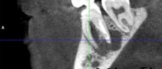
- Comprehensive X-ray examination.
- CT and MRI.
- Cytological examination of discharge obtained during puncture, sinus lavage, as well as nasal discharge.
- Histological examination of pieces of the neoplasm.
Treatment of nasal tumors
- Surgical removal of the tumor (if it is of significant size with preliminary embolization of the vessels feeding the tumor).
- Cryodestruction of the tumor.
- Sclerosis therapy.
- Radiation therapy in combination with surgical removal (for tumor malignancy).
Bleeding polyps of the cartilaginous part of the nasal septum are red, round in shape with a smooth surface. They are characterized by frequent bleeding. Treatment is surgical.
Fibromas, myomas, lipomas are rarely found in the cavity and paranasal sinuses. Angiofibroma, fibromyoma, osteofibroma, adenofibroma, neurofibroma, histiocytoma may occur. The treatment of these tumors is surgical.
Osteomas. They are most common among benign tumors of the paranasal sinuses, in people 20-40 years old. According to some authors, osteomas are localized in the area of bone sutures in 50% of patients. Histologically, three forms of osteomas are distinguished: compact (ivory), spongy and compact-spongy. Osteomas are extremely slow growing and predominantly asymptomatic. They are often discovered incidentally during X-ray or CT scanning. With large tumors or localization in the main sinus, a headache may be observed; with osteomas of the frontal sinus, exophthalmos, diplopia, and visual impairment may be observed.
Treatment of osteomas is exclusively surgical. The sooner it is carried out, the better success will be achieved. Mandatory histological examination of the removed tumor. Sometimes, with compact osteomas of the main sinus, when there is a risk of serious complications (damage to the cavernous sinus, internal carotid artery), surgical intervention should be carried out jointly with neurosurgeons.
Chondromas. They develop from the remains of the primordial cartilaginous skeleton and can be attributed to developmental anomalies. Tumors are characterized by slow expansive growth with penetration into the orbit or cranial cavity. They often recur and, in addition, can become malignant. Osteochondromas can occur in the nasal cavity. Neurogenic tumors
Paragangliomas (glomus tumors, chemodectomas) grow from small cell groups of the nasal mucosa. Rarely found in the nasal cavity. The tumor has infiltrative growth, often recurs, and malignantly degenerates. In such cases, it often grows into the skull and brain.
Tumor-like lesions
Tumor-like diseases are described in the relevant sections of the reference book, but fibrous dysplasia deserves special attention.
Fibrous dysplasia. Refers to tumor-like lesions of the nose and paranasal sinuses and is a defect in the formation of osteogenic mesenchyme. Characterized by the replacement of bone with fibrous tissue. Some authors consider the disease to be a developmental anomaly of unknown origin. Diagnosis of the disease is radiological but mainly histological confirmation is the main motive for choosing treatment. This lesion is also known as fibrous osteodystrophy, osteitis fibrosa. The bones of the upper jaw, main and frontal bones are predominantly degenerated. Treatment is surgical. The affected bone areas are removed. Relapses are possible. Sometimes such patients are treated in dental hospitals. V.
Malignant tumors of the nose and paranasal sinuses
The incidence of malignant tumors is steadily increasing by 2.6-3.0% per year. Malignant tumors of the nose and paranasal sinuses account for about 0.5% of cancer incidence statistics. In 2002, per 100 thousand population of Ukraine, the incidence of malignant tumors of this localization was 0.59, 287 patients were diagnosed for the first time. For comparison, in Belarus in the same year it was 0.7, 70 patients were identified. In Russia, 832 patients were detected for the first time, the incidence was 0.6. In the United States, 2,000 patients with cancer of the nasal cavity and paranasal sinuses are diagnosed annually.
Localization of tumors in the nose and paranasal sinuses, which are a system of narrow thin-walled cavities rich in nerves, blood and lymphatic vessels, promotes growth and rapid spread to adjacent formations, which significantly complicates treatment and leads in many cases to death.
Among the factors that cause malignant growth in this area are wood dust, petroleum products, professions associated with exposure to potent compounds - benzenes, acids, alkalis, nickel ores, varnishes and smoking. Among the factors that contribute to the growth of these tumors, chronic diseases are also noted - rhinitis, polypous sinusitis, ethmoiditis, frontal sinusitis, viral agents. The Epstein-Bar virus is considered the etiological factor of malignant lymphomas, papillomovirus types 11, 16, leads to the occurrence of transitional cell carcinoma and papillomas in the area of the nasal epithelium.
In most cases, the disease affects people of working age. Early clinical manifestations in people with tumors of this localization are insignificant and therefore the disease is more often detected in stages III – IV, when the walls of the cavities are already destroyed and penetration into adjacent anatomical areas begins.
According to most authors, malignant tumors are most often localized in the maxillary sinus (75-80%), in second place are the cells of the ethmoidal labyrinth and the nasal cavity (10-15%), less often the sphenoid and frontal sinuses are affected (1-2%).
When treating people with malignant tumors of the nose and paranasal sinuses, surgical, radiation and chemotherapy methods are used, but the results remain unsatisfactory, while the number of patients with this pathology does not decrease.
It should be noted that the palette of morphological forms of malignant tumors of the nose and paranasal sinuses is diverse.
Basal cell carcinoma occurs more often in various areas of the skin of the nose. Trichoepithelioma also belongs to basal cell malignant neoplasms.
Melanoma is observed in 2-3% of malignant tumors of this location. Most often it is located in the nasal cavity. The tumor is characterized by polymorphism. However, the sarcomatous form predominates. Early symptoms include bleeding and difficulty breathing. Melanoma metastasizes in 60% of cases to the cervical lymph nodes.
Clinical picture of tumors in the nose
Clinical manifestations of malignant tumors depend on the stage of the disease, localization, growth form, as well as the morphological structure of the tumor.
Symptoms:
- Pain. They appear sharply when localized in the upper jaw along the n. trigeminus. When the tumor is localized or grows into the area of the pterygopalatine fossa, shooting pain appears in the area of the eyeball or temple, which indicates irritation of the n.auriculotemporalis. Pain as the first sign of the disease was observed in 17% of patients.
- Impaired nasal breathing. When the tumor was localized in the nose, similar complaints were noted in 62.8% of patients. Patients whose tumor did not spread beyond the paranasal sinuses, or those whose tumor grew towards the cheek, temporal bone, alveolar process, or orbit did not complain of nasal breathing disorder.
- Nasal discharge. More often they are unilateral mucopurulent with an admixture of blood.
- Headache of various types, often with various paresthesias in the facial area on the side of the tumor. Often in such cases neuralgia is diagnosed. If they are not treatable, you should always remember about the possible development of a malignant disease.
- Pain in the teeth, deformation of the face, nose, lacrimation, exophthalmos, deformation of the hard and soft palate - these symptoms can occur to varying degrees with malignant tumors of the nose and paranasal sinuses.
Malignant tumors of the mucous membrane of the maxillary sinus occur without symptoms for a long time. Clinical manifestations depend on the starting point and size of the tumor. Tumors arise on all walls of the sinus, from the superomedial part of the sinus they quickly cause bleeding, swelling of the eyelids, lacrimation, exophthalmos; when they grow forward, they grow into the anterior wall and then in the cheek area they can be identified by palpation. As the tumor grows downward, it grows into the hard palate and changes its configuration. The growth of a tumor from the medial wall into the nasal cavity makes breathing difficult and leads to the formation of polyps. If the tumor grows predominantly in the direction of the posterior wall with germination into the fossa, the nasal part of the pharynx, then it causes pain.
When a tumor destroys the orbital wall, the process spreads to the orbital tissue, which is manifested by exophthalmos, decreased visual acuity and diplopia. Malignant tumors of the ethmoid labyrinth. Isolated lesions of the labyrinth cells are observed in 10% of cases.
When the tumor spreads towards the orbit, the thin wall of the latter is destroyed and tumor growth into the orbit is observed.
From the cells of the labyrinth, neoplasms can also spread to the maxillary, sphenoid sinuses, anterior and middle cranial fossa, and nasal pharynx.
Tumors of the sphenoid (main) sinus. Primary cancer of this location is extremely rare. Typically, these are tumors that extend from the maxillary sinus or ethmoid. The earliest symptoms are ocular, often damage to the efferent nerve, less often - decreased vision. Patients seek help from an ophthalmologist or neurologist. Characteristic orbital-superior syndrome (damage to the vessels and nerves that pass through the optic foramen and the superior orbital fissure), visual impairment, up to blindness, ptosis, diplopia, pain in the occipitotemporal and occipital regions.
Tumors of the frontal sinus are rare and have symptoms when affected, which do not extend beyond the sinus, similar to the symptoms of chronic frontal sinusitis. In recent years, CT and MRI have made it possible to differentiate this disease from mucocele, osteomyelitis and sinusitis. Symptoms depend on the direction of tumor growth. Germination downwards and medially leads to penetration into the orbit, ethmoidal labyrinth, exophthalmos appears, germination posteriorly - the anterior cranial fossa is affected, then a sharp headache, convulsions, and mental disorders are possible. Penetration of the tumor through the anterior wall leads to swelling and discoloration of the skin in the frontal area of the head.
Overview video of ENT surgery in Moscow, Kurkino and Khimki:
Diagnostics
For pain in the nose, a visit to an otolaryngologist is indicated. The doctor receives information valuable for making a diagnosis when collecting complaints, visual examination and palpation of the paranasal area, assessing the amount and nature of discharge. A diagnostic search in otolaryngology requires instrumental or laboratory research methods, the most informative of which are:
- Rhinoscopy.
When examining the nasal passages, pay attention to the color and presence of swelling of the mucous membrane, atypical growths or foreign bodies, and deformation of the nasal septum. To clarify the diagnosis, a nasal endoscopy is performed using modern equipment. - Radiography.
An X-ray of the nasal bones and paranasal sinuses is the standard method for identifying sinusitis, which is visualized as darkening and narrowing of the affected sinus. In case of traumatic injury, an X-ray or computed tomography of the facial skeleton is additionally performed. - Bacteriological research
. If there are signs of inflammation, the release of cloudy mucus or pus when sneezing, a smear test for microflora is required. When a boil is detected, a smear is taken from the top of the pustule to diagnose the type of bacterial flora and determine its sensitivity to antimicrobial agents. - Lab tests
. A clinical blood test shows nonspecific signs of inflammation (leukocytosis with a shift to the left, increased ESR); with massive nosebleeds, a decrease in hemoglobin and red blood cells is possible. Blood biochemistry is done in case of severe intoxication, for example, with an extensive burn complicated by a purulent process.
If the doctor suspects malignant transformation of cells, an endoscopic biopsy of the formation is performed, followed by histological examination. In case of multiple injuries to the bones of the skull and nose, the patient requires examination by a neurologist or neurosurgeon. If the pathological process affects the organ of vision, an ophthalmologist is involved in the diagnostic search.
For nasal pain, consultation with an otolaryngologist is indicated.