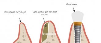
Osteoplasty in dentistry is the augmentation of bone tissue necessary for successful implantation. After a person has a tooth removed, the hard tissue tends to dissolve, and the new artificial tooth must be held securely in the jaw. Bone augmentation for dental implantation successfully solves this problem, but requires additional costs and more time (than, for example, with one-stage implantation, when the tissue volume is sufficient).
Why is bone augmentation necessary?
Bone augmentation (osteoplasty, sinus lift, bone replenishment) is the process of replenishing hard bone in the area where a dental root-shaped implant is subsequently planned to be installed.
Why is it important to build bone tissue? Because due to the absence of a tooth in the socket for a long time, the bone gradually decreases, and future implantation, in fact, loses its meaning. If there is not enough bone tissue, over time the implant loses its stable position, and the risk of rejection is high - all the work on its installation will have to be redone, again incurring financial losses and experiencing moral and physical risks.
Note! Sometimes bone grafting is required due to the physiological characteristics of the human jaw structure. Also, due to the fact that the bone decreases, inflammation of the soft tissues occurs (periodontitis, periodontitis).
The roots are located in the so-called alveolar processes, that is, the spongy part of the bone. Tooth roots constantly keep this area in good shape, active blood circulation occurs. In the absence of a tooth, the pressure on the jaw stops; accordingly, the bone tissue dissolves, resorption (loss) of the jaw bone occurs, and atrophy of the alveolar process occurs.
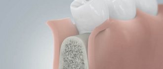
Bone tissue augmentation involves supplementing it with material that activates natural growth. Thus, the new bone is strengthened, its volume increases to the desired size.
Dentistry for those who love to smile
+7
Make an appointment
Expert opinion
Igor Yurievich Malinovsky
Maxillofacial surgeon, implantologist
Experience: more than 11 years
Osteoplastic surgery often precedes dental implantation. Especially in cases where the tooth was removed more than a year ago. Without load, the bone sags, atrophies, and its volume becomes insufficient to install an implant. This is why it is so important to restore teeth immediately after their removal. If there are a large number of missing teeth, the only way to do without osteoplasty may be basal implantation with deeper installation of implants into hard layers of bone tissue.
Main types of bone augmentation
Depending on the projection of the atrophic process, there are:
- Bone tissue growth in case of horizontal atrophy, when the alveolar process resolves along its width or horizontally;
- Building up the block during vertical atrophy, when the alveolar process resolves along its length or vertically;
- Bone tissue growth in case of combined atrophy, when resorption occurs both in width and length.
Depending on the chosen augmentation technique, there are several types of bone augmentation:
- classical bone grafting;
- sinus lift;
- transplantation of a donor drug.
Note! Resorption of bone tissue in different areas of the jaw occurs differently. For example, if a tooth is missing at the top, atrophy begins within a couple of months. For the bone atrophy to begin, a little more time must pass below, that is, from 4 months, respectively.
Autogenous transplantation
involves the use of the patient's own tissue. It is considered one of the most effective operations, since your own material takes root better and faster.
Alloplastic transplantation
involves the use of synthetic drugs. The implant surgeon makes an incision in the gum, implants an artificial composition, and covers it with a membrane. A few months after successful engraftment, you can proceed to implantation.
Xenogeneic or allogeneic transplantation
- an operation that involves the use of natural bone material. For example, the composition is of animal origin or donor bone tissue from another patient.
General overview
Bio Oss is made from bovine bone material. The drug is available in several dosage forms:
- Bio-Oss Spongiosa. Granules in bottles are suitable for various types of surgical interventions.
- Bio-Oss Pen. Granules in a syringe are used when the working area is located in a hard-to-reach place.
- Bio-Oss Block. Blocks are used to fill in significant anomalies.
- Bio-Oss Collagen. A combined preparation consisting of 90% granules and only 10% pork collagen. Demonstrates high adhesive, regenerative and manipulation properties.
The company produces Geistlich Bio-Gide as an auxiliary material. The composition allows you to create a barrier between the sinus and the bone, accelerates tissue regeneration. The drug is recommended for use in all types of bone grafting: in maxillofacial surgery, implantology, periodontology and surgical dentistry.
The drug exceeds autogenous bone grafts in terms of healing and regenerative capabilities.
Advantages of the drug:
- Possibility of individual use or combination with autogenous bone.
- Stabilizing the blood clot and preventing bleeding in the socket.
- The presence of many hydro- and macropores ensures hydrophilicity.
- Stability and preservation of the volume of the formed bone.
- The variability of forms allows the material to be used in various clinical cases.
The technique of using the drug is determined by the clinical situation and dosage form. General Features:
- Soaking the composition with saline solution or blood.
- Delivery of medicine to the work area and distribution according to pathology.
- Closing the surgical wound with a mucosal flap, suturing, applying a Bio-Gide membrane.
Bone augmentation of the lower jaw
The operation also has its own specifics, since in this projection there is a large accumulation of nerves, the mandibular canal, the temporomandibular joint, etc. It is important to place the bone carefully, without touching or crushing the nerve endings.
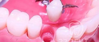
The lower jaw is characterized by the transplantation of a donor composition, which is taken from the area of the wisdom teeth or the chin. The donor block is fixed with metal screws, and the gaps are filled with bone chips. After a few months, once the block has taken root, the screws are removed and you can proceed to the implantation procedure.
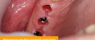
The most common question is: is it possible to do without replenishing bone volume? Yes, in some cases it is possible. For example, with express implantation, when a root-shaped implant is implanted immediately after the extraction of its tooth. In almost all other cases, even if a month has passed since the tooth was removed, it is impossible to do without bone tissue augmentation. But this operation guarantees a stable position of the implant - reliability, durability and an ideal result of implantation and prosthetics.
Examples of work “Before” and “After”
Restoration of chewing teeth of the lower jaw
Case: destruction of the chewing group of teeth of the lower jaw on the right under a bridge.
Basal implantology with instant loading
Case: The patient complained of an uncomfortable removable denture.
Restoration of all teeth on the upper and lower jaw - basal implantation
Case: partial absence of teeth on the upper and lower jaws, complicated by a severe form of periodontitis (tooth mobility).
Simultaneous implantation in the lateral sections of the upper and lower jaw
Case: tooth decay under crowns in the lateral sections of the upper and lower jaws.
Which material is better?
Recently, operations to transplant one's own bone are used less frequently, since taking the material is another operation, a load on the body and general stress. However, dentists do not exclude this method, but are gradually moving to BMR technologies (based on Bone Morphogenetic Protein), which do not require the collection of a donor composition. We are talking about a group of morphogenetic protein tissue growth stimulators that activate processes, and after some time a sufficient volume of bone is formed. The composition is based on molecules that attract natural cells to areas where it is necessary to compensate for bone deficiency. There are no synthetic components or donor bone blocks - exclusively natural mechanisms of tissue regeneration.
Application in dentistry
According to statistics, most patients require bone grafting before restoring their dentition. Bone atrophy is a common pathological process that is both a cause and a consequence of premature tooth loss. The decrease in volume and acquired fragility of bone tissue leads to the fact that it is almost impossible to effectively implant teeth without prior bone augmentation. The consequences of tooth loss and thinning of the jaw bone tissue are:
- violation of chewing function and, as a result, problems with the digestive tract;
- violation of the location of other teeth;
- changes in the configuration of the facial part of the skull, premature appearance of wrinkles;
- speech disorder;
- the formation in the oral cavity of conditions conducive to the rapid spread of pathological processes.
To avoid the above-mentioned unpleasant consequences of tooth loss, you should promptly contact your dentist and consult about options for restoring your dentition. Dental implantation is the most reliable way to regain a healthy and radiant smile, however, if there is a deficiency of bone tissue, you must first undergo bone grafting.
There are different types of osteoplasty, and the choice of one of them depends on the clinical picture, the presence of medical indications and contraindications for the patient. Thanks to the development of science and technology, modern bone grafting is minimally invasive and has a low risk of postoperative complications. The main purpose of this intervention is to fill the bone deficiency by installing an autograft or introducing special synthetic bone materials. Bone grafting using synthetic bone materials is a popular procedure and is not inferior in effectiveness to bone grafting using biological materials. There are various synthetic preparations for osteoplasty on the modern market, each of which has its own advantages and indications for use.
Efficiency of the procedure
The average period of engraftment of bone blocks is three to six months, sometimes more. It all depends on the individual characteristics of the patient’s body, as well as on the skill of the doctor. If the doctor did everything correctly, the grafted bone soon becomes one with the patient’s own bone. The implant can be securely fixed in it.
However, if the grafting of the bone block was performed poorly and the blood supply to the structure is not normalized, a serious inflammatory reaction may begin. In this case, the graft will have to be removed and the affected area sanitized. Rejection of bone blocks by the body can also occur in the event of biological incompatibility of the implanted materials.
The safety and effectiveness of the operation also depends on the patient’s compliance with the doctor’s recommendations, otherwise complications may develop. Thus, with careless oral care or the traumatic effects of solid food, the protective membrane can be exposed. This will provoke the occurrence of purulent infectious processes that are difficult to treat.
How is bone grafting surgery performed?
Like any surgical intervention, bone grafting requires accuracy and precision from the surgeon, and compliance from the patient with all the doctor’s recommendations. Preparation for jaw bone augmentation surgery consists of the following steps:
- 1. Sanitation of the oral cavity and removal of all foci of inflammation to eliminate complications after surgery.
- 2. Preliminary intake of antibiotics and anti-inflammatory drugs, designed to further facilitate the rehabilitation period.
- 3. If necessary, selection of nicotine replacement therapy for smokers, since smoking is prohibited for several days after the operation.
When all preparatory procedures have been completed and the patient is ready, the operation can begin. Let's take a closer look at how bone grafting is done:
- 1. The area of the jaw where the intervention will be performed is anesthetized. As a rule, anesthesia is not performed during bone grafting surgery - local anesthesia is quite enough to insure the patient from unpleasant sensations.
- 2. The surgeon makes an incision in the gum and periosteum, exposing the area of bone that requires augmentation.
- 3. Using a special instrument, the doctor splits the bone, places bone material into the resulting cavity and secures it with miniature screws.
- 4. The operation area is covered with a barrier membrane, which protects the bone material from displacement and protects the operation area from the penetration of bacteria.
- 5. The gum is sutured. Typically, surgeons use self-absorbing materials, so further suture removal is not required.
After the operation, the doctor gives the patient recommendations for recovery, prescribes antibiotics and painkillers.
Stages of bone grafting on the lower jaw
- Gum incision and flap removal
- Bone grafting
- Stitching
- Subsequent implant placement
- Features of bone grafting of the upper jaw
Sinus lifting is a common method of bone grafting of the upper jaw, which involves placing bone material in the patient’s maxillary sinus. During the operation, the doctor carefully moves the bottom of the sinus and inserts a graft into the resulting cavity, which, after engraftment, creates a reliable “foundation” for the implant.
3 techniques for increasing the bone of the upper jaw:
The open technique is used when it is necessary to build up a large volume of bone, and involves a rather serious surgical intervention: the surgeon dissects the gum and cuts a large hole in the bone for further manipulation. This operation requires a long healing period, and the installation of a dental implant is delayed for several months.
If the bone thickness is sufficient, then a closed sinus lift can be done. This is a much less traumatic procedure that is performed through a small hole designed to accommodate a dental implant. The implant itself is implanted immediately after bone grafting to the upper jaw, which, of course, is very convenient for the patient.
Balloon sinus lift g is the most advanced technology for bone grafting of the alveolar process of the upper jaw, which allows you to instantly create a “foundation” and install an implant even on the thinnest bone. As with a closed sinus lift, all manipulations are carried out through an opening for installing an artificial root: a special balloon is inserted into the maxillary sinus, which is filled with liquid and, expanding, carefully separates the soft tissue from the bone. Bone is then placed into the cavity and an implant is installed.
When is the transplant performed?
The collection and replanting of bone blocks is a rather traumatic intervention. Therefore, experienced implantologists prefer to restore teeth using implants of a special design, without resorting to bone grafting. But in certain clinical situations, bone block transplantation is an inevitable procedure.
For example, it is performed when it is impossible to split the bone ridge - due to the fact that the ridge is uniformly narrow throughout its entire height or has a curved shape, for example, in the form of a moon or an hourglass. The extremely low height of the gingival bone is also an indication for its augmentation using bone blocks.
Extranasal operations play a significant role in surgery of the paranasal sinuses [1, 2]. However, after extranasal operations on the paranasal sinuses, significant bone defects remain into which soft tissue grows. The result is stagnation of secretions in the sinus, proliferation of fibrous tissue and the development of scarring, which ultimately leads to a decrease in sinus volume and thickening of the bony walls of the sinus [3, 4]. The tendency to obliteration is most clearly manifested after Caldwell-Luc surgery in childhood [5]. In addition, retraction of the soft tissues of the face appears in the area of the defect [6], and neuralgic pain occurs due to compression of the branches of the trigeminal nerve by scar tissue [7].
Currently, most clinicians are of the opinion that it is necessary to close postoperative defects in the walls of the paranasal sinuses. However, despite the successes achieved in the use of drainage operations, in the practice of an otorhinolaryngologist such pathological conditions of the frontal-ethmoidal zone are observed when the preservation of the frontal sinus as an air cavity is not possible, and there is a need to obliterate the affected frontal sinuses [8-13].
For the purpose of obliteration, both auto- and allografts and adhesive osteoplastic compositions are used [14–19]. During surgery on the frontal sinus to obliterate it, all contents and all the mucous membrane are carefully removed from the sinus. Complete isolation of the sinus and graft from the external environment is achieved, and the contours of the frontal region are restored [20]. This technique has its drawbacks. The literature [19] describes cases of melting of the allograft with the formation of a cavity and purulent fistula of the frontal sinus, cases of the formation of a secondary pyomucocele and limited osteomyelitis of the anterior wall of the frontal sinus [19].
In search of another way to close defects in the walls of the sinuses, the most rational solution to the problem is plastic restoration of the walls of the paranasal sinuses using various materials. Today there is a huge amount of frame materials on the world market. Bone grafts are any implanted materials that, alone or in combination with other materials, promote bone formation by providing local osteoconductive, osteoinductive, or osteogenic activity.
Osteoconductive materials promote the attachment, proliferation and differentiation of poorly differentiated cells into osteoblasts with subsequent bone formation on their surface. Osteoconductive osteogenesis is considered to be “creeping replacement,” i.e., primary resorption of the implant with secondary subsequent ingrowth of supporting tissues from the maternal bed. In these cases, the implant plays the role of a scaffold for the germination of blood vessels. Then the ingrowth of cells from the maternal bed occurs. This mechanism combines the processes of resorption and the formation of new supporting tissue, starting from the borders of the defect. Osteoinductive materials contain biologically active substances that induce recipient bed cells to differentiate into osteoblasts [21]. Osteogenic materials (autografts and materials enriched with cultured autogenous bone cells) contain living host cells capable of differentiating into osteoblasts.
Any bone grafts must have the following properties: a) be completely biocompatible and bioinert; b) be porous; c) serve as a matrix on the surface of which recipient cells are fixed (osteoconductivity); d) gradually be resorbed and replaced by newly formed bone (creeping replacement); e) have a density and physical properties similar to the fabric where it is placed, as well as the ability to resist mechanical stress; f) be easily accessible, well preserved and sterilized; g) in preparation for transplantation, it is easy to process to obtain the desired shape; h) not to be rejected [22].
Currently, both synthetic and natural materials are used for plastics. Main types of bone implants:
1. Autograft - the donor is the patient himself.
These can be bone flaps, crushed or preserved autologous bone or cartilage. Since one's own bone or cartilage tissue is implanted, very good compatibility is ensured with the area in which the tissue is implanted, as well as with all other adjacent tissues. The essence of osteoplastic methods used in rhinology is to create various modifications of the osteoperiosteal plastic flap, which is “broken out” from the wall of the frontal or maxillary sinus before surgery and placed in its original place at the end of the operation [23-25]. Autocartilage of the nasal septum is also used to close defects in the anterior wall of the frontal sinus [26] and the upper wall of the maxillary sinus [27]. This method also has disadvantages. The collection of autologous bone or cartilage may be accompanied by complications: damage to blood vessels and nerves, the formation of hematomas, and the development of an infectious and inflammatory process. In addition, autografts are often resorbed faster than their integration and restoration of the bone defect occurs [28].
2. Allograft - the donor is another person.
We are talking about a bone graft taken from a donor. Allografts include fetal anlage or bone, fresh allogeneic bone, ground allogeneic bone, preserved bone, and demineralized bone matrix. The recipient's body is able to transform this type of bone tissue into its own tissue, and therefore, after implantation, the foreign bone tissue takes root in the body, turning into its own. Bone allografts are characterized by slow osseointegration; their use carries the risk of developing histoincompatibility reactions and chronic granulomatous inflammation [29].
Among biological materials, in recent decades, demineralized bone grafts, called “bone matrix” by some authors, have become widespread in otorhinolaryngology to cover various defects of bone structures. Prof. A.G. Volkov was one of the first in his specialty to use demineralized bone graft - DBT, which is a specially processed cadaveric bone, to restore extensive bone defects that arise after radical surgical treatment for tumors of the frontal sinuses. Demineralized bone matrix was used in plastic surgery of the external nose and nasal septum [22, 30], to restore the lower wall of the orbit destroyed during a fracture [31], and fractures of the walls of the frontal sinuses [32]. A similar bone plastic material with the commercial name “Perfoost” is a demineralized lyophilized perforated plate prepared from long and flat bones of allogeneic origin. The Perfoost allo-implant is successfully used in reconstructive plastic surgeries on the nasal septum that require compensation for defects in both the cartilaginous and bony parts of the nasal septum [21].
A number of authors [22] drew attention to the possibility of using fetal bone (brefomatrix). Due to its pronounced osteoinductive properties, embryonic matrix is successfully used in patients with various defects of the external nose and nasal septum.
3. Xenograft is an animal source. This is a bone or cartilage graft taken from the animal's body. When the graft is in the body, the process of its replacement with human tissue occurs, and it serves as a “framework” for the formation of a person’s own bone tissue. The disadvantage of xenografts is that they are resorbed and replaced by new bone tissue extremely slowly. In rhinology, preserved porcine costal cartilages were used for rhinoplasty, for obliteration of the frontal sinuses and plastic surgery of the fronto-orbital region after extranasal removal of osteoma of the ethmoidal labyrinth [33].
4. Synthetic source.
This implant is an inert, synthetic, artificial substance that is manufactured in laboratories. The use of artificial materials avoids preliminary implantation near the defect and does not limit the surgeon in the amount of material.
According to the literature, synthetic implantable materials include explants (metals, polymers, porous carbon compounds, ceramics). Depending on the manufacturing method and the type of material, bone substitutes are classified as “absorbable” or biodegradable and “non-absorbable”. In other words, our body may or may not replace synthetic material with its own bone tissue, depending on the type of material. If these implants have pores with a diameter greater than 100 µm, bone ingrowth may occur, which ensures its attachment to the material (biological anchorage).
The main disadvantage of synthetic materials, in contrast to auto-, allo- and some xenomaterials, is their lack of osteoinductive properties. The osteoinductive properties of bone replacement materials include their ability to stimulate bone tissue regeneration. For this purpose, composite materials were created. Composite materials are a combination of an osteoconductive matrix with bioactive agents that provide osteoinductive and/or osteogenic properties, which makes these materials equal in bioactive properties to auto-, allo-, and xenografts. This biological activity may be due to the inclusion of sulfated glycosaminoglycans, amino acids, growth factors and morphogens in the bone-replacing material [34].
A number of calcium phosphate materials, such as hydroxyapatite, tricalcium phosphate, some compositions of silicate glass and glass ceramics, are bioactive materials similar in composition to human bone tissue. The latter contribute to the formation of bone on their surface and the formation of strong chemical bonds with the latter (bioactive fixation). These bioactive materials are an osteoconductive matrix that causes adhesion of morphogenetic proteins, osteoblast precursor cells, their proliferation and differentiation into osteoblasts. Synthetic materials based on artificial hydroxyapatite are superior to hydroxyapatite of animal origin in a number of characteristics. They eliminate the possibility of transmitting infectious diseases and allow you to regulate the rate of resorption due to the peculiarities of synthesis and various substitutions of phosphate and hydroxyl groups in the structure of apatite. This characterizes synthetic hydroxyapatite as a promising osteoplastic material for use in all areas of osteoplastic surgery.
In the literature under study [25], there are works on the use of hydroxyapatite to close a defect in the upper wall of the maxillary sinus [35], postoperative defects in the walls of the paranasal sinuses after removal of osteomas. One of the disadvantages of calcium phosphate ceramics is the low mechanical strength of this type of implant.
Corundum ceramics, which is bioinert and biocompatible, and has a high degree of strength, turned out to be suitable for plastic surgery of the walls of the paranasal sinuses in terms of its physicochemical properties [36]. But, unfortunately, full modeling of corundum ceramic plates based on the shape of a complex defect in the walls of the frontal sinuses is impossible [37].
Titanium mesh implants are very widely used for plastic surgery of postoperative and post-traumatic bone defects of the paranasal sinuses and skull [38–41]. In the literature studied, there was work where porous titanium nickelide was used to close perforations of the nasal septum and postextraction fistulas [42]. Structures made of these alloys implanted into the body are deformed in accordance with the laws of elastic behavior of body tissues and ensure long-term harmonious functioning. In addition, porous titanium nickelide, which has physical and mechanical properties close to the parameters of bone tissue (pore sizes, permeability, general patterns of elastic behavior), provides favorable conditions for the ingrowth of tissue structures into the pores and its fusion with the recipient bone [43, 44] . However, even titanium implants can provoke rejection reactions. Therefore, in order to isolate titanium from adjacent living tissues of the body, a coating of biositall, a bioactive material, is applied to the titanium mesh to improve osseointegration and biocompatibility [16, 45, 46].
Of great clinical interest are data on tissue reaction and the course of the wound process during implantation of polymer materials [47, 48]. Thus, for the purpose of plasticizing a bone defect in osteomas of the paranasal sinuses, polyphosphazene endoprostheses [49] and protacryl [26] were successfully used; to close a post-traumatic defect in the upper wall of the maxillary sinus, silicone [50] or Teflon [51] were used. Plastic material made of polyester - “Knitted material for the restoration of bone and cartilage structures” was used to correct the contours of the nasal septum and the walls of the paranasal sinuses [52].
Currently, combined synthetic materials are of greatest clinical interest. Combined forms consist of a polymer matrix and nano-hydroxyapatite as a filler. The emergence of composites of synthetic hydroxyapatite in the form of powders, granules and gels in combination with polysaccharides, chitosan, alginate, hyaluronic acid, collagen protein, peptides, drugs and other drugs has expanded the possibilities of bone tissue restoration [53-55].
In this regard, the development and introduction into the clinic of new materials for plastic surgery of bone walls is relevant.
Benefits of demineralized lyophilized bone with proven osteoinductive activity
Among all the materials on the market, one can highlight a product called AlloGro from the largest tissue bank in the United States. This preparation is a demineralized lyophilized bone implant whose osteoinductive activity is determined by testing. The need for this test lies in the fact that the inductive properties of donor tissue are highly pronounced, and conducting a biotest eliminates about 10% of donors for the production of material for implantation. Today, in order to save money, to test for osteoinductive activity, laboratories use cell cultures of the SAOS-2 (osteogenic human sarcoma) type, into which thymidine and donor CMP are placed. As a result, the activity of cell division and the rate of thymidine excretion are determined. These data allow the osteoinductive efficiency to be calculated.
As a result of these clinical studies, the company was able to label the product as “Proven Osteogenic Activity.” One of the risks with allogeneic transplants is transmission of a viral infection such as hepatitis or AIDS. But to date, there have been no recorded cases of infection with any virus, even those infections whose development zone is the dura mater. Today, DLK is the most economical and effective osteogenic material.
Oral care after surgery
After surgery to graft bone blocks, the patient is prescribed a course of antibiotics to prevent bacterial complications. During this period, it is extremely important for the patient to maintain oral hygiene: not only brush your teeth with a soft brush twice a day, but also rinse your mouth with an antiseptic after each meal.
It is also necessary to strictly follow the diet: do not eat hard, tough foods that can damage soft tissues and transplanted bone blocks. During the rehabilitation period, you should chew food on the side opposite to the operation site. And if discomfort in the mouth increases, bleeding and wounds appear on the gums, you should consult your doctor as soon as possible.