Russian – Latin dictionary
| A | |
| acromion, humeral process | acromion, in |
| alveolus | alveŏlus, im |
| alveolar | alveolaris, e |
| anatomical | anatomycus, a, um |
| aorta | aorta, ae f |
| aortic | aorticus, a, um |
| aperture, hole | aperture, ae f |
| aponeurosis | aponeurosis, is f |
| appendix, appendage, appendage | appendix, ĭcis f |
| arterial | arteriōsus, a, um |
| artery | arteria, ae f |
| atlas | atlas, antis m |
| B | |
| drum | tympănum, in |
| drum | tympanĭcus, a, um |
| hip | femur, ŏris n |
| bifurcation | bifurcatio, onis f |
| block | trochleāris, e |
| side | lateralis, e |
| tibia | Tibia, ae f |
| tibial | tibiālis, e |
| large (half st.) | magnus, a, um |
| big (comparative) | major, jus |
| big brain | cerebrum, in |
| large molar (tooth) | molaris, e |
| furrow | sulcus, im |
| eyebrow | supercilium, in |
| peritoneum | peritonaeum, in |
| tubercle | tuber, ěris n |
| tubercle | tuberclum, in |
| tuberosity | tuberosĭtas, ātis f |
| IN | |
| impression | impressio, ōnis f |
| eyelid | palpĕbra, ae f |
| vein | vena, ae f |
| coronary | coronarius, a, um |
| venous, venous | venōsus, a, um |
| spit | trochanter, ēris m |
| maxillary | maxillaris, e |
| upper | superior, ius |
| upper jaw | maxilla, ae f |
| apex; tip (of nose, tongue) | apex, ĭcis m |
| branch | ramus, im |
| temporal | temporalis, e |
| vagina | vagīna, ae f |
| interior | internus, a, um |
| water pipes | aquaeductus, us m |
| elevation | jugum, in |
| gates | hilus, im |
| gates | porta, ae f |
| tenderloin | incisūra, ae f |
| projection | protuberantia, ae f |
| neck | nucha, ae f |
| G | |
| ganglion, ganglion | ganglion, in |
| pituitary | hypophysis, is f |
| eye | ocŭlus, im |
| eye socket | orbita, ae f |
| orbital | orbitalis, e |
| pharynx | pharynx, yngis m |
| pharyngeal | pharyngēus, a, um |
| deep | profundus, a, um |
| shin, leg | crus, cruris n |
| head, head | caput, itis n |
| brain | encephalon, in; cerĕbrum, in |
| horizontal | horizontalis, e |
| larynx | larynx, yngis m |
| crest | crista, ae f |
| sternal | sternalis, e |
| chest, chest | thorax, ācis m |
| chest | pectoralis, e |
| chest | thoracĭcus, a, um |
| lip | labium, in |
| spongy | spongiōsus, a, um |
| D | |
| deltoid | deltoideus, a, um |
| gum | gingiva, ae f |
| longest | longissimus, a, um |
| long | longus, a, um |
| bottom | fundus, im |
| additional | accessorius, a, um |
| lobular | lobātus, a, um |
| share | lobus, im |
| slice | lobulus, im |
| dorsal, dorsal, dorsal | dorsālis, e |
| tree, wood | arbor, ōris f |
| arc | arcus, us m |
| AND | |
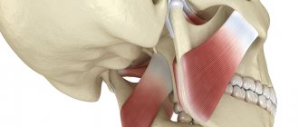
| chewing | masseterĭcus, a, um |
| gland | glandŭla, ae f |
| yellow | flavus, a, um |
| stomach | gaster, tris f |
| stomach, ventricle (heart) | ventriclus, im |
| gastric | gastrĭcus, a, um |
| belly, belly | abdōmen, ĭnis n |
| life | vita, ae f |
| fatty, fatty | adipōsus, a, um |
| Z | |
| rear | posterior, ius |
| anus | anus, im |
| damper | valvŭla, ae f |
| occipital | occipitalis, e |
| pharynx | fauces, ium f (pl) |
| visual | optĭcus, a, um |
| tooth | dens, dentis m |
| wisdom tooth | dens sapientiae (dens serotĭnus) |
| dental | dentalis, e |
| serrated | serrātus, a, um |
| AND | |
| And | et |
| bend | flexūra, ae f |
| or | seu (s.) |
| TO | |
| rocky | petrōsus, a, um |
| channel | canālis, is m |
| tubule | canaliclus, im |
| capillary | capillaris, e |
| capsule | capsŭla, ae f |
| cystic | cystōsus, a, um |
| chest cavity | thorax, ācis m |
| sphenooccipital | sphenooccipitalis, e |
| wedge-shaped | sphenoidalis, e |
| clew | glomus, ěris n |
| fang | dens canīnus |
| leather | cutis, is f |
| cutaneous | cutaneus, a, um |
| collateral, lateral | collateralis, e |
| annular | anularis, e |
| end, end | terminatio, ōnis f |
| limb, body member | membrane, in |
| constrictor (squeezing muscle) | musculus constrictor (ōris m) |
| coccyx | coccyx, ýgis m |
| coccygeal | coccygēus, a, um |
| bark, cortex | cortex, ĭcis m |
| root | radix, īcis f |
| short | brevis, e |
| oblique | obliquus, a, um |
| bone | osseus, a, um |
| bone marrow (ossium – bones, r.p. plural) | medulla ossium |
| bone | os, ossis n |
| edge | margo, ĭnis m |
| red | ruber, bra, brum |
| sacral | sacrālis, e |
| circulatory | sanguineus, a, um |
| blood | sanguis, ĭnis m |
| round | rotundus, a, um |
| round | teres, ětis |
| circular | orbicularis, e |
| wing | ala, ae f |
| pterygoid | pterygoideus, a, um |
| wing | alāris, e |
| roof, cover, tire | tegmen, ĭnis n |
| L | |
| labyrinth | labyrinthus, im |
| palmar | palmaris, e |
| lateral, lateral | laterālis, e |
| left | sinister, tra, trum |
| lung | pulmo, ōnis m |
| pulmonary | pulmonalis, e |
| staircase | scalēnus, a, um |
| lymphatic | lymphatĭcus, a, um |
| line | linea, ae f |
| facial | faciālis, e |
| face | facies, ēi f |
| frontal | frontalis, e |
| spatula | scapŭla, ae f |
| bulb | bulbus, im |
| radius | radius, im |
| M | |
| fibula | fibŭla, ae f |
| peroneal | fibularis, e |
| peroneal | peronaeus, a, um |
| small | minor, us |
| small | parvus, a, um |
| small molar (tooth), premolar | praemolāris, e |
| weight | Massa, ae f |
| uterus | utěrus, im |
| medial | medialis, e |
| interalveolar | interalveolaris, e |
| interlobar | interlobularis, e |
| interroot | interradicularis, e |
| interosseous | interosseus, a, um |
| interspinous | interspinōsus, a, um |
| intercostal | intercostalis, e |
| interball | interglobularis, e |
| little finger | digitus minimus |
| meninges | mater, tris f |
| medulla | medulla, ae f |
| cerebral | cerebrālis, e |
| cerebellum | cerebellum, in |
| horny | callōsus, a, um |
| breast | mamma, ae f |
| baby (falling off) tooth | dens deciduus |
| molar, molar | dens molaris |
| wisdom | sapientia, ae f |
| muscular | muscularis, e |
| muscle | musclus, im |
| rotator cuff | muscle rotator |
| tensor muscle | muscle tensor |
| depressor muscle | muscle depressor |
| abductor muscle | muscle abductor |
| levator muscle | musclus levātor |
| adductor muscle | muscle adductor |
| extensor muscle | muscle extensor |
| dilator muscle | muscŭlus dilatātor |
| corrugator muscle | muscŭlus corrugātor |
| soft | pius, a, um |
| N | |
| adrenal | glandŭla suprarenālis |
| adrenal | suprarenalis, e |
| Name | nomen, ĭnis n |
| greatest | maxĭmus, a, um |
| highest | suprēmus, a, um |
| least | minimus, a, um |
| outer | externus, a, um |
| palatovaginal | palatovaginalis, e |
| palatine | palatinus, a, um |
| sky | palātum, in |
| nerve | nervus, im |
| nervous | nervōsus, a, um |
| mandibular | mandibularis, e |
| lower | inferior, ius |
| lower jaw | mandibŭla, ae f |
| leg, foot | pes, pedis m |
| nail | unguis, is m |
| leg | pediŭlus, im |
| nose | nasus, im |
| nasal | nasālis, e |
| ABOUT | |
| region | regio, ōnis f |
| colon | colĭcus, a, um |
| shell | tunĭca, ae f |
| general | communis, e |
| oval | ovalis, e |
| ending | terminatio, onis f |
| base | basis, is f |
| spinous | spinōsus, a, um |
| axis, 2nd cervical vertebra | axis, is f |
| awn | spina, ae f |
| hole | forāmen, ĭnis n |
| falling off, milk (tooth) | deciduus, a, um |
| shoot | processus, us m |
| P | |
| sinus, sinus | sinus, us m |
| finger | digitus, im |
| partition | septum, in |
| front | anterior, ius |
| cross | chiasma, ătis n |
| membrane | membrane, ae f |
| membranous | membranaceus, a, um |
| rooster | gallus, im |
| liver | hepar, ătis n |
| cavernous | cavernōsus, a, um |
| pyramid | pyrămis, ĭdis f |
| nutritious | nutricius, a, um |
| esophagus | oesophagus, im |
| plate | lamĭna, ae f |
| pleural | pleuralis, e |
| brachial | humerālis, e |
| shoulder | Brachĭum, in |
| flat | planus, a, um |
| surface | superficialis, e |
| surface, face | facies, ēi f |
| chin | mentalis, e |
| iliac | iliăcus, a, um |
| mobile | mobilis, e |
| pancreas | pancreas, ătis n |
| sublingual | sublingualis, e |
| sublingual (bone) | hyoideus, a, um |
| vertebra | vertebra, ae f |
| vertebrate | vertebrālis, e |
| late (tooth), wisdom tooth | serotĭnus, a, um |
| cavity | cavĭtas, ātis f |
| semilunar | semilunaris, e |
| hollow | cavus, a, um |
| transverse | transversus, a, um |
| renal | renālis, e |
| bud | ren, renis m |
| belt | cingŭlum, in |
| lumbar | lumbalis, e |
| right | dexter, tra, trum |
| vestibule | vestibŭlum, in |
| atrium | atrium, in (cordis) |
| prostate | prostata, ae f |
| premolar, small molar | dens praemolāris |
| pyloric, pyloric | pyloricus, a, um |
| longitudinal | longitudinalis, e |
| pronator | musculus pronator |
| simple | simplex, icis |
| space | spatium, in |
| duct | ductus, us m |
| rectum | rectum, in |
| straight | rectus, a, um |
| R | |
| extensor (muscle) | extensor, ōris m |
| bifurcation, bifurcation | bifurcatio, ōnis f |
| sink | concha, ae f |
| costal | costālis, e |
| edge | costa, ae f |
| cutter | dens incisīvus |
| incisive | incisīvus, a, um |
| ciliary | ciliāris, e |
| lattice | ethmoidālis, e |
| horn, horn | cornu, us n |
| mouth | os, oris n |
| hand (hand) | manus,us f |
| WITH | |
| sagittal, sagittal | sagittalis, e |
| free | liber, ěra, ěrum |
| vault, arch | fornix, ĭcis m |
| bunch | ligamentum, in |
| flexor (muscle) | flexor, ōris m |
| saddle | sella, ae f |
| spleen | lien, ēnis m |
| cordial | cardiăcus, a, um |
| heart | cor, cordis n |
| retina | retĭna, ae f |
| net | rete, is n |
| sympathetic | sympathicus, a, um |
| synovial | synovialis, e |
| sinus, sinus | sinus, us m |
| sinusoid | sinusoideus, a, um |
| synchondrosis | synchondrōsis, is f |
| system | systēma, ătis n |
| skeleton | skelěton, in (scelěton, in) |
| fold | plica, ae f |
| zygomatic | zygomatĭcus, a, um |
| tearful | lacrimalis, e |
| mucous membrane | mucōsa, ae f |
| slimy | mucōsus, a, um |
| difficult | composĭtus, a, um |
| layer | stratum, in |
| canine, canine (tooth) | canīnus, a, um |
| own | proprius, a, um |
| carotid artery | carōtis, idis f |
| sleepy | carotĭcus, a um |
| nipple, papilla | papilla, ae f |
| vessel | vas, vasis n |
| mastoid | mastoideus, a, um |
| opener | vomer, ěris m |
| back | dorsum, in |
| spinal | cerebrospinalis, e (spinalis, e) |
| plexus | plexus, us m |
| average | medium, a, um |
| trunk, torso | truncus, im |
| glassy | vitreus, a, um |
| wall | paries, ĕtis m |
| pillar | columna, ae f |
| foot, leg | pes, pedis m |
| sagittal, sagittal | sagittalis, e |
| stroma, skeleton | stroma, ătis n |
| string | chоrda, ae f |
| bag | bursa, ae f |
| arch support | musculus supinātor (ōris m) |
| joint | articulatio, ōnis f |
| articular | articularis, e |
| tendon | tendo, inis m |
| sphincter | musculus sphincter (ēris m) |
| T | |
| pelvis | pelvis, is f |
| talus | talus, im |
| solid | durus, a, um |
| body | corpus, ŏris n |
| parietal | parietalis, e |
| thymus, thymus gland | thymus, im |
| trapezoidal | trapezoideus, a, um |
| trachea | trachēa, ae f |
| trigeminal | trigeminalis, e |
| dorsal, dorsal, dorsal | dorsālis,e |
| U | |
| corner | angŭlus, im |
| holder | retinaclum, in |
| node | nodus, im |
| nodule | nodŭlus, im |
| snail | cochlea, ae f |
| snail | cochlearis, e |
| compacted | indurātus, a, um |
| mouth, hole | ostium, in |
| ear | auris, is f |
| ear | auricularis, e |
| F | |
| fascia | fascia, ae f |
| fibrous, fibrous | fibrōsus, a, um |
| X | |
| move, passage | meātus, us m |
| cartilage | cartilāgo, ĭnis f |
| C | |
| central | centralis, e |
| H | |
| part, side | pars, partis f |
| scull | cranium, in |
| cranial | craniālis, e |
| scaly | squamōsus, a, um |
| wonderful | mirabĭlis, e |
| Sh | |
| cervical | cervicalis, e |
| neck | collum, in |
| neck, cervix | cervix, īcis f |
| wide | latus, a, um |
| widest | latissimus, a, um |
| suture (bone suture) | sutūra, ae f |
| seam (on soft tissues) | raphe, es f |
| SCH | |
| gap | fissūra, ae f |
| crevice, crevice | hiātus, us m |
| thyroid | thyreoideus, a, um |
| I | |
| gluteal | glutaeus, a, um |
| core | nucleus, im |
| language | lingua, ae f |
| lingual | lingualis, e |
| pit (roundish in outline) | fovea, ae f |
| fossa (elongated) | fossa, ae f |
| dimple | foveŏla, ae f |
| jugular | jugularis, e |
Latin pharmaceutical terminology and formulation
| Introduction to Latin pharmaceutical terminology. Rules for the design of pharmaceutical terms in Latin | Class |
1
Digestive system. Names in Latin
Chapter 5. Splanchnology (internal organs)
| SYSTEMA DIGESTORIUM | DIGESTIVE SYSTEM |
| Os | Mouth |
| Cavitas oris | Oral cavity |
| Tunica mucosa oris | Oral mucosa |
| Vestibulumoris | Vestibule of the mouth |
| Rima oris | Mouth gap |
| Labia oris | Lips of the mouth |
| Labium superius | Upper lip |
| Philtrum | philtrum |
| Tuberculum | Tubercle |
| Labium inferius | Underlip |
| Frenulum labii suorioris | Upper lip frenulum |
| Frenulum labii inferioris | Frenum of the lower lip |
| Commissura labiorum | Lip commissure |
| Anqulusoris | Angle of mouth |
| 8ucca | Cheek |
| Corpus adiposum buccae | Cheek fat pad |
| Orqanum iuxtaorale | Perioral organ |
| Papilla ductus parotidei | Parotid duct papilla |
| Cavitas oris propria | The oral cavity itself |
| Palatum | Sky |
| Palatum durum | Solid sky |
| Palatum molle; Velum palatinum | Soft sky; velum |
| Raphe palati | Palate seam |
| Plicae palatinae transversae; Rugae palatinae | Transverse palatal folds |
| Papilla incisiva | Incisive papilla |
| Ginqiva | Gum |
| Marqoqinqivalis | Gingival edge |
| Papilla quinqivalis; Papilla interdentalis | Gingival papilla; interdental papilla |
| Sulcus qinqivalis | Gingival sulcus |
| Caruncula sublinqualis | Sublingual papilla |
| Plica sublinqualis | Sublingual fold |
| Glandulaeoris | Glands of the mouth |
| Glandulae salivariae majores | Major salivary glands |
| Glandula parotidea | Parotid gland |
| Pars superficialis | Surface part |
| Pars profunda | Deep part |
| Glandula parotidea accessoria | Accessory parotid gland |
| Ductus parotideus | Parotid duct |
| Glandula sublinqualis | Sublingual gland |
| Ductus sublinqualis maior | Greater hypoglossal duct |
| Ductus sublinquales minores | Small sublingual ducts |
| Glandula submandibularis | Submandibular gland |
| Ductus submandibularis | Submandibular duct |
| Glandulae salivariae minores | Minor salivary glands |
| Glandulae labiales | Labial glands |
| Glandulae buccales | Buccal glands |
| Glandulae molares | Molar glands |
| Glandulae palatinae | Palatine glands |
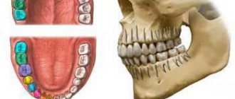
| Dentes | Teeth |
| Arcus dentalis maxillaris; Arcus dentalis superior | Maxillary dental arch; upper dental arch |
| Arcus dentalis mandibularis; Arcus dentalis | Mandibular dental arch; lower |
| inferior | dental arch |
| Dens mcisivus | Cutter |
| Dens caninus | Fang |
| Dens premolaris | Premolar; premolar |
| Dens molaris | large molar; molar |
| Dens molaris tertius; Dens serotinus | Third major molar; third molar; wisdom tooth |
| Corona dentis | Crown of the tooth |
| Cusoisdentis | Tooth tip |
| Apex cuspidis | Tip tip |
| Cuspis accessoria | Additional point |
| Tuberculum dentis | Tubercle of tooth |
| Christa transversalis | Transverse scallop |
| Crista triancularis | Triangular comb |
| Christa obliqua | Oblique comb |
| Fissura occlusalis | Occlusal gap |
| Fossa occlusalis | Occlusal fossa |
| Cuspis buccalis | Buccal cusp |
| Cuspis palatinalis | Palatal tubercle |
| Cuspis linqualis | Lingual tubercle |
| Cuspis mesiobuccalis | Buccal mesial tubercle |
| Cuspis mesiopalatalis | Palatomesial tubercle |
| Cuspis mesiolinqualis | Lingual-mesial tubercle |
| Cuspis distobuccalis | Buccal distal tubercle |
| Cuspis distopalatinalis | Palatodistal tubercle |
| Cuspis distolinqualis | Lingual-distal tubercle |
| Cuspis distalis | Distal tubercle |
| Corona clinic | Clinical crown |
| Cervix dentis | Tooth neck |
| Radix dentis | Tooth root |
| Apex radicis dentis | Apex of tooth root |
| Radix clinic | Clinical root |
| Facies occlusalis | Occlusal surface |
| Facies vestibularis | Vestibular surface |
| Facies buccalis | Buccal surface |
| Facies labialis | Labial surface |
| Facies linqualis | Lingual surface |
| Facies palatinalis | Palatal surface |
| Facies mesialis | Mesial surface |
| Facies distalis | Distal surface |
| Facies approximalis | Approximal surface |
| Area continences | Contact zone |
| Cinqulum | Belt |
| Christa marqinalis | Marginal scallop |
| Maroo incisalis | Cutting edge |
| Cavitas dentis; Cavitas pulparis | Dental cavity, pulp cavity |
| Cavitas coronae | Crown cavity |
| Canalis radicis dentis | Tooth root canal |
| Foramen apicis dentis | Tooth apex hole |
| Pulpa dentis | Dental pulp |
| Pulpa coronalis | Crown pulp |
| Pulpa radicularis | Root pulp |
| Papilla dentis | Dental papilla; gingival papilla |
| Dentinum | Dentine |
| Enamelum | Enamel |
| Cementum | Cement |
| Periodontium | Periodontium |
| Mammillae | Papillae |
| Stria canina; Sulcus caninus | Fang strip; canine groove |
| Fossa canina | Canine fossa |
| Fovea mesialis | Mesial fossa |
| Fovea distalis | Distal fossa |
| Radix buccalis | Buccal root |
| Radix palatinalis | Palatine root |
| Radix mesialis | Mesial root |
| Radix distalis | Distal root |
| Radix mesiobuccalis | Buccal mesial root |
| Radix mesiolinqualis | Glossomesial root |
| Radix accessoria | Accessory root |
| (Tuberculum anomale) | (Abnormal lump) |
| Cuspis paramolaris; Tuberculum paramolare | Perimolar tubercle |
| Tuberculum molare | Molar cusp |
| Alveolus dentalis | Dental alveolus |
| Curvea occlusalis | Occlusal curvature |
| Dentesdecidui | Baby teeth |
| Dentes permanentes | Permanent teeth |
| (Diastema) | (Diastema) |
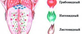
| Lingua | Language |
| Corpus linquae | Body of tongue |
| Radix linquae | Root of tongue |
| Dorsum linquae | Dorsum of tongue |
| Pars anterior; Pars presulcalis | Front end; pre-furrow part |
| Pars posterior; Pars postsulcalis | Rear end; post-furrow part |
| Facies inferior linquae | Underside of the tongue |
| Plica fimbriata | Fringed fold |
| Marqo linquae | Edge of tongue |
| Apex linquae | tip of tongue |
| Tunica mucosa linquae | Mucous membrane of the tongue |
| Frenulum linguae | Tongue frenulum |
| Papillae Imquales | Tongue papillae |
| Papillae filiformes | Filiform papillae |
| Papillae fungiformes | Fungiform papillae |
| Papillae vallatae | circumvallate papillae |
| Papillae foliatae | Leaf-shaped papillae |
| Sulcus medianus linquae | Median sulcus of tongue |
| Sulcus terminalis linquae | Border groove of tongue |
| Foramen caecum linquae | Blind opening of the tongue |
| (Ductus thyroqlossalis) | (Thyroglossal duct) |
| Tonsilla linqualis | Lingual tonsil |
| Noduli lymphoidei | Lymphoid nodules |
| Septum linquae | Tongue septum |
| Aponeurosis linquae | Aponeurosis of the tongue |
| Musculi linguae | Muscles of the tongue |
| M. qenioqlossus | Genioglossus muscle |
| M.hyoqlossus | Hypoglossus muscle |
| M.chondroqlossus | Cartilaginous muscle |
| M. ceratoqlossus | Cornoglossus muscle |
| M. stvloqlossus | Styloglossus muscle |
| M. lonqitudinalis superior | Superior longitudinal muscle |
| M. lonqitudinalis inferior | Inferior longitudinal muscle |
| M. transversus linguae | Transverse tongue muscle |
| M. verticalislinquae | Vertical tongue muscle |
| M. palatoqlossus | Palatoglossus muscle |
| Fauces | Zev |
| Isthmus faucium | Isthmus of pharynx |
| Palatum molle; Velum palatinum | Soft sky; velum |
| Uvula palatina | Uvula |
| Arcus palatoglossus; Plica anterior faucium | Palatoglossus arch; anterior throat fold |
| (Plica triancularis) | (Triangular fold) |
| Arcus palatopharyngeus; Plica posterior | Palatopharyngeal arch; back fold |
| faucium | pharynx |
| (Plica semilunaris} | (Semilunate fold) |
| Fossa tonsillaris; Sinus tonsillaris | Tonsil fossa; tonsillar sinus |
| Fossa supratonsillaris | Supramygdaloid fossa |
| Tonsilla palatina | Palatine tonsil |
| Capsula tonsillae | Almond capsule |
| (Fissura tonsillaris; Fissura intratonsillaris) | (Amygdaloid fissure; intratonsilal fissure) |
| Fossulae tonsillae | Almond dimples |
| Cryptae tonsillae | Almond crypts |
| Musculi palati mollis et faucium | Muscles of the soft palate and pharynx |
| Aponeurosis palatina | Palatal aponeurosis |
| M. levator veh palatini | Levator palatine muscle |
| M. tensor veli palatini | Muscle that tenses the velum palatine |
| M. uvulae | Uvula muscle |
| M. palatoqlossus | Palatoglossus muscle |
| M. palatopharynqeus | Velopharyngeal muscle |
| Fasciculus anterior | Front bun |
| Fasciculus posterior; M. sphincter palatopha-fyngeus | Posterior bundle; velopharyngeal sphincter |
| Pharynx | Pharynx |
| Cavitas pharynqis | Pharyngeal cavity |
| Pars nasalis pharyngis | Nasal part of the pharynx |
| Fornix pharynqis | Vault of the pharynx |
| Hypophysis pharynqealis | Pharyngeal pituitary gland |
| Tonsilla pharynqealis | Pharyngeal tonsil |
| Fossulae tonsillae | Almond dimples |
| Cryptae tonsillae | Almond crypts |
| Noduli lymphoidei pharynqeales | Pharyngeal lymphoid nodules |
| (Bursa pharynqealis) | (pharyngeal bursa) |
| Ostium pharyngeum tubae auditivae; Ostium pharynqeum tubae auditoriae | Pharyngeal opening of the auditory tube |
| Torus tubarius | Pipe roller |
| Plica salpinqopharynqea | Tubalopharyngeal fold |
| Plica salpinqopalatina | Tupalatal fold |
| Torus leva tone | The levator soft palate muscle |
| Tonsilla tubaria | Tubal tonsil |
| Recessus pharynqeus | Pharyngeal pocket |
| Crista palatopharynqea | Velopharyngeal ridge |
| Pars oralis pharyngis | Oropharynx |
| Vallecula epiqlottica | Epiglottis fossa |
| Plica qlossoepiqlottica mediana | Median lingual epiglottis fold |
| Plica glossoepiglottica lateralis Pars larynqea pharynqis | Lateral lingual epiglottis fold |
| Laryngeal part of the pharynx | |
| Recessus piriformis | Pear-shaped pocket |
| Plica nervi larynqei superioris | Fold of the superior laryngeal nerve |
| Constrictio pharynqooesophaqealis | Pharyngeal-esophageal constriction |
| Fascia pharynqobasilaris | Pharyngobasilar fascia |
| Tela submucosa | Submucosa |
| Tunica mucosa | Mucous membrane |
| Glandulae pharynqeales | Pharyngeal glands |
| Musculi pharyngis; Tunica muscularis pharynqis | Muscles of the pharynx; muscular layer of the pharynx |
| Raphe pharyngis | Suture of the pharynx |
| Raphe ptervqomandibularis | Pterygomandibular suture |
| M. constrictor pharynqis superior | Superior pharyngeal constrictor |
| Pars pteryqopharynqea | Pterygopharyngeal part |
| Pars buccopharynqea | Buccal-pharyngeal part |
| Pars mylopharvnqea | Maxillopharyngeal part |
| Pars qlossopharynqea | Glossopharyngeal part |
| M. constrictor pharynqis medius | Middle pharyngeal constrictor |
| Pars chondropharynqea | Cartilaginous part |
| Pars ceratopharynqea | Cornopharyngeal part |
| M. constrictor pharynqis inferior | Inferior pharyngeal constrictor |
| Pars thyropharynqea; M. thyropharynqeus | Thyropharyngeal part |
| Pars cricopharynqea; M. cricopharvnqeus | Cricopharyngeal part |
| M.stylopharynqeus | Stylopharyngeal muscle |
| M. salpmqopharynqeus | Tubalopharyngeal muscle |
| M. palatopharyngeus | Velopharyngeal muscle |
| Fascia buccopharynqealis | Buccal pharyngeal fascia |
| Spatium penpharynqeum | Periopharyngeal space |
| Spatium retropharynqeum | Retropharyngeal space |
| Spatium lateropharyngeum; Spatium pha-rynqeum laterale; Spatium parapharynqeum | Lateral parapharyngeal space |
| Oesophagus | Esophagus |
| Parscervicalis: Pars colli | Neck part |
| Pars thoracica | Thoracic part |
| Constrictio partis thoracicae; Constrictio bronchoaortica | Narrowing of the chest; bronchoaortic narrowing |
| Constrictio phrenica; Constrictio diaphragmatica | Diaphragmatic constriction |
| Pars abdominalis | Abdominal part |
| Tunica serosa | Serosa |
| Tela subserosa | Subserosal membrane |
| Tunica adventitia | Adventitia |
| Tunica muscularis | Muscularis |
| Tendo cncoesophaqeus | cricoesophageal tendon |
| M. bronchoesophaqeus | Bronchoesophageal muscle |
| M. pleurooesophaqeus | Pleuroesophageal muscle |
| Tela submucosa | Submucosa |
| Tunica mucosa | Mucous membrane |
| Lamina muscularis mucosae | Muscular plate of the mucous membrane |
| GASTER | STOMACH |
| Paries anterior | Front wall |
| Paries posterior | Rear genka |
| Curvatura major | greater curvature |
| Curvatura minor | Small curvature |
| Incisura anqualis | Corner cut |
| Cardia;Parscardiaca | Cardia; cardiac part |
| Ostium cardiacum | Cardiac foramen |
| Fundus quastricus | Fundus of the stomach |
| Fornix qastricus | Gastric vault |
| Incisura cardialis | Cardiac notch |
| Corpus quastricum | Body of stomach |
| Canalis qastricus | Stomach channel |
| Pars pvlorica | Pyloric (pyloric) part |
| Antrum pyloricum | Gatekeeper Cave |
| Canalis pyloricus | Gatekeeper channel |
| Pylorus | Gatekeeper (pylorus) |
| Ostium pyloricum | Pyloric hole |
| Tunica serosa | Serosa |
| Tela subserosa | Subserosal base |
| Tunica muscularis | Muscularis |
| Stratum lonqitudmale | Longitudinal layer |
| Stratum circulare | Circular layer |
| M. sphincter pyloricus | Pyloric sphincter |
| Fibrae obliquae | Oblique fibers |
| Tela submucosa | Submucosa |
| Tunica mucosa | Mucous membrane |
| Plicae qastricae | Folds of the stomach |
| Lamina muscularis mucosae | Muscular plate of the mucous membrane |
| Areae qastricae | Gastric fields |
| Plicae villosae | Villous folds |
| Foveolae qastricae | Gastric dimples |
| Glandulae qastricae | Stomach glands |
| Intestinum tenue | Small intestine |
| Tunica serosa | Serosa |
| Tela subserosa | Subserosal base |
| Tunica muscularis | Muscularis |
| Stratum longitudinale; Stratum helicoidale lonqiqradus | Longitudinal layer; long pitch spiral layer |
| Stratum circulare; Stratum helicoidale brevis qradus | Circular layer; short pitch spiral layer |
| Plicae circulares | Circular folds |
| Tela submucosa | Submucosa |
| Tunica mucosa | Mucous membrane |
| Lamina muscularis mucosae | Muscular plate of the mucous membrane |
| Villi intestinales | Intestinal villi |
| Glandulae intestinales | Intestinal glands |
| Noduli lymphoidei solitarii | Single lymphoid nodules |
| Noduli lymphoidei aqqreqati | Group lymphoid nodules |
| DUODENUM | DUODENUM |
| Pars superior | Top part |
| Ampulla; 8ulbus | Ampupa; button |
| Flexura duodeni superior | Superior flexure of the duodenum |
| Pars descendens | Descending part |
| Flexura duodeni inferior | Lower and lower flexure of the duodenum |
| Pars horizontalis; Pars inferior | Horizontal part; Bottom part |
| Pars ascendens | Rising part |
| Flexura duodenojejunalis | Duodenum-jejunum |
| bend | |
| Pars tecta duodeni | Hidden part of the duodenum |
| M. suspensorius duodeni; Lig. suspensorium duodeni | Duodenal suspensory muscle; duodenal suspensory ligament |
| Pars phrenicocoeliaca | Diaphragmatic-celiac part |
| Pars coeliacoduodenalis | Celiac-duodenal part |
| Plica longitudinalis duodeni | Longitudinal fold of the duodenum |
| Papilla duodeni major | Major duodenal papilla |
| GUTS | |
| Papilla duodeni minor | Lesser duodenal papilla |
| Glandulae duodenales | Duodenal glands |
| Ileum | Ileum |
| Pars terminalis | End part |
| (Oiverticulum ilei) | (Ileal diverticulum) |
| Intestinum crassum | Colon |
| Tunica serosa | Serosa |
| Tela subserosa | Subserosal base |
| Tunica muscularis | Muscularis |
| Tela submucosa | Submucosa |
| Tunica mucosa | Mucous membrane |
| Lamina muscularis mucosae | Muscular plate of the mucous membrane |
| Glandulae intestinales | Intestinal glands |
| Caecum | Cecum |
| Papilla ilealis | Ileointestinal papilla |
| Ostium ileale | Ileum intestinal orifice |
| Frenulum ostii ilealis | Frenum of the ileo-intestinal foramen |
| Labrum ileocolicum; Labrum superius | Ileocolic lip; upper lip |
| Labrum ileocaecal; Labrum inferius | Ileocecal lip; underlip |
| Appendix vermiformis | Appendix, appendix |
| Ostium appendicis vermiformis | Foramen of the appendix |
| Noduli lymphoidei aqqreqati | Group lymphoid nodules |
| (Fascia precaecocolica) | (Prececal fascia) |
| Colon | Colon |
| Colon ascendens | Ascending colon |
| Flexura coli dextra; Flexura coli hepatica | Right flexure of the colon; hepatic flexure of the colon |
| Colon transversum | Transverse colon |
| Flexura coli sinistra; Flexura coli splenica | Left flexure of the colon; splenic flexure of the colon |
| Colon descendens | Descending colon |
| Colon siqmoideum | Sigmoid colon |
| Plicae semilunares coli | Semilunate folds of the colon |
| Haustra coli | Gaustra of the colon |
| Appendices omentales; Appendices adiposae coli; Appendices epiploicae | Omental processes |
| Tunica muscularis | Muscularis |
| Stratum lonqitudmale | Longitudinal layer |
| Taeniae coli | Colon tapes |
| Taenia mesocolica | mesenteric band |
| Taenia omentalis | Oil seal tape |
| Taenia libera | Free Tape |
| Stratum circulare | Circular layer |
| Rectum | Rectum |
| Flexura sacralis | Sacral flexure |
| Flexurae lateralis | Lateral bends |
| Flexura superodextra lateralis; Flexura superior lateralis | Upper right lateral flexure; superior lateral bend |
| Flexura intermediosmistra lateralis; Flexura intermedia lateralis | Intermediate left lateral flexure; lateral intermediate bend |
| Flexura inferodextra lateralis; Flexura inferior lateralis | Inferior right lateral flexure; inferior lateral bend |
| Plicae transversae recti | Transverse folds of the rectum |
| Ampulla recti | Rectal ampoule |
| Tunica muscularis | Muscularis |
| Stratum lonqitudmale | Longitudinal layer |
| M. rectococcyqeus | Rectococcygeus muscle |
| Mm. anorectoperineales; Mm. recto-urethrales | Anal-rectal-perineal muscles; rectourethral muscles |
| M. rectopermealis; M. rectourethralis superior | Rectoperineal muscle; superior rectourethral muscle |
| M. anopermealis; M. rectourethralis inferior | Anal-perineal muscle; inferior rectourethral muscle |
| M. rectovesicalis | Rectovesical muscle |
| Stratum circulare | Circular layer |
| Liq. recti laterale | Lateral ligament of the rectum |
| Canalis analis | Anal canal; anal canal |
| Flexura anorectalis; Flexura perinealis | Anal-rectal flexure; perineal curve |
| Junctio anorectalis | Anal-rectal junction |
| Columnae anales | Anal pillars; anal poles |
| Valvulaeanales | Anal valves; anal valves |
| Sinus anales | Anal sinuses; anal sinuses |
| Zona transitionalis analis | Anal transition zone; anal transition zone |
| Linea anocutanea | Anal-cutaneous line; anal cutaneous line |
| Linea pectinata | Comb line |
| Pecten analis | Anal ridge; anal comb |
| M. sphincter ani intemus | Internal anal sphincter |
| Sulcus intersphinctericus | Intersphincteric groove |
| M. sphincter ani externus | External anal sphincter |
| Pars profunda | Deep part |
| Pars superficialis | Surface part |
| Pars subcutanea | Subcutaneous part |
| Anus | Anus |
| Hepar | Liver |
| Facies diaphragmatica | Diaphragmatic surface |
| Pars superior | Top part |
| Impressiocardiaca | Cardiac depression |
| Pars anterior | Front end |
| Pars dextra | Right part |
| Pars posterior | Rear end |
| Area nuda | Extraperitoneal field |
| Sulcus venae cavae | Groove of the inferior vena cava |
| Fissura liqamenti venosi | Venous ligament gap |
| Liq. venosum | Venous ligament |
| Facies visceralis | Visceral surface |
| Fossa vesicae biliaris; Fossa vesicae felleac | Gallbladder fossa |
| Fissura liqamenti teretis | Round ligament gap |
| Liq. teres hepatis | Round ligament of the liver |
| Porta hepatis | Gate of the liver |
| Tuber omentale | Omental tubercle |
| Impressio oesophaqeale | Esophageal depression |
| Impressioqastrica | Gastric depression |
| Impressio duodenalis | Duodenal depression; duodenal depression |
| Impressio juice | Colon indentation |
| Impressio renalis | Renal depression |
| Impressio suprarenalis | Adrenal depression |
| Marqo inferior | Bottom edge |
| Incisura liqamenti teretis | Cutting of the round ligament |
| Lobus hepatis dexter | Right lobe of the liver |
| Lobus hepatis sinister | Left lobe of the liver |
| Appendix fibrosa hepatis | Fibrous process of the liver |
| Lobus quadratus | Square fraction |
| Lobus caudatus | Caudate lobe |
| Processus papillaris | Papillary process |
| Processus caudatus | Caudate process |
| Segmentatio hepatis: lobi, paries, divisions et segmenta | Liver segmentation: lobes, parts, sections and segments |
| Fissura umbilicalis | Umbilical shel |
| Fissura portalis principalis | Main gateway |
| Fissura portalis dextra | Right portal slit |
| Pars hepatis sinistra | Left side of the liver |
| Divisio lateralis sinistra | Left lateral area |
| Segmentum posterius laterale sinistrum; Segmentum II | Posterior left lateral segment; segment II |
| Segmentum anterius laterale sinistrum; Segmentum III | Anterior left lateral segment - segment III |
| Divisio medialis sinistra | Left medial area |
| Seqmentum mediale sinistrum; Seqmentum IV | Left medial segment, segment IV |
| Pars posterior hepatitis; Lobus caudatus | Posterior part of the liver: caudate lobe |
| Segmentum posterius; Lobus caudatus; Seqmentum 1 | Posterior segment; caudate lobe; segment 1 |
| Pars hepatis dextra | Right side of the liver |
| Divisio medialis dextra | Right medial area |
| Segmentum anterius mediale dextrum; Seqmentum V | Anterior right medial segment; segmentU |
| Segmentum posterius mediale dextrum; Seqmentum VIII | Posterior right medial segment; segment VIII |
| Divisio lateralis dextra | Right side section |
| Segmentum anterius laterale dextrum; Seqmentum VI | Anterior right lateral segment; segment VI |
| Segmentum posterius laterale dextrum; Seqmentum VII | Posterior right lateral segment; segment VII |
| Tunica serosa | Serosa |
| Tela subserosa | Subserosal base |
| Tunica fibrosa | Fibrous membrane |
| Capsula fibrosa perivascularis | Perivascular fibrous capsule |
| Lobuli hepatis | Liver lobules |
| Aa. interlobu lares | Interlobular arteries |
| Vv. interlobu lares | Interlobular veins |
| Vv. centrales | Central veins |
| Ductus bihferi interlobulares | Bile interlobular ducts |
| Ductus hepaticus communis | Common hepatic duct |
| Ductus hepaticus dexter | Right hepatic duct |
| R. anterior | Anterior branch |
| R. posterior | Posterior branch |
| Ductus hepaticus sinister | Left hepatic duct |
| R. lateralis | Lateral branch |
| R. medialis | Medial branch |
| Ductus lobi caudati dexter | Right duct of the caudate lobe |
| Ductus lobi caudati sinister | Left duct of the caudate lobe |
| Vesica biliaris; Vesica fellea | Gallbladder |
| Fundus vesicae biliaris; Fundus vesicae felteae | Gallbladder bottom |
| Infundibulum vesicae biliaris; Infundibulum vesicae felleae | Funnel of the gallbladder |
| Corpus vesicae biliaris; Corpus vesicae felleae | Body of the gallbladder |
| Collum vesicae biliaris; Collum vesicae felleae | Neck of gallbladder |
| Tunica serosa | Serosa |
| Tela subserosa | Subserosal base |
| Tunica muscularis | Muscularis |
| Tunica mucosa | Mucous membrane |
| Plicae mucosae; Ruqae | Folds of the mucous membrane |
| Ductus cysticus | Cystic duct |
| Plica spiralis | Spiral fold |
| Ductus choledochus; Ductus biliaris | Common bile duct |
| M. sphincter ductus choledochi; M. sphincter ductus biliaris | Sphincter of the common bile duct |
| M. sphincter superior | Upper sphincter |
| M. sphincter inferior | Lower sphincter |
| Ampulla hepatopancreatica; Ampulla bili- aropancreatica | Hepatic-pancreatic ampulla |
| M. sphincter ampullae | Sphincter ampulla |
| Glandulae ductus choledochi; Glandulae ductus biliaris | Glands of the common bile duct |
| Pancreas | Pancreas |
| Caput pancreatis | Head of the pancreas |
| Processus uncinatus | Uncinate process |
| Incisura pancreatis | Pancreatic Tenderloin |
| Collum pancreatis | Neck of the pancreas |
| Corpus pancreatis | Body of the pancreas |
| Facies anterosuperior | Anterosuperior surface |
| Facies posterior | Rear surface |
| Facies anteroinferior | Anteroinferior surface |
| Marqo superior | Top edge |
| Marqo anterior | Front edge |
| Marqo inferior | Bottom edge |
| Tuber omentale | Omental tubercle |
| Cauda pancreatis | Tail of the pancreas |
| Ductus pancreaticus | Pancreatic duct |
| M. sphincter ductus pancreatici | Pancreatic duct sphincter |
| Ductus pancreaticus accessorius | Accessory duct of the pancreas |
| (Pancreas accessorium) | (Accessory pancreas) |
| Insulae pancreaticae | Pancreatic islets |
| Cavitas abdominis; Cavitas abdominalis | Abdominal cavity; abdomen |
| Cavitas pelvis; Cavitas pelvina | Pelvic cavity; pelvic cavity |
| Spatium extraperitoneale | Extraperitoneal space |
| Spatium retroperitoneale | Retroperitoneal space |
| Spatium retropubicum | Retropubic space |
| Peritoneum | Peritoneum |
| Tunica serosa | Serosa |
| Tela subserosa | Subserosal base |
| Peritoneum parietale | Parietal peritoneum |
| Peritoneum viscerate | Visceral peritoneum |
| Mesenterium | Mesentery of the small intestine |
| Radix mesenterii | Root of the mesentery of the small intestine |
| Mesocolon | Mesentery of the colon |
| Mesocolon transversum | Mesentery of the transverse colon |
| (Mesocolon ascendens) | (Mesentery of the ascending colon) |
| (Mesocolon descendens) | (Mesentery of the descending colon) |
| Mesocolon siqmoideum | Mesentery of the sigmoid colon |
| Mesoappendix | Mesentery of the appendix (appendix) |
| Omentum minus | Small seal |
| Liq. hepatophrenicum | Hepatophrenic ligament |
| Liq. hepatoesophaqeale | Hepatoesophageal ligament |
| Liq. hepatoqastricum | Hepatogastric ligament |
| Liq. hepatoduodenal | Hepatoduodenal ligament |
| (Liq. hepatocolicum) | (Hepatocolic ligament) |
| Omentum majus | Big seal |
| Liq. qastrophrenicum | Gastrophrenic ligament |
| Lig. qastrosplenicum; Lig. gastroliennale | Gastrosplenic ligament |
| Plica presplenica | Presplenic fold |
| (Liq. qastrocohcum) | (Gastrocolic ligament) |
| Liq. phrenicosplenicum | Phrenosplenic ligament |
| Liq. splenorenal; Liq. lienorenale | Splenorenal ligament |
| Lig. pancreaticosplenicum | Pancreasplenic ligament |
| Liq. pancreaticocolicum | Pancreatocolic ligament |
| Liq. splenocolicum | Splenocolonic ligament |
| Liq. phrenicocolicum | Phrenocolic ligament |
| Ligamenta bepatis | Ligaments of the liver |
| Lig. coronarium | Coronary ligament |
| Liq. falciforme | Falciform ligament |
| Liq. tnanqulare dextram | Right triangular ligament |
| Liq. trianqulare sinistrum | Left triangular ligament |
| Liq. hepatorenal | Hepatorenal ligament |
| Recessus, fossae et plicae | Depressions, pits and folds |
| Bursa omentalis | Omental bag |
| Foramen omentale; Foramen epiploicum | Stuffing box hole |
| Vestibulum | vestibule |
| Recessus superior | Upper recess |
| Recessus inferior | Bottom recess |
| Recessus splenicus; Recessus lienalis | Splenic recess |
| Plica qastropancreatica | Gastropancreatic fold |
| Plica hepatopancreatica | Hepatopancreatic fold |
| Plica duodenalis superior; Plica duodenoje- junalis | Superior duodenal fold; duodenal-jejunal fold |
| Recessus duodenalis superior | Superior duodenal recess |
| Plica duodenalis inferior; Plica duodenome- socolica | Inferior duodenal fold; duodenomesenteric fold |
| Recessus duodenalis inferior | Inferior duodenal recess |
| (Plica paraduodenalis) | (Paraduodenal fold) |
| (Recessus paraduodenalis) | (Paraduodenal recess) |
| (Recessus retroduodenalis) | (Retroduodenal recess) |
| Recessus intersiqmoideus | Intersigmoid recess |
| Recessus ileocaecalis superior | Superior ileojecal recess |
| Plica caecalis vascularis | Vascular cecal fold |
| Recessus ileocaecalis inferior | Inferior ileocecal recess |
| Plica ileocaecalis | Ileocecal fold |
| Recessus retrocaecalis | Retrocolic recess |
| Plicae caecales | Cecal folds |
| Sulci paracolici | Paracolic grooves |
| Recessus subphrenicus | Subphrenic recess |
| Recessus subhepaticus | Subhepatic recess |
| Recessus hepatorenalis | Hepatorenal deepening |
| Triqonum cystohepaticum | Vesicohepatic triangle |
| Plica umbilicalis mediana | Median umbilical fold |
| Fossa supravesicalis | Supravesical fossa |
| Plica umbilicalis medialis | Medial umbilical fold |
| Fossa inquinalis medialis | Medial inguinal fossa |
| Trigonum inguinale | Inguinal triangle |
| Plica umbilicalis lateralis; Plica epiqastrica | Lateral umbilical fold |
| Fossa inquinalis lateralis | Lateral inguinal fossa |
| Peritoneum urogenitale | Urogenital peritoneum |
| Fossa paravesicalis | Paravesical fossa |
| Plica vesicalis transversa | Transverse vesical fold |
| Excavatio vesicouterina O | Vesicouterine cavity O |
| Liq. latum uteri 0 | Broad ligament of uterus O |
| Mesometrium 0 | Mesentery of the uterus 0 |
| Mesosalpinx 0 | Mesentery of the fallopian tube O |
| Mesovarium 0 | Mesentery of the ovary O |
| Triqonum panetale laterale pelvis 0 | Triangle of the lateral wall of the pelvis O |
| Fossa ovarica 0 | Ovarian fossa O |
| Liq. suspensorium ovarii 0 | Ovarian suspensory ligament |
| Plica rectouterina 0 | Rectal uterine fold O |
| Excavatio rectouterina 0 | Rectumuterine recess O |
| Excavatio rectovesicalis 0 | Rectovesical recess O |
| Fossa pararectalis | Periacolic fossa |
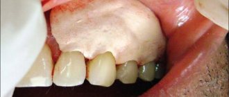
Teeth (dentes)
Located
in the dental alveoli of the upper and lower jaws using a continuous connection - impaction, gomphosis.
Tooth structure:
- Corona dentis - tooth crown
Facies lingualis - lingual surface
Facies vestibularis (facialis) – vestibular (facial) surface
Facies occlusalis [masticatoria] – closure surface [masticatory]
- Radix dentis - tooth root
Apex radicis dentis – apex of the tooth root
Foramen apicis dentis - opening of the apex of the tooth root
- Cervix dentis – neck of the tooth
- Cavitas dentis [pulparis] – dental cavity
Cavitas coronalis [coronae] – crown cavity
Canalis radicis dentis – tooth root canal
Pulpa dentis - tooth pulp
Tooth substance:
- Dentinum - dentin
- Enamelum – enamel
- Cementum - cement
Types of teeth:
- Dentes decidui – milk teeth
Complete dental formula of primary teeth:
| 2 0 1 2 | 2 1 0 2 |
| 2 0 1 2 | 2 1 0 2 |
2. Dentes permanentes - permanent teeth
Complete dental formula of an adult
| 3 2 1 2 | 2 1 2 3 |
| 3 2 1 2 | 2 1 2 3 |
- Dentes incisivi – incisors
- Dentes canini – fangs
- Dentes premolares – small molars (premolars)
- Dentes molares – large molars (molars)
- Dens serotinus – wisdom tooth
Function of teeth
– grasping, separating, crushing, chewing, grinding food, forming speech.
Text of the book “Fundamentals of clinical dental morphology: a textbook”
3.2.2. Mandibular canine
The lower canine is smaller in size than the upper one. The contact surfaces of the crown converge to the base of the crown to a lesser extent than those of the upper canine, and the root is more flattened in the mesial-distal direction. All signs of lateralization are quite pronounced.
In vestibular and lingual norms
the crown is narrower than that of the maxillary canine, and its contours blend relatively smoothly into each other (Fig. 24).
Rice. 24. Canine of the lower jaw, right.
a – vestibular norm; b – language norm
The contour of the main tubercle is represented by two segments (slopes), which, in place of the apex of the tubercle, can form an acute, close to right, or obtuse angle. The mesial segment, as a rule, is shorter than the distal one and is located less vertically. The apex of the distal angle of the crown is closer to the neck than the mesial one. The distal angle is larger than the mesial angle.
The mesial and distal contours of the crown weakly converge towards the neck of the tooth (sometimes they are located almost parallel).
The line of the enamel-cementum border, both in the vestibular and lingual norms, is convex towards the root with the most prominent points located approximately along the USV,
The transition of the contact contours of the crown into the same contours of the cone-shaped root is well defined and more prominent on the distal side.
The root is usually deviated distally (a sign of root position), although the root apex may be curved either mesially or distally.
On the vestibular surface of the crown, from the neck to the main tubercle, there is a noticeable median ridge, which protrudes less than that of the antagonist of the same name. The vertical grooves separating the median ridge from the mesial and distal ones are weakly expressed (the mesial groove is somewhat deeper than the distal one). On the lingual surface of the crown there are well developed marginal ridges (the distal one is better expressed than the mesial one), a median ridge (located from the lingual to the main tubercle), and a lingual tubercle.
The lingual surface of the root is already vestibular, so both approximal surfaces of the root are visible from the lingual side.
In mesial and distal norms
the shape of the crown approaches a triangle with the most acute angle at the site of the main tubercle (Fig. 25). The base of the crown is significantly narrower in the vestibular-lingual direction than that of the maxillary canine.
Rice. 25. Canine of the lower jaw, right.
a – mesial norm; b – distal norm.
The occlusal contour is less extensive than that of the maxillary canine, and is often shifted to the lingual side from the USV.
The vestibular and lingual contours of the crown transform into the occlusal contour near the USV. The vestibular contour of the crown is convex with the most prominent point near the border of the cervical and middle thirds.
The lingual contour of the crown, reflecting the shape of the lingual tubercle and marginal ridges, is convex in the cervical third and concave throughout the rest of the crown.
The enamel-cementum border is curved towards the main tubercle, more so on the mesial surface than on the distal surface. The most prominent point of the enamel-cementum border is located approximately along the USV.
The transitions of the vestibular and lingual contours of the crown into the same contours of the root are weakly noticeable.
The contours of the cone-shaped root evenly converge towards its apex. The vestibular contour of the root is convex, the lingual contour can be convex or straight. The apex of the tooth root is located near the USV.
On the approximal surfaces of the root there are vertical grooves, approximately equal in depth.
In occlusal norm
the crown can be compared in shape to an irregular quadrangle (Fig. 26, a). Just like the upper canine, the vestibular contour has a slope in the mesial-distal direction (a sign of crown curvature). The point of greatest convexity of the vestibular contour of the crown is displaced mesially from the USV, and the point of greatest convexity of the lingual contour is located closer to the distal contour of the crown.
Rice. 26. Canine of the lower jaw, right.
a – occlusal norm and section at the level of the base of the crown; b – tooth cavity.
On horizontal sections of the root, the vestibular-lingual dimension prevails to a greater extent over the mesial-distal dimension than in the root of the upper canine.
Tooth cavity
resembles its external shape (Fig. 26, b). The crown cavity has recesses for the pulp horns in the area of the corners and tubercles and passes into the root canal without a noticeable border.
Anatomical options.
The shape of the crown is determined by the degree of differentiation of the tooth and may be similar to the shape of the crown of the incisors or premolars of the lower jaw.
In the vestibular norm
The sharpness of the main cusp and the degree of convergence of the contact contours of the crown to the neck of the tooth can vary.
On the lingual surface
The marginal ridges and lingual tubercle may be differently expressed.
In contact standards
The vestibular contour of the crown can be convex to varying degrees, sometimes it can approach a straight line.
The nature of the root contours is quite variable. Sometimes the root can be “split” into two – vestibular and lingual, with different ratios of their sizes.
The root canal is usually single and straight. In cross section, it has an oval shape with a predominant vestibular-lingual size. The root canal can be deviated in both the vestibular and lingual directions. There are variants of the tooth cavity with two channels.
The height of the tooth is from 16.1 to 34.5 mm, while the height of the crown is 6.8-16.4 mm, the height of the root is 9.5-22.2 mm.
The mesial-distal size of the crown between the contact points ranges from 5.7 to 8.6 mm, the neck - from 4.1 to 6.4 mm. The size of the crown in the vestibular-lingual direction in the equator region ranges from 6.4 to 9.5 mm, in the cervical region - from 5.8 to 9.4 mm. Test tasks
In the maxillary canine, the longer one is:
a – mesial slope of the “tearing tubercle”;
b – distal slope of the “tearing tubercle”.
The vertical groove of the maxillary canine root is more pronounced:
a – on the mesial surface of the root;
b – on the distal surface of the root.
The largest of the fangs is:
a – canine of the upper jaw;
b – canine of the lower jaw.
The contact contours of the crown converge towards the neck to a greater extent:
a – at the canine of the upper jaw;
b – at the canine of the lower jaw.
The lingual tubercle is more pronounced:
a – at the canine of the upper jaw;
b – at the canine of the lower jaw.
The root position sign is expressed:
a – at the canine of the upper jaw;
b – at the canine of the lower jaw; c – all permanent canines.
The antimer for the right permanent maxillary canine is:
a – right permanent canine of the lower jaw;
b – left permanent canine of the lower jaw;
c – left permanent canine of the upper jaw;
d – right primary canine of the upper jaw.
The antagonist of the left permanent mandibular canine is:
a – right permanent canine of the lower jaw;
b – left permanent canine of the lower jaw;
c – left permanent canine of the upper jaw;
d – left primary canine of the lower jaw.
The enamel-cement border on the approximal surfaces of the canines is more convex:
a – on the medial (mesial) surface;
b – on the distal surface.
The diameter of the neck of the fangs predominates:
a – in the mesial-distal direction;
b – in the vestibular-lingual direction.
Answers to test tasks:
1
b;
2
a;
3
a;
4
a;
5
a;
6
in;
7
in;
8
in;
9
a;
10
b.
3.3. Group of small molars (Premolars)
Small molars (dentes premolares) –
teeth with a bicuspid occlusal surface, located in the dental arch in front of the molars (fourth and fifth positions) and are designed for crushing and crushing food. Premolars occupy the middle part of each half of the dental arch. Humans have 8 premolars:
– first and second premolars
of the upper jaw
(right and left);
– first and second premolars
of the lower jaw
(right and left).
A common feature in the anatomy of small molars is the presence of an occlusal surface with two tubercles - vestibular and lingual. The first premolar of the upper jaw, as a rule, has a bifurcated root. The remaining premolars often have a single root. The upper premolars are larger than the lower ones. The largest is the first premolar of the upper jaw, the smallest is the first premolar of the lower jaw.
In the upper premolars, the contours of the crown in the occlusal norm are oval, in the lower premolars they are rounded.
Crown angle sign
in the premolars of the lower jaw it is determined, but in the upper premolars it is not convincing.
Sign of crown curvature
“reverse” in the premolars of the upper jaw, is inconclusive in the second premolar of the mandible and is determined only in the first lower premolar.
Sign of root position
expressed in the first premolar of the mandible, unconvincing in the second premolar of the mandible and uninformative in the premolars of the upper jaw.
3.3.1.
The first small molar of the upper jaw The first premolar of the upper jaw, unlike the adjacent canine, has an occlusal surface with two cusps. The root is wider, but shorter than that of the canine, and, as a rule, is forked. The sign of crown angle is unreliable, the sign of crown curvature is “reverse”, and the sign of root position is inconsistent.
In the vestibular norm
the crown is shaped like the crown of a canine tooth in the upper jaw (Fig. 27, a).
Rice. 27. First premolar of the upper jaw, right.
a – vestibular norm; b – language norm.
The occlusal contour consists of two fragments (slopes) running from the tip of the tip of the vestibular tubercle (more often it is located near the USV) to the mesial and distal contours of the crown. The mesial fragment of the occlusal contour is longer and less vertical than the distal one.
The contours of the approximal surfaces converge towards the neck of the tooth, while the mesial contour slopes more towards the USV than the distal one. At the neck of the tooth, the approximal contours are connected by a line of the enamel-cement border, curved towards the root.
The transition of the contact contours of the crown to the corresponding contours of the root is more distinct on the distal side.
The root is flattened in the mesial-distal direction. The root apex is usually located distal to the USV.
On the vestibular surface of the crown there are vertical vestibular ridges. The median enamel ridge is separated from the mesial and distal ridges by vertical grooves, of which the mesial one is usually better expressed than the distal one.
In the language norm
due to the contours of the lingual tubercle, the contours of a larger vestibular tubercle are visible (Fig. 27, b).
The occlusal contour of the lingual tubercle is formed by two fragments (slopes) extending from the apex of the lingual tubercle. This apex is often shifted to the mesial side of the USV. This sign can be used as an additional criterion to determine whether a tooth belongs to the right or left side of the dental arch. The mesial slope of the lingual tubercle is shorter and gentler than the distal one.
The approximal contours of the lingual surface of the crown converge towards the neck of the tooth. The distal contour is shorter than the mesial one.
The enamel-cementum border is curved towards the root, and to a greater extent than on the vestibular side.
The transition of the contours of the crown into the corresponding contours of the root, as in the vestibular norm, is more pronounced on the distal side. The apex of the tooth root is often displaced distally from the USV.
In the lingual norm, in contrast to the vestibular relief, the crown is smoothed. The lingual surface of the crown is narrower than the vestibular surface, the contact surfaces diverge in the vestibular direction.
In mesial and distal norms
the crown is shaped like a non-convex polygon with an inward curved occlusal contour line (Fig. 28).
Rice. 28. First premolar of the upper jaw, right.
a – mesial norm; b – distal norm.
The vestibular tubercle, as a rule, is higher than the lingual one, and the point of connection of the slopes of the occlusal contour of these tubercles is slightly shifted to the lingual side from the USV. Often the junction of the mentioned slopes is hidden by a well-defined mesial transverse ridge.
The vestibular contour of the crown is convex with the most prominent point near the border of the cervical and middle thirds of the crown.
The lingual contour is shorter than the vestibular one and has a greater curvature. The point of greatest convexity of the lingual contour is located in the middle third of the crown.
The line of the enamel-cementum border, as a rule, has two convexities in the occlusal direction. The convexity at the level of the vestibular tubercle is more pronounced. The enamel-cementum border is more curved on the mesial surface than on the distal surface.
The root is conical. The vestibular contour of the root is convex, and the lingual contour is somewhat flattened. When the root is not split, its apex is located on the lingual side of the USV. If the root is forked, its apices are located on both sides of the USV.
The transition of the contours of the crown into the corresponding contours of the root is quite noticeable both from the vestibular and lingual sides.
On the approximal surfaces of the root there are vertical grooves, running from the base of the crown to the apex of the root (or to the level of bifurcation of the root when it is split).
In occlusal norm
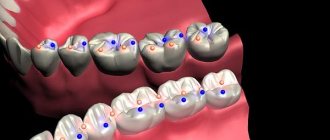
the crown is elongated in the vestibular-lingual direction (Fig. 29, a).
Rice. 29. First premolar of the upper jaw, right.
a – occlusal norm and section at the level of the base of the crown; b – tooth cavity.
There are two tubercles on the chewing surface - the larger vestibular and the smaller lingual. The tubercles are separated from each other by a deep intertubercular groove. This groove does not reach the contact surfaces of the crown, but connects with the grooves that separate the transverse ridges from the vestibular and lingual cusps. The relief furrow of the first order takes the shape of the letter H.
The intersections of the grooves are called mesial and distal fossae.
Transverse ridges are located along the mesial and distal contours of the occlusal surface. In relation to the intertubercular groove, two fragments are identified in the transverse ridges: a longer vestibular one and a smaller lingual one. In the occlusal norm, the slope of the enamel of the vestibular surface is determined in the distal-mesial direction (“reverse” sign of crown curvature).
The root in transverse sections of the cervical part is flattened in the mesial-distal direction, and is often bifurcated in the apical direction.
Tooth cavity
resembles its external shape (Fig. 29, b). In the cavity of the crown there are depressions that correspond to the tubercles of the occlusal surface. The vestibular recess is more pronounced than the lingual one.
The bottom of the tooth crown cavity is usually located at the level of its neck.
The cavity of the tooth crown smoothly passes into the root canals.
According to the division of the root into vestibular and lingual, the vestibular and lingual canals are formed, which, as a rule, begin in the cervical part of the root.
Anatomical options.
The first premolar of the upper jaw, depending on the degree of its differentiation in shape, may resemble a canine or molar.
In the vestibular norm
The contours of the crown are more often shaped like the crown of a canine tooth in the upper jaw. The mesial and distal parts of the occlusal contour of the vestibular tubercle have different lengths. More often than not, the mesial part of the occlusal contour is shorter than the distal part. Often the opposite ratio of their sizes is found.
There are options for the shape of the crown, when its distal or mesial angle is more rounded (the sign of the crown angle is not informative). The severity of vertical enamel ridges (mesial and distal) on the vestibular surface varies. In some cases, with significant differentiation of the tooth, there is a longitudinal groove on the vestibular surface of the root, which can “split” the vestibular root into two (three-rooted premolars).
In the language norm
depending on the severity of the lingual tubercle, the ratio of its size to the size of the vestibular tubercle may be different.
On the occlusal surface
the depth of the intertubercular groove varies (shallow, medium and deep). The furrow may cross the transverse ridges. The severity of transverse ridges also varies. Between the vestibular and lingual cusps, near the mesial and distal contours of the crown, there may be additional intermediate cusps - mesial and distal. The intermediate distal tubercle is less common.
In mesial and distal norms
The vestibular and lingual contours of the crown can be convex or straight. Features of the crown shape are determined by the amount of convexity of the lingual and vestibular contours. As a rule, the lingual contour is more convex than the vestibular one.
The occlusal contour is formed by the slopes of the vestibular and lingual cusps. The size ratios of the tubercles may be different. As a rule, the vestibular tubercle is larger. Less commonly, the tubercles are approximately equal in size. The contours of the tubercles, converging to the intertubercular groove, form an obtuse angle (usually closer to the unfolded one).
The mesial and distal transverse ridges are well defined. Often the scallops are “split” by grooves, more often on the mesial surface than on the distal surface.
The shape of the enamel-cementum border is variable. As a rule, on the mesial surface the curvature of the enamel-cementum border is better expressed than on the distal surface. Near the IVS, enamel may leak toward the root bifurcation.
The level of root bifurcation varies. When the root bifurcation is located close to the crown, the bottom of the crown cavity is well defined. The root canals (from one to three) are often curved and have additional branches.
The height of the tooth ranges from 15.5 to 28.9 mm, while the height of the crown is 7.1-11.1 mm, the height of the root is 8.3-19.0 mm.
The mesial-distal size of the crown between the contact points ranges from 5.5 to 9.4 mm, the neck - from 3.6 to 8.5 mm. The size of the crown in the vestibular-lingual direction in the equator region varies from 6.6 to 11.2 mm, in the cervical region - from 5.0 to 9.4 mm. 3.3.2.
The second small molar of the upper jaw The second upper small molar is identical to the first premolar, but is somewhat smaller in size. The root is usually single. The sign of the crown angle and the sign of the root are not convincing; the sign of the curvature of the crown is “reverse”.
In the vestibular norm
the crown is similar in shape to the crown of the first premolar (Fig. 30, a).
Rice. 30. Second premolar of the upper jaw, right.
a – vestibular norm; b – language norm.
The line of the occlusal contour is represented by two fragments (slopes) running at an obtuse angle from the top of the vestibular tubercle, which, as a rule, is located on the USV. The slopes of the vestibular tubercle are almost indistinguishable both in length and location. The mesial angle of the crown is somewhat sharper than the distal one.
The contours of the approximal surfaces of the crown converge towards the neck of the tooth, while the mesial surface slopes somewhat more noticeably towards the USV than the distal one.
The line of the enamel-cementum border is curved towards the root. The most prominent point is located approximately along the USV.
The transition of the contact contours of the crown into the same contours of the root is more prominent on the distal side.
The root is cone-shaped, its apex is located along the USV or is displaced distally.
The enamel ridges of the vestibular surface are weakly expressed.
In the language norm
the occlusal contour of the lingual surface is formed by two segments converging to the most protruding point of the lingual tubercle (Fig. 30, b). The mesial segment is shorter than the distal one, the tip of the tip of the lingual tubercle is often displaced mesially from the USV. This anatomical feature can be used as an additional sign of tooth lateralization. The apex of the lingual tubercle is more rounded than the vestibular one. Because of the contours of the occlusal surface of the lingual cusp, the occlusal contour of the higher vestibular cusp is noticeable.
The contact contours of the lingual surface of the crown converge towards the neck of the tooth.
The enamel-cementum border is curved towards the root and has approximately the same curvature on the vestibular and lingual sides. The most prominent point of the enamel-cementum border is located near the USV or is displaced distally.
The approximal contours of the crown merge into the corresponding contours of the root with a fairly noticeable border.
The root apex, as in the vestibular norm, is located near the USV or somewhat distal.
In the lingual norm, the surface of the crown is narrower than in the vestibular norm, so that both contact surfaces of the crown are visible from the lingual side. The relief lingual surface is smoothed.
In mesial and distal norms
the shape of the crown, like that of the first premolar, has the appearance of a non-convex polygon (Fig. 31).
Rice. 31. Second premolar of the upper jaw, right.
a – mesial norm; b – distal norm.
The line of the occlusal contour is curved towards the neck of the tooth, the degree of its curvature is determined by the severity of the vestibular and lingual cusps. The vestibular tubercle is somewhat larger in size than the lingual tubercle. The slopes of the tubercles, converging towards each other, form an obtuse angle. The projection of the intertubercular groove is located approximately in the middle of the occlusal contour.
The vestibular contour of the crown is curved towards the vestibular side with the point of greatest convexity approximately at the border of the cervical and middle thirds. The lingual contour of the crown is more convex than the vestibular one, and its most protruding point is located approximately in the middle third of the crown.
The line of the enamel-cementum border is slightly curved, as a rule, with two convexities in the occlusal direction at the level of the bases of the vestibular and lingual cusps. The curvature of the vestibular part of the enamel-cementum border is slightly greater than that of the lingual part. The curvature of the enamel-cementum border on the mesial and distal surfaces differs little.
The transition of the vestibular and lingual contours of the crown into the same contours of the root is more noticeable on the lingual side.
The root is conical. The groove on the distal surface of the root is usually more pronounced than on the mesial surface.
In occlusal norm
(Fig. 32, a) the crown is oval with a predominance of the vestibular-lingual size over the mesial-distal one. The angle between the vestibular contour of the crown and its distal contour is less than between the vestibular and mesial contours (“reverse” sign of crown curvature).
The intercuspal groove runs closer to the middle of the occlusal surface than that of the first premolar. As with the first premolar, near the mesial and distal contours of the crown there are well-defined transverse ridges, as well as mesial and distal fossae.
Rice. 32. Second premolar of the upper jaw, right.
a – occlusal norm and section at the level of the base of the crown; b – language norm.
The intertubercular groove reaches the transverse grooves on the mesial and distal sides. Taken together, the relief furrows of the first order form a figure in the form of the letter H.
The root in transverse sections is as flattened in the mesial-distal direction as in the first premolar. The contours of the approximal root surfaces are concave.
Tooth cavity
it is widest at the level of the neck, then, gradually narrowing, it passes into the root canal. (Fig. 32, b). The cavity of the crown has recesses towards the vestibular and lingual cusps.
Anatomical options.
In
the vestibular norm,
the shape of the crown varies. The vestibular surface of the crown can have the shape of an oval or pentagon. The curvature of the occlusal contour of the vestibular tubercle can be varied. The tubercle can be round or pointed. The position of the most protruding point of the vestibular tubercle in relation to the contact contours of the crown is variable. More often than not, this point is located near the IVDS. The degree of convergence of the contact contours of the crown changes towards the neck of the tooth - from their almost parallel arrangement to a pronounced deviation towards the USV. Enamel ridges can be expressed to varying degrees, which determines the nature of the relief of the vestibular surface. The root may have noticeable bends both distally and mesially.
In approximate norms
the point of connection between the slopes of the vestibular and lingual tubercles may be closer to the middle of the chewing surface or be shifted to the lingual contour. The severity of the tubercles on the chewing surface can vary in height and width of their bases.
In occlusal norm
the vestibular and lingual cusps may be “split.” Between these tubercles, near the contact contours of the crown, additional tubercles are found.
There are tooth options with two or three roots. The number of channels in the root ranges from one to three.
When there are two canals, they often end in a single hole at the root apex.
The height of the tooth ranges from 15.2 to 28.4 mm, while the height of the crown is 5.2-10.5 mm, the height of the root is 8.0-20.6 mm. The mesial-distal size of the crown between the contact points ranges from 5.5 to 8.9 mm, the neck - from 4.0 to 5.8 mm. The size of the crown in the vestibular-lingual direction in the equator region varies from 6.9 to 11.6 mm, in the cervical region - from 5.8 to 10.5 mm.