The baby has a facial cleft - how to care for him and rehabilitate him after surgery
We continue a series of publications devoted to neurosurgical diseases. The project was prepared jointly with the MBOO for helping children with neurosurgical diseases “He Needs You.” We want these “terrible” diagnoses not to put pressure on parents and not to take away their hope. In our publications, the best specialists and experienced parents will talk about ways to cure and overcome, give recommendations and show that life can go on with any, even such complex, diseases.
In our first publication on facial clefts, we highlighted the types of clefts and talked about the modern surgical approach to their treatment. In the second part, we will talk about the simplest things related to nursing a baby, which present certain difficulties for babies with clefts.
FREE CONSULTATIONS!
We are pleased to announce the start of free admission at the Konstanta Clinic for patients under 18 years of age by maxillofacial surgeon, pediatric surgeon L.A. Eremeyshvili. for questions:
- Congenital facial pathologies: cleft lip and palate;
- Benign neoplasms of the face and oral cavity;
- Diseases of the temporomandibular joints;
- Pathology of the frenulum of the oral cavity: lips and tongue.
Correction (plasty) of tongue frenulum is provided free of charge for children under 1 year of age.
*Valid for residents of Yaroslavl and the Yaroslavl region.
You can make an appointment by phone or through the website (4852) 37-00-85 Daily from 8:00 to 20:00
Sign up for a consultation
Congenital cleft palate or cleft palate is a fairly common malformation of the facial region. This pathology is ranked 4th among all developmental defects, and it is diagnosed with a frequency of 1:700 births. Often, a cleft palate is not an independent deviation, but part of a hereditary syndrome.
If the child has a pathology
A diagnosis of “cleft palate” in a child is not a death sentence, and parents need to remember that the pathology responds well to treatment if it is started early. During the treatment period, the main role is given not only to surgical intervention, but also to the patience of parents, their responsibility and willingness to deal with the health of their own child. You must be prepared to carefully implement all recommendations.
Once a diagnosis of cleft palate is made, the child is taken to a surgeon who can create a treatment plan.
In our Clinic, such defects are dealt with by Sergey Nikolaevich Bessonov and Levan Avtandilovich Eremeyshvili, who are maxillofacial surgeons. Appointment with doctors S.N. Bessonov and L.A. Eremeyshvili takes place over the phone.
Thanks to the help of a charitable foundation, surgery to correct a cleft palate can be completely free. All you need to do is prepare the documents from the list and wait for an individual call to a specialist.
Our patients
Milan
| BEFORE AFTER | Diagnosis: rhinocheiloplasty Attending doctor: Bessonov Sergey Nikolaevich |
Vasilina
| BEFORE AFTER | Diagnosis: rhinocheiloplasty, uranoplasty Attending doctor: Bessonov Sergey Nikolaevich |
Violations associated with vice
A cleft of the soft and hard palate leads to the appearance of a communication between the oral and nasal cavities. A cleft of the soft palate leads to improper attachment of muscle structures and deformities of the pharynx in the middle section. The soft palate is also significantly shortened.
A cleft palate leads to the fact that the child cannot normally perform sucking and swallowing movements, his breathing processes are disrupted, and his bite is formed incorrectly. With age, the deformation intensifies, and problems with speech appear.
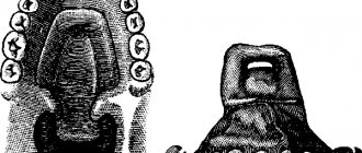
Cleft palate
Cleft lip
Rice. 1a
Rice. 1b
Rice. 2a
Rice. 2b
Rice. 2s
Rice. 2d
Rice. 3a
Rice. 3b
Rice. 3s
Rice. 4a
Rice. 4b
Rice. 4s
Rice. 5a
Rice. 5b
Rice. 5s
Rice. 6a
Rice. 6b
Rice. 6s
Rice. 7a
Rice. 7b
Rice. 7s
Rice. 7d
Rice. 7e
Rice. 7f
Rice. 7g
Rice. 7h
Rice. 7i
Rice. 7j
Rice. 7k
Rice. 8a
Rice. 8b
Rice. 8s
Rice. 8d
Rice. 9a
Fig 9b
Rice. 9s
Rice. 9d
Rice. 10a
Rice. 10b
Rice. 10s
Rice. 11a
Rice. 11b
Rice. 12a
Rice. 12b
Rice. 12s
Rice. 13a
Rice. 13b
Rice. 13s
Rice. 14a
Rice. 14b
Rice. 14s
Rice. 15a
Rice. 15b
Rice. 15s
Rice. 15d
Rice. 15th
Rice. 15f
Rice. 16a
Rice. 16b
Rice. 16s
Rice. 16d
Congenital clefts of the upper lip and palate are among the most common defects in humans and account for 12–30% of all deformities. This pathology ranks second, second only to cardiovascular pathology, among all congenital malformations of the body. In the structure of lesions in the craniofascial and maxillofacial areas, it accounts for up to 90%. According to the literature, the incidence of births of children with congenital cleft palate varies in different regions of our planet from 1 in 300 to 1 in 2000. Statistical studies conducted in recent years indicate an increase in their number. The severity of these conditions is determined not only by external facial disfigurement, pronounced functional disorders of the dentofacial apparatus and ENT organs (breathing, nutrition, speech, facial expressions, hearing), social impairment of children in the family, preschool and school groups, but also by a number of somatic disorders, leading to impaired growth and development of the body.
Etiology of congenital clefts of the upper lip and palate
- I. Exogenous causes
Physical factors: mechanical, thermal, radiation
Chemical factors: hypoxia, malnutrition, hormonal imbalances, teratogenic poisons
Biological factors: viruses, bacteria and their toxins, protozoa
Mental factors
- II. Endogenous causes
Heredity
Biological inferiority of germ cells
Influence of parental age
Classification of cleft lip and palate
1. By nature and prevalence:
- single or double sided
- complete or incomplete
- hidden
- By localization:
- upper lip
- upper lip and alveolar process in/jaw
- upper lip, alveolar process of the jaw and hard palate
- upper lip, alveolar process in the jaw, hard and soft palate
- alveolar process in the jaw, hard and soft palate
- hard and soft palate
- soft palate
The problem of rehabilitation of patients with clefts of the upper lip, hard and soft palate continues to be relevant to this day, despite the large number of developments devoted to its various aspects. In the rehabilitation of this category of patients, one of the key points that directly affects its outcome is the surgical method of treatment. Recently, the timing of cheilorhinoplasty has been more clearly defined. Most leading centers for the treatment of such patients recommend performing it for unilateral nonunion starting at 2–3 months, and for bilateral nonunion at 5–8 months. The indicated timing is due to the fact that by this age physiological jaundice is eliminated, the immunity acquired from the mother at the time of birth, lost after 2 weeks of age, is replenished, albeit minimally, and parts of the upper lip are minimally formed.
As for the timing of uranostaphyloplasty, these issues are not always possible to solve unambiguously due to the variety of variants of this defect and the complex dependencies of its cause-and-effect pathogenetic aspects. In the last thirty years, there has been a clear tendency of domestic and foreign surgeons to perform surgical interventions at an earlier date. This circumstance is caused by the following points: the suffering of children in the period from 2-3 to 6-7 years of age with palatal defects due to deformations of the dentition, speech incomprehensible to the interlocutor and subsequent social inconveniences associated with this; an increase in background diseases every year, which causes a delay in medical rehabilitation. A number of experts consider it necessary to first perform veloplasty (suturing of the soft palate) at the age of 6–9 months, then, at the age of 12–18 months, uranoplasty, which is often associated with the eruption of the upper temporary teeth necessary for fixing the artificially manufactured protective palatal plate on wound healing period, justifying this by the fact that early suturing of the soft palate promotes the development of the muscles of the soft palate, which in turn improves speech function in this category of patients. For the most part, we can agree with this opinion.
The goal of uranoplasty is to restore the anatomical features of all parts of the palate and create the prerequisites for achieving adequate closing function of the velopharyngeal ring. After various types of interventions, postoperative defects often occur in the area of the anterior, middle and other parts of the hard palate or at the border of the hard and soft palate. According to various literature sources, their number reaches more than 75%. The main reasons for such complications should be considered improper planning and non-compliance with the surgical technique; use of local tissues for plastic surgery in cases of medium or large defect sizes, when it is not possible to bring mucoperiosteal flaps together without tension; necrosis of mucoperiosteal flaps due to disruption of their nutrition; failure of sutures due to inflammatory infiltrates or suppuration of wounds associated with hematomas or the occurrence of acute infectious diseases in the immediate postoperative period; improper postoperative management of patients.
Our own technique for eliminating congenital cleft palate.
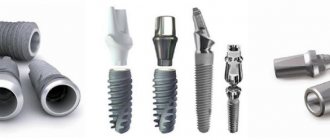
In order to eliminate the formation of postoperative defects in various parts of the hard palate, as well as to simplify the technique of uranoplasty by eliminating the application of eversion sutures to the mucous membrane of the bottom of the nasal passages in this category of patients, a technology for surgical treatment using tissue implants made of titanium nickelide has been developed
(RF Patents No. 2239374 , 2240062, 2254078, 2284772, 2286730, 2289336, 2352275)
.
The edges of the cleft are refreshed, in the area of the lateral sections of the hard palate the mucous membrane and periosteum are dissected according to Langenbeck and incisions are made in the anterior and middle sections of the hard palate to form additional mucoperiosteal flaps, using one of the well-known techniques (Zausaev V.I., 1953; Dubov M. D., 1960; Kabakov B. D., 1964; Muratov I. V., Kotov G. A., 1998). In patients with bilateral clefts in the anterior and middle sections, mucoperiosteal flaps are mobilized on the right and left sides of the defect and subsequently mutually moved, taking into account the overlap of the cleft, formed by the mucous membrane and periosteum of the premaxillary bone, palatine and alveolar processes of the upper jaws. Mucoperiosteal flaps are separated from the palatine and alveolar processes of the upper jaws, as well as the horizontal plates of the palatine bones. The neurovascular bundles are removed from the large palatine canals after resection of the posterior walls of the latter and their preparation from flaps is performed. Next, the flaps are separated from the nasal mucosa in the area of the border of the hard and soft palate forward, the mucous membrane and submucosal layer are dissected in the retromolar zones to the lingual surface of the alveolar part of the lower jaw and the hooks of the pterygoid processes of the main bone are exposed. The flaps from the latter are separated in the layer of interfascial space from the inner surface of the inner plate of the main bone to the attachment of m. pharingopalatini, without changing the place of attachment of m. tensor veli palatini, and if there is insufficient tissue mobility, the upper pole of the tendons of these muscles is crossed. The flaps of the soft palate are sutured together in three layers. Wounds in the areas of the pterygomandibular spaces, taking into account the retransposition of the palate, are tightly sutured with interrupted sutures. Titanium nickelide fabric is placed between the mucoperiosteal flaps and the bony part of the hard palate so that the width of this implant corresponds to the total width of the mucoperiosteal flaps. In the anterior and middle sections of the hard palate, nickelide-titanium fabric is installed below the bony part of the cleft, the other part of the tissue is fixed to the border of the mucous membrane of the palate and nose with a nickelide-titanium thread 40-50 microns thick, followed by isolation from the oral cavity with tipping mucoperiosteal flaps , adjacent to the tissue, areas of which are de-epithelialized. Mucoperiosteal flaps are fixed with interrupted sutures. The surgical wound is isolated from external influences using an iodoform bandage; additional fixation of the mucoperiosteal flaps is carried out with a protective dental palatal plate.
Depending on the indications, paracentesis of the eardrums is performed, followed by evacuation of mucous or mucopurulent contents from the tympanic cavities and shunting of the latter. In cases of reduced volume of the lower nasal meatus caused by hypertrophy of the inferior turbinate, to prevent difficulty in nasal breathing, laser radiation from an Nd-Yag laser with a wavelength of 0 is applied to the medial surface of the anterior end of the inferior turbinate using the tip of a laser quartz light guide with a diameter of 0.5 mm. 89 microns with a power of 20 W in continuous mode, with the advancement of the light guide in the submucosal layer of the inferior turbinate parallel to the bone skeleton and mucous membrane along the entire length of the inferior turbinate at a speed of 2 cm per second, then a hemostatic tampon is inserted into the nasal cavity. Deviation of the nasal septum is eliminated by resection of the area protruding to the side or by dissection at the base, in the middle and upper part of the quadrangular cartilage. Hypertrophic changes in the pharyngeal tonsil are excised completely or only in the projection of the mouths of the auditory tubes (in order to preserve the function of the velopharyngeal seal). (RF Pat. No. 2368337; authors: A. A. Radkevich, S. G. Vakhrushev, V. A. Ivanov, V. I. Kolga).
Replacement of iodoform tamponade and treatment of the wound is carried out at intervals of 24 hours; sutures from the oral cavity are removed on the 10th day. The vault of the palate, if necessary, is formed by layering the thermoplastic mass “Stens” or others onto the protective palatal plate for a month at weekly intervals.
The advantages of the proposed technology over previously known ones are that the installed titanium nickelide fabric between the mucoperiosteal flaps, the bony part of the hard palate and the bottom of the nasal cavity, due to biochemical and biomechanical compatibility with body tissues, fluid retention properties, ensures restoration of the mucous membrane membranes of the bottom of the nasal passage, reliably isolates the oral and nasal cavities in cases of divergence of mucoperiosteal sutures, necrosis of part of the flaps. In these situations, the missing tissue is replaced along the implant material by marginal epithelization of the oral and nasal mucosa on the exposed areas of the implant. The connective tissue from the recipient areas will grow through the cellular structure of the implant to form a single connective tissue regenerate in the area of the former defect. The absence of the need to apply reversible sutures to the mucous membrane of the bottom of the nasal cavity significantly reduces the time characteristics of the operation, prevents hypertrophic cicatricial deformation and narrowing of the lower nasal passage. The immediate elimination of pathological conditions from the organs of the nasal cavity ensures the normalization of nasal breathing and prevents the development of rhinosinusitis. Segmental adenotomy makes it possible to normalize the functional characteristics of the auditory tubes without leading to functional insufficiency of the velopharyngeal seal.
Clefts of the alveolar processes of the upper jaws.
A defect in the alveolar process of the upper jaw is a common form of manifestation of congenital pathology - cleft lip and palate; according to the literature, this combination accounts for 70-80%. Some authors believe that in almost all forms of cleft lip there is an alveolar ridge cleft, which can be hidden, incomplete (at the level of only the apical base) or complete. In all forms of cleft of the alveolar process of the upper jaw, the apical base of the upper jaw is underdeveloped or has a bone defect significantly larger than in the area of the crest of the alveolar process. The presence of a defect in the area of the basal part of the alveolar process leads to instability of the results of surgical and orthodontic treatment. In addition, attention is drawn to the fact that in the group of children with congenital malformations of the maxillofacial localization the most severe forms of clefts have increased, including bilateral clefts of the upper lip, hard and soft palate, accompanied by clefts of the alveolar process of the upper jaw, in which pronounced protrusion is most often observed premaxillary bone and medial displacement of the lateral fragments, which creates unfavorable conditions for the healing of the surgical wound, since under conditions of pronounced tissue tension, local hypoxia is observed, creating a threat of suture dehiscence.
Complete social rehabilitation of such patients is possible only if full surgical treatment is carried out at an early stage of their development. Instability of the position of two or three bone fragments of the upper jaw leads to the development of severe deformations of the facial skeleton and reduces the stability of the teeth located at the edges of the alveolar defect. Uncorrected defect of the alveolar process and oronasal anastomosis are the reasons for a number of unsatisfactory functional and cosmetic results of treatment of patients with congenital clefts of the upper lip and palate. The constant reflux of oral contents into the nasal cavity contributes to the development of chronic rhinitis, maxillary sinusitis, and is also a complicating factor in reconstructive cheilorhinoplasty. The correct shape and size of the basal part of the upper jaw is a necessary condition for obtaining stable results of orthodontic treatment, formation of the position of the teeth and bite. The asymmetrical contours of the upper lip and the displacement of the base of the nasal wing on the side of the cleft, caused by the presence of a bone defect in the alveolar process and the deformation of the anterior outer surface of the upper jaw and the edges of the pyriform foramen, do not allow for effective reconstructive surgery for deformities of the upper lip and nose. Additional air leakage through the oronasal anastomosis, unstable and dystopic position of the teeth of the upper jaw have a negative impact on the speech function of patients. In connection with the above, elimination of the defect of the alveolar process of the upper jaw in patients with clefts, which can be achieved by performing osteoplastic surgery for these types of congenital defects, creates a normal basis of the alveolar process, preventing or reducing the possibility of developing orthodontic pathology, and contributes to the elimination of general somatic changes body, creates conditions for the eruption of impacted teeth. The continuity of the alveolar process of the upper jaw improves the conditions for the eruption and formation of the anterior teeth, the anterior part of the upper jaw, and optimizes the correct growth and development of the upper lip and nose. Many surgeons observed the displacement of tooth buds and their eruption through the thickness of the autograft.
Technique for eliminating congenital defects of the alveolar processes of the upper jaws.
Vestibular mucoperiosteal flaps are cut out and mobilized in the projection of the cleft, taking into account their use to cover the defect of the oral mucosa and periosteum, pathological tissues are excised between the bony edges of the fragments of the upper jaw, blind sutures are applied to the mucous membrane and the skin part of the bottom of the nasal passage in the projection of the cleft. , a bone autograft is collected, which is combined with finely granulated porous titanium nickelide with a particle size of 1–500 microns, in a ratio of 3:1 and placed in the area of the bone defect by dense tamponade until complete replacement, under the mucous membrane of the bottom of the nasal cavity and on the anterior side Nickelide-titanium tissue is installed in the bone autograft, taking into account the overlap of the cleft, the mucoperiosteal flaps are moved until the defect is completely covered, the wound is sutured tightly. In cases where it is necessary to reconstruct a defect in the mucous membrane and periosteum on the palatal side, nickelide-titanium tissue is installed on the posterointernal side of the autograft under the palatal mucoperiosteal flaps, taking into account the overlap of the bone defect, after their formation, mobilization and movement to completely eliminate the defect. The wound is sutured tightly.