Oral fibroma is a benign neoplasm on the mucous membrane of the oral cavity. It looks like a small nodular seal, rising above the plane with the help of a long flat stalk and consists of connective tissue fibers. Typical places for fibroids to occur are the inner surface of the cheek, the mucous membrane of the lips, the soft palate, gums and tongue. Most often, this type of neoplasm appears in children between the ages of six and fifteen years.
One of the common factors influencing the appearance of fibroids is the habit of biting the lip or tongue. The gums are most often injured by the sharp edge of a broken tooth or an incorrectly installed denture. Patients often have a history of chronic inflammation, gingivitis, stomatitis, periodontal disease, glossitis of various origins, etc.
What it is
Oral fibroma is a benign formation, similar to a small nodule on a stalk, which consists of connective tissue. Most often it appears on the mucous membrane of the gums, lips, cheeks or palate, less often on the tongue. Children aged six to fifteen years are more susceptible to the pathological process.
Fibroma at the initial stage of its formation is completely invisible, it does not bring any discomfort to the patient. As it grows, it thickens and swells.
Despite the fact that fibroma is not so dangerous, it must be treated. If the formation becomes larger in size and is constantly injured, a wound forms, which becomes infected. In such a situation, treatment will take longer and will be much more difficult.
In most cases, surgical excision of the fibroid is required.
Signs of the disease
Fibroma in the oral cavity has the appearance of a spherical formation, painless to the touch. The neoplasm rests on a low stalk, so it protrudes above the surface. The tumor has a smooth surface and is completely free of growths, erosions or any visible changes in the mucous membrane. Fibroids, as a rule, grow and increase in size quite slowly and relatively imperceptibly for the host. If the tumor has not been injured, it may remain “quiet” for some time, maintaining its normal size. If the traumatic factor has not been eliminated, over time a malignant tumor may form in its place.
Reasons for the growth of education
Scientists have not identified the exact reasons for the growth of fibroids in the oral cavity. But there is a list of factors that provoke the appearance of an anomaly:
- hereditary factor (as a rule, fibroma appears in very young patients aged from one to ten years);
- constant injury (regular biting of one area of soft tissue, injury from the sharp edges of a filling, crown or orthodontic structure, eating very hard food);
- taking certain medications (epileptic drugs, some hormonal pills, drugs that correct vascular tone);
- inflammatory processes in the oral cavity (stomatitis, periodontal disease, glossitis).
It is impossible to exclude a genetic predisposition, but you can reduce the risk of the problem if you follow basic preventive measures:
- do not neglect oral hygiene;
- clean the oral cavity from soft and hard plaque using professional hygiene;
- treat the soft tissues of the oral cavity with care (avoid temperature changes when eating, do not abuse solid foods);
- promptly treat diseases of teeth and gums;
- do not self-medicate;
- undergo preventive examinations with a dentist once every six months.
Treatment of oral fibroids
To effectively and permanently get rid of a tumor in the oral cavity, surgical intervention is necessary. The fibroma is excised either using a laser or radio wave method using local anesthesia.
If the fibroma has a pedicle, then it must be removed using two bordering incisions. The base of the fibroma is removed using an arcuate incision. To excise a fibroma located on the inner surface of the lip mucosa, a perpendicular dissection is used through the fibers of the orbicularis oris muscle. If the fibroma is large, it is necessary to prevent deformation of the mucosa. To do this, the defect remaining after removal is covered with a V-shaped flap from nearby tissue.
After removing fibroids from the oral cavity, the specialist prescribes wound-healing drugs or auxiliary procedures. In most cases, the prognosis for recovery is favorable due to successful removal of the tumor.
What are fibroids?
The formations are divided into several types, differing in their density, nature of origin and how severe the clinical manifestations of the neoplasm are. Dense education. It is characterized by a thick consistency and is most often localized on the gums and palate. Consists of soft fibers of connective tissue. Typically found on the inside of the tongue and cheeks.
Formation arising from irritation. A chronic disease caused by constant trauma to one area of the mouth. The cause may be installed fillings or prostheses, as well as piercings.
Fibrous epulis. A dense pink formation that does not cause any discomfort to the patient. It has very clear boundaries and an asymmetrical shape. Localized on the vestibular region of the gums or in the interdental spaces.
Diagnosis of oral fibroma
To determine the presence of fibroids, the dentist conducts a thorough examination of the oral cavity, palpating the tumor. If there is a suspicion of tumor growth into neighboring tissues, an ultrasound scan is prescribed. In some cases, if there are inflammatory changes or the presence of ulcers on the surface of the fibroma, a tumor biopsy is required. After removal of the fibroma, a histological analysis of its tissue is carried out in the laboratory.
A specialist needs to diagnose the cause of the neoplasm, so additional examination measures are carried out, such as:
- periodontograms;
- radiovisiography;
- orthopantomograms;
- radiography.
If the patient uses dentures, then he needs to consult with an orthopedic dentist to prevent tissue injury from this device.
Differential diagnosis of a neoplasm is carried out if the following is found in the patient’s oral cavity:
- lipoma;
- papilloma;
- epulis;
- neuroma.
If the fibroma is localized on the tongue, then first it is necessary to exclude cancer of the patient’s tongue and possible other tumors, but of a benign nature.
Diagnosis and treatment
Diagnostics consists of interviewing and examining the patient; if necessary, additional studies and radiography are performed.
The most common and effective method is surgery. The formation is removed using a laser; the procedure lasts about thirty minutes.
If the disease is caused by taking medications, they should be excluded and replaced with similar drugs. After stopping the medication, the appearance of the mucous membrane can be restored without any intervention. But this does not apply to cases where the pathology is advanced.
You can also do without intervention in case of traumatic effects of a prosthesis, filling or braces. Elimination of the cause of the formation leads to the disappearance of fibroids.
Thus, fibroids in the oral cavity do not pose a danger only if proper measures are taken in a timely manner. You should not take any action on your own. If a problem is detected, you should immediately contact a specialist.
Among benign formations of the oral cavity, the most common is fibroma.
According to a study conducted in 2008 in Spain, out of 300 patients with a histologically confirmed diagnosis of a benign tumor of the oral mucosa, 53% of the subjects were diagnosed with fibroma of the oral mucosa (Fig. 1).
Figure 1. Percentage of benign lesions according to a 2008 study in Spain.
Fibroma is a benign formation of mature fibrous connective tissue.
A patient approached the specialized department of the Department of General and Aesthetic Dentistry of Moscow State Medical University with complaints of a formation on the mucous membrane of the lower lip on the right.
According to the patient: 5 years ago I discovered a small formation that was slowly growing. I did not seek professional help.
External examination without any features.
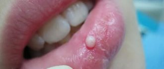
When examining the oral cavity, on the mucous membrane of the lower lip on the right there is a tumor-like formation of a round shape, rising on a wide base with a diameter of about 5 mm; on the surface of the formation, the mucous membrane in color is not changed; on palpation: soft-elastic consistency, painless (Fig. 2).
Figure 2. View of the formation on the mucous membrane of the lower lip.
Based on the survey and objective examination data, a diagnosis was made: fibroma of the mucous membrane of the lower lip on the right.
Due to the fact that chronic trauma to this formation can cause malignancy, excision of the formation within healthy tissue was recommended.
Brilliant green was used to determine the boundaries of the intervention (Fig. 3).
Figure 3. Appearance of the formation after determining the boundaries of the intervention using brilliant green.
Under infiltration anesthesia Sol. Artiphrini 4% 1:100,000, 1.7 ml (Fig. 4)
Figure 4. Type of formation after infiltration anesthesia.
two converging semilunar incisions were made and the mass was excised (Fig. 5-7).
Figure 5. Making semilunar converging incisions.
Figure 6. Excision of the lesion.
Figure 7. View of the wound after excision.
The wound was then sutured with three interrupted sutures using catgut (Fig. 8).
Figure 8. View of the wound after suturing.
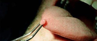
To confirm the diagnosis, the removed lesion was sent for cytological examination (Fig. 9).
Figure 9. View of remote education.
The patient was given the following recommendations:
- follow a gentle diet, excluding rough, spicy, hot foods for a week;
- oral baths with chlorhexidine solution;
— application of dental adhesive paste solcoseryl;
— for pain, take Nurofen forte 400 mg.
When re-examining the patient on the 4th day after surgery: there are no stitches, the wound is covered with fibrinous plaque, palpation is painless. An antiseptic treatment was carried out with a chlorhexidine solution and the dental adhesive paste solcoseryl was applied (Fig. 10).
Figure 10. View of the wound on the 4th day after surgery.
The patient is under dynamic observation, the wound is in the regeneration stage, healing by secondary intention.
Thus, timely diagnosis and treatment of benign formations, such as fibroma, reduces the risk of malignancy.
Symptoms
The development of the tumor process is accompanied by moderate pain and a feeling of fullness. In 55–60% of cases, the pathology is asymptomatic. The likelihood of developing specific signs of fibromatosis depends on the type and location of the tumor. Fibromatosis of cartilaginous tissue is characterized by increasing symptoms - from mild pain when performing movements to complete loss of mobility of the upper or lower limb.
Specific symptoms develop when fibroma affects the patient’s internal organs. Thus, fibrosis of the uterus can cause bleeding that is not related to a girl’s menstrual cycle. Fibrous lesions of the lungs provoke difficulty breathing and shortness of breath in a child or adult after short physical activity.
Fibroma: origin and essence of the disease
The causes of dermatofibroma and other forms of this type of tumor are not known. Some researchers believe that fibroids can form as a localized tissue reaction after minor trauma. Sometimes fibroids can have a genetic component, especially in people of Northern European descent. Some medications, including beta blockers, have been reported to cause changes in fibrous tissue. In addition, some fibroids may be influenced by hormonal imbalances or pathologies of endocrine organs, including problems with the thyroid and pancreas.
Hyperhidrosis (increased sweating), inflammatory processes on the skin, especially chronic ones, as well as the influence of prolonged UV radiation, poor nutrition and bad habits can have a certain impact.
Types, characteristics of fibroids
There are more than 10 different morphological variants. Pathologists give a description of what each form looks like and how it differs after histological examination - elastrophibroma, soft, dense fibroma.
- Dermatofibroma is a single or multiple formation of intradermal localization, mobile and painless when palpated with unchanged color.
- Desmoid fibroma has another name - aggressive fibrosis, refers to mesenchymal tumors originating from excess collagen fibers and differentiated fibroblasts. They often progress and recur, forming in soft tissues, the retroperitoneum or on the abdominal wall.
- Chondriomyxomas are rare tumors that affect bone tissue - the edges of long bones, pelvis, ribs, vertebrae, feet, metatarsus.
- Angiofibriomas in the cheek and nose area, containing small tubercles with connective tissue fibers.