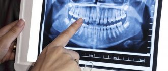
An x-ray is the dentist’s main tool in making the correct diagnosis. However, a conventional orthopantomogram or targeted photograph has limited diagnostic potential and does not provide complete data on the condition of the teeth and maxillofacial area. But technologies are constantly being modernized and today, more informative technology has come to the aid of conventional radiography - dental computed tomography (CT).
What does a 3D dental x-ray show?
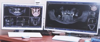
3D dental tomography is a highly accurate diagnostic method that makes it possible to obtain a three-dimensional image of the dental system in different projections. Volumetric images obtained with CT allow the specialist to enlarge, rotate and examine the area of interest from all sides and at different depths:
- The entire maxillofacial apparatus.
- A dentition or an individual tooth.
- Paranasal sinuses.
- Bone and periodontal tissues.
Dental CT allows you to detect inflammation, assess the homogeneity of the filling material and check the quality of installation of a filling, crown or implant, see the number of dental roots and their fragments, identify neoplasms, assess the degree of curvature of teeth, determine the exact parameters of bone tissue (height, width, density, etc.). d.). The information obtained allows the doctor to optimize treatment measures and predict the result.
Features of treatment of individual groups of teeth
The treatment tactics for incisors, canines, premolars, molars of the upper and lower jaws are different. Each tooth carries a different load. One part of the row is visible when you smile, while the other remains invisible to others. Let's look at the features of treatment for each area of the oral cavity.
Treatment of wisdom teeth
Eights are the outermost molars in the row. They erupt later than others, practically do not participate in chewing food, and regularly cause trouble. 1, 16, 17, 32 teeth can erupt incorrectly, displacing the entire row, injuring the gums, and causing the formation of a soft tissue hood. This increases the risk of plaque accumulation and tooth decay.
| Features of wisdom teeth treatment | |
| Question | Dentist recommendation |
| Do I need to remove wisdom teeth? | In most cases, this is necessary and recommended. |
| Is it possible to treat caries on the 8th tooth? | If they do not disturb the structure of the dentition or cause discomfort, then after consultation with a doctor you can do this. |
In most cases, eights must be removed. If they grow at an angle, disrupt the bite, displace other molars, cause discomfort, or have underdeveloped enamel, it is recommended to get rid of them as quickly as possible. Extraction of teeth 1, 16, 17, 32 does not affect the chewing function or appearance of the patient.
Is caries of eights curable? If they have a normal structure, do not cause discomfort, and are involved in chewing, superficial caries can be treated using the traditional method - drilling, installing a filling. The procedure is performed at the request of the patient. If a person refuses conservative therapy, the extreme molars are removed.
If there is significant damage to the visible part of the figure eights, their removal is indicated. Artificial crowns are not installed on them due to their location and anatomical features. For acute pain, the only way to solve the problem is extraction. The figure eights are located in the far corners of the oral cavity, which makes depulpation and endodontic treatment impossible. Lack of visibility and access to instruments prevents nerve removal and canal filling.
Extreme molars are unreliable and often destroyed, so they are not recommended for use as a support for bridges. If eights are lost, prosthetics are not performed; there is no such need or possibility.
Treatment of molars
2, 3, 14, 15, 18, 19, 30, 31 teeth are molars that bear the main load when chewing. They are large, with a wide crown, have several roots, so they are firmly held in the jaw. They are not visible when you smile, but the defeat or absence of one of them causes severe discomfort to the patient. When treating this group, they pay attention to strength and strive for complete restoration of function.
| Features of treatment of molars | |
| Question | Dentist recommendation |
| What treatment methods are there if a molar is severely damaged? | In case of severe destruction of molars, ceramic inlays are most often used. |
| Which crowns are best for molar prosthetics? | More often, patients are recommended to install metal-ceramic crowns in the molar area. |
| What to do if you lose molars? | The best option in this case is implantation; patients also consider removable dentures. |
The main method of treating caries of sixes and sevens is drilling and filling. If the process does not affect the canal with the neurovascular bundle, the doctor drills out the affected areas and closes the defect with a filling made of composite materials. If the affected area is large, it is advisable to use ceramic inlays. They are made from impressions for each patient and close the cavity formed after treatment. The use of inlays allows you to increase the service life of the molar and ensures its resistance to chewing loads.
If the visible part of the six or seven is significantly destroyed, but the root is preserved, restoration is carried out using a crown. The best material for prosthetics of 2, 3, 14, 15, 18, 19, 30, 31 teeth is metal ceramics. Such designs are very durable and allow you to chew even hard food. A thin strip of metal that is visible between the metal-ceramic crown and the gum is not a disadvantage when restoring molars. It is completely invisible because these teeth are not located in the smile area. You can choose crowns made of zirconium dioxide, but they will cost more than metal-ceramics. Ceramic structures are not installed on chewing teeth.
The loss of molars affects the condition of the gastrointestinal tract and leads to displacement of the entire dentition, so prosthetics are necessary. To replace 6-k bridges, bridges are often used. If nearby units can serve as support, this is the fastest, least expensive, painless option for restoring mastication. For multiple defects, removable clasp and plate prostheses are used.
The optimal way to replace molars is implantation. The condition of adjacent teeth does not matter. With the help of titanium structures, it is possible to restore both one and several units. In most cases, root-shaped prostheses are used for prosthetics. They are able to withstand heavy loads. Such implants can serve as a support for artificial crowns and bridges.
Treatment of premolars
Teeth 4, 5, 12, 13, 20, 21, 28, 29 are premolars. They also perform the function of chewing, but are not as large and massive as molars. Some premolars are located in the smile zone, so when treating them, not only strength and functionality are important, but also beauty and naturalness.
| Features of treatment of premolars | |
| Question | Dentist recommendation |
| What to do if premolars are destroyed? | In case of significant destruction, ceramic inlays or crowns are used. The best material for premolar restoration is zirconium dioxide. |
| What to do if there are no premolars? | In this case, there are several treatment options - bridge structures, butterfly prosthesis or implantation of a missing tooth. |
The most common lesion of fours and fives is caries. Destruction occurs especially often in patients with deep fissures and narrow interdental spaces.
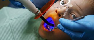
Due to the location of small molars, caries on them can be noticed in the initial stages. Sometimes even patients themselves pay attention to the problem and seek dental help. If a carious lesion is detected at the spot stage, modern clinics carry out treatment without drilling using the ICON system. Careful removal of damaged enamel using an etching gel allows you to save the tooth, and sealing the cavity with a special compound stops further destruction. For deeper lesions, destroyed tissue is removed using a drill or laser. Small defects are restored with durable photopolymer materials. You can choose the shade of the filling that perfectly matches the color of the enamel. In case of significant destruction, leading dentists recommend using ceramic inlays, which are made in a dental laboratory using individual impressions.
The destroyed outer part of the premolar is an indication for the installation of crowns. If only the root remains, a stump tab is installed to securely secure the prosthesis. The choice of crown material depends on the financial capabilities of the patient. The best choice is zirconium dioxide. Such designs are aesthetic and resistant to chewing loads. Their only drawback is their high price. Metal ceramics are available to everyone. It is strong and durable, but the strip of metal under the gum can be very noticeable for some. Ceramics are not suitable for fours and fives. This material is too fragile; with intensive chewing, chips will quickly appear on it.
The absence of premolars in a person is immediately noticeable to others. When they are lost, most turn to dentistry for prosthetics. If you want to quickly restore chewing and restore an attractive smile, installing an inexpensive bridge will help. The design consists of several interconnected crowns made of metal ceramics or zirconium dioxide. The outer crowns are placed on the supporting teeth, and the middle ones cover the defect in the dentition. The main disadvantages of this type of prosthetics are the need to grind healthy teeth and uneven distribution of load on the jaw.
If installing a bridge is not possible, the prosthetist will suggest a removable clasp prosthesis. Typically, the design is used when several teeth are missing. This method saves the budget and has virtually no contraindications. Lightweight butterfly prostheses are used to temporarily replace the defect. They last no more than six months, but this is enough for patients who are awaiting the manufacture of a permanent prosthesis or implantation.
The best method of replacing a lost premolar is implantation. If there are no contraindications and your budget allows, it is better to choose this modern method. A titanium rod is inserted into the jaw bone tissue and becomes a support for an artificial crown. To install such a prosthesis, you do not need to grind down the adjacent teeth. In addition, the load during chewing is distributed physiologically. Externally, an artificial premolar is difficult to distinguish from your own. Even a dentist will not always do this at first glance.
When treating 4- and 5-k, it is important to maintain a balance between practicality and aesthetics. For this reason, this group is the most difficult for dentists.
Treatment of fangs
There are four fangs in the oral cavity. These are 6, 11, 22, 27 teeth according to the universal numbering system. They have a different shape from other teeth, so during restoration it is necessary to have experience working with this group.
| Features of canine treatment | |
| Question | Dentist recommendation |
| What treatment options are there for lost fangs? | In this case, implantation of the lost canine is recommended. |
| How can you correct abnormally growing fangs? | After consultation, the patient is offered options for either orthodontic treatment or the installation of veneers or crowns. |
The fangs do not bear the chewing load. They help you bite off small pieces of food. In the process of evolution, the role of fangs in humans was lost; we do not use them to hold food or tear it into fragments. That is why the treatment of triplets is aimed at restoring their natural appearance.
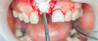
In case of carious lesions, the preparation of fangs is carried out carefully, trying to preserve healthy tissue as much as possible. This will allow for high-quality restoration. With superficial caries, it is possible to preserve the volume of hard tissue necessary for high-quality filling, but deep lesions serve as an indication for installing a crown. To maintain a natural smile, it is recommended to use structures made of ceramics or zirconium dioxide.
If 6, 11, 22, 27 teeth are lost, implantation is recommended. Manufacturers offer titanium prostheses of a special configuration, taking into account the anatomical features of the jaw. Such prostheses are already classic, they have a special thread that allows you to securely fasten the structure in the narrow alveolar process.
A popular dental service is canine shape correction. Some patients have teeth that are too long, pointed, or significantly narrower than the incisors, which makes the smile less attractive. The best way to give the fangs the ideal shape is to use ceramic onlays made from individual impressions, as well as modeling with composite materials.
Why are dental x-rays prescribed?
A 3D photograph of teeth is performed if the following indications exist:
- Injuries of the maxillofacial area.
- Preparation for endodontic treatment (structure of root canals, pathological processes in the periodontium, degree of pulp damage, etc.).
- Diagnosis of neoplasms (cysts, abscesses, granulomas, tumors).
- Anomalies of development and deformation of the maxillofacial apparatus.
- Quality control of filling and implant installation.
- Planning of orthodontic treatment (identification of impacted and dystopic teeth, analysis of the condition of the tissues around each tooth, etc.).
- Detection of hidden periodontal cavities and pockets.
- Implantation planning (assessment of jaw bone parameters, indications for sinus lift or osteoplasty, modeling the result of implantation).
- Endogenous pathologies of the maxillary sinuses.
Three-dimensional x-ray examination is the gold standard when planning any complex dental procedure or surgery. CT allows you to quickly make an accurate diagnosis, competently plan treatment or dental prosthetics, and monitor the results.
Situations in which it is necessary to remove an installed crown
Unfortunately, there may be circumstances in which an existing crown may need to be removed:
- An active inflammatory process under the prosthesis, most often resulting from poor-quality endodontic treatment. To eliminate inflammation, the prosthesis is removed, the tooth canals are re-treated, after which the prosthesis is reinstalled;
- The crown is inconvenient for everyday use and causes serious discomfort to the patient. The reason for this phenomenon is always errors made during the production process of the crown.
Crowns are also removed for repairs and planned replacement, if complications arise after the restoration.
In order for the crown installation process to be successful, and for the structure to serve you flawlessly for many years, while maintaining functional and aesthetic characteristics, the prosthetic procedure should be carried out in a modern dental clinic, with an experienced and competent specialist.
Our Unident clinic in St. Petersburg is equipped with high-tech equipment that allows us to perform dental prosthetics quickly, efficiently and with long-lasting results. In their work, the clinic’s specialists actively use innovative technologies and materials from the world’s best manufacturers, and therefore by contacting us you can have no doubt about the quality and safety of treatment!
Branches of our clinic are located at the metro stations: Akademicheskaya, Vyborgskaya and Pionerskaya and you can choose the branch that will be most convenient for you to get to. Make an appointment with us by phone or through the interactive form on the website and come to Unident for a beautiful smile and healthy teeth!
What equipment is used
To carry out 3D diagnostics, a three-dimensional computed tomograph SOREDEX Scanora 3D with advanced functionality is used. This is the latest generation equipment, which allows you to obtain three-dimensional images of the anatomical structures of the maxillofacial region in a few seconds, with the least radiation exposure for the patient.
The program analyzes the obtained multiplanar sections and builds them into a 3D model, thanks to which the specialist is able to accurately assess the condition of the dental system, detect all pathological processes occurring in this area and competently plan a treatment regimen.
A virtual 3-dimensional model of the scanned area can be recorded on any digital media (CD, flash drive), which allows the attending physician, if necessary, to view diagnostic data or involve related specialists in the analysis of the received information.
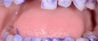
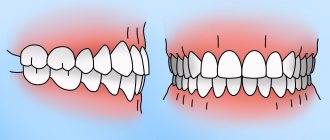
Microdentia
The size of teeth in microdentia is less than anatomical standards. The list of reasons for deviation includes exposure to radiation, premature removal of a baby tooth, narrow jaw, and infectious diseases. There are several types of anomalies:
- isolated - a single violation affecting the lateral incisors;
- relative - the teeth are of normal size, but look smaller due to the enlarged jaw, as a result of which interdental spaces and diastema are formed;
- generalized - the defect covers a group of teeth.
Possible harm
Cone beam dental computed tomography is the safest and fastest diagnostic method. Thanks to the use of a conical X-ray beam, the radiation dose received during the study is 10 times less than when using spiral CT. And the pulsating mode of the X-ray beam further reduces the radiation dose. The three-dimensional computed tomograph SOREDEX Scanora 3D is one of the safest devices in terms of X-ray radiation dose - only 0.035 m3v.
However, despite the safety of the study, CT also has contraindications. If we just talk about dental tomography, it is not performed during pregnancy (in the 1st trimester). 3D dental x-rays with contrast are prohibited for pregnant and lactating women, patients with endocrine disorders (diabetes mellitus, thyroid pathologies), renal failure and intolerance to iodine-containing drugs.
Advantages of the method
- The ability to rotate, enlarge, and examine images in any projection and section, which is impossible with conventional 2-dimensional scanning.
- The examination lasts only a few seconds (8-20 seconds).
- Complete diagnostic information.
- Maximum security.
- Digital information format.
- Detection of any pathological processes at an early stage.
- No prior preparation required.
- 3D reconstruction without distortion or artifacts.
- A wide range of purposes - from endodontic dental treatment and implantation to maxillofacial operations.
Is there an alternative to CT
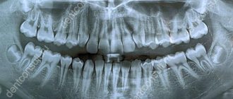
There are many other diagnostic imaging methods (x-ray, orthopantomogram, ultrasound, etc.), but only CT provides the possibility of highly accurate, separate images of all types of tissue at different angles and to different depths. Although a panoramic dental photograph remains an equally important diagnostic tool for a dentist today, it can only provide a general overview. In turn, a 3D tomogram allows you to obtain not a single flat image of the jaw, but a whole series of sequential multiplanar images in different projections and without the distortions inherent in a panoramic image.
Example:
due to the different density of bone structures exposed to X-ray radiation, it is impossible to see less dense bone in a 2-dimensional image; accurate information is provided by a 3- D image of the teeth.
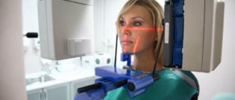
How does the procedure and decoding work?
To take a 3D photograph of teeth, a standing or sitting patient needs to bite a special plate and fix his position in the device using a fixing stand. During the entire scanning time, you must remain absolutely still.
The tomograph sensor makes a series of revolutions around the patient’s head for 8-20 seconds, producing about 200 images in different projections. Processing digital data takes 5-15 minutes, after which the information is written to a disk or flash drive. No preparation is required, you just need to remove all metal jewelry from your neck, ears, and hair before the procedure.
Oral hygiene products
To effectively care for your teeth at home, you need to purchase several items. Toothpaste and a brush alone are not enough to clean the interdental space. If you neglect additional means, pathogenic bacteria and food debris will accumulate there, causing problems (including tooth loss).
Toothbrushes
You should select a toothbrush individually, taking into account several factors. First of all, this is the stiffness of the bristles and the size of the handle. The bristles are of medium hardness and are universal. Soft brushes are recommended for gum disease and bleeding, while hard brushes are recommended for very strong teeth or for dentures. The latter must be used with caution, because improper brushing with it will lead to serious damage to the gums and enamel.
The optimal length of a toothbrush handle is about 30 mm.
Effective dental care begins with choosing the right toothbrush. It is equally important to store it correctly. It is important that the brush dries quickly because moisture promotes the growth of bacteria. In addition, it is recommended to store brushes without a case.
The quality and effectiveness of teeth cleaning depends not only on the stiffness of the bristles, but also on the shape. Dentists do not have a consensus on this indicator. For example, brushes with fibers that form a V-shape clean the V-shape more effectively. Bristles of different heights remove plaque from the cervical area, as they effectively penetrate into the gingival sulcus.
The brush needs to be changed every 3 months.
Toothpastes
Another essential oral care product. Depending on the condition of the teeth, you can choose a therapeutic or prophylactic agent. Textures also vary - from gel-like to creamy. Most toothpastes contain fluoride, but its percentage varies. If you can choose a prophylactic paste yourself, based on reviews and advice from friends, then a therapeutic paste can be chosen only on the recommendations of doctors.
For one brushing of your teeth, it is not necessary to squeeze out a large amount of paste - a pea-sized volume is enough.
You should change your toothpaste every few months to ensure comprehensive oral care. Anti-bleeding agents can be alternated with strengthening and whitening agents. For example, in the morning you can use pastes against caries, and in the evening against inflammation. Before you buy baby toothpaste, study the special lines of famous brands.
Dental floss
Dental floss (or floss) is necessary to completely clean the interdental space. There are several varieties: flat, round, twisted. The threads may be coated with wax.
It is necessary to select a specific floss taking into account individual characteristics. First of all, it is worth considering the distance between the teeth. Without dental floss, comprehensive oral care is impossible; only it can completely remove food debris and prevent the proliferation of pathogenic bacteria.
Tongue cleaners
It is necessary to cleanse not only your teeth and gums of bacteria, but also your tongue. The plaque that forms on it causes bad breath and promotes the development of bacteria. To remove plaque, you can use special toothbrushes, which are equipped with a special rubber ribbed surface on the back. There are also separate tools for this procedure. It is necessary to clean the tongue by making careful movements in the direction from the base to the tip.
Rinse aids
You should use mouthwash at least twice a day, after each brushing of your teeth. It is also useful to apply them after each meal to avoid the growth of bacteria in rotting pieces of food.
Regular use of fluoride rinses for 30 seconds helps to further clean the teeth and mucous membranes. They help remove plaque, bacteria, and waste products from hidden and hard-to-reach areas of the oral cavity. These products contain various useful components, for example, purified essential oils, menthol, which reduce the concentration of microbes, protect the enamel from caries, reduce inflammation and bleeding of the gums, and remove bad breath.