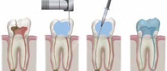
Fissure sealing is an extremely effective method of caries prevention and the main etiotropic method for the prevention of fissure caries. It involves the prevention of caries of the chewing surface of molars by filling fissures and other anatomical recesses of healthy teeth with adhesive materials - sealants in order to create a barrier to external cariogenic factors (microorganisms and carbohydrates)
Sealing tasks:
— eliminating the local risk of caries;
— creating conditions for the death of microorganisms remaining in the deep areas of the fissures; — elimination of potential reservoirs for the accumulation of cariogenic microorganisms; — acceleration of enamel mineralization in the fissure area when using glass ionomer cements.
Currently, glass ionomer cements, compomers, and composites are used for sealing.
Based on their chemical composition, sealants can be divided into groups:
1. Composite sealants: - classic, - sealants-ormokers, - flowable composite materials. 2. Compomers. 3. Glass ionomer cements Composite sealants can be: self-hardening and photo-hardening, for example: Estiseal LC, Fissurit, Helioseal.
Composite sealants and compomers are used to seal the fissures of teeth that have completely erupted.
Glass ionomer cements can be used for prophylactic coating of fissures in teeth at the eruption stage (Fuji VII, Fuji Triage).
Techniques for using sealants: - cleaning teeth with a regular brush, - cleaning with a circular brush with antiseptics, - applying material to fissures, - correction of teeth closure, - insulating the glass ionomer surface with varnish, - monitoring the safety of the sealant every 3-6 months.
Toothpastes for children
Requirements for children's toothpastes: Fluoride content. Daily use of fluoride toothpaste in combination with proper brushing techniques is the basis for caries prevention! The use of low fluoride toothpastes containing less than 500 ppm F in the formula as a preventive measure is considered unsatisfactory. One of the problems associated with the use of fluoride toothpaste in children is the ingestion of the toothpaste by children with the subsequent risk of dental fluorosis.
Parents are advised to monitor the amount of toothpaste and the process of brushing teeth in children under seven years of age. Low abrasiveness. For temporary teeth (baby teeth) and newly erupted permanent teeth, the use of gel pastes is optimal. The RDA value for baby toothpastes should not exceed 50.
The absence of flavoring additives that can make a child want to eat pasta. It is preferable to use neutral, mint or fruit flavors that do not cause rejection in the child.
Examples of toothpastes for children aged 2 to 3 years: - Blend-a-med (0.005% NaF-250 ppmF) - My first Colgate (NaF) - Elmex enfant (aminofluoride - 250) - Lacalut Baby (aminofluoride – 250 ppm F, vitamins A, E) – Lacalut Kids 4-8 (aminofluoride – 500 ppm F, vitamins A, E) – Oral-B Stages fruity ( NaF – 500 ppm F) – Pokemon ( Na2PO3F- 500 ppm F)
Toothpastes for children from 2 to 6 years old: - Blend-a-med Junior Gel - Colgate junior (0.15% NaF- 680 ppm F-) - Colgate junior super star (0.76% Na2PO3F - 1000 ppm million F-) - Lacalut Kids 4-8 (aminofluoride - 500 ppm F-) - Lacalut Teens 8+ (aminofluoride and NaF -1400 ppm F-) - Dental dream for children (0.5% Na2PO3F -660 ppm F-) – Four Fruit – Mildfresh junior (0.76% Na2PO3F – 1000 ppm F) – Sanino Junior – New Pearl Junior 7-12 years (0.76% Na2PO3F – 1000 ppm F, oil tea tree) – Children's pearl complex – Prodent for teenagers
Palatine blind fossae
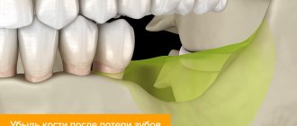
6. Histopographic studies of edentulous jaws have shown that: a) the prosthetic bed is constantly changing due to bone resorption that occurs after the loss of teeth, and continues even after the disappearance of the alveolar process (type III), although the intensity of atrophy in the latter case slows down;
b) the apex of the alveolar process is covered with a mucous membrane with a thick layer of fibrous tissue, poor in blood vessels, and tightly fused with the periosteum. On the slopes of the alveolar process, the layer of mucous membrane is thinner, and closer to its base the amount of loose connective tissue rich in small blood vessels increases. After complete atrophy of the alveolar process, the excess fibrous tissue at its apex disappears and the mucous membrane covering the bone becomes thinner;
c) atrophy of the bone substance of the alveolar process is of the lacunar type, and of the hard palate is of the smooth type of resorption:
d) on the hard palate, starting from the zone of the former first premolars, distally to the soft palate, the thickness of the soft tissues gradually increases due to mucous glands, loose connective tissue and an increase in the vascular network. A small dense vascular network is located laterally between the median suture and the base of the palatine slope of the alveolar process, which allows selective compression to be applied to these areas when taking a functional impression.
7. The palatine blind fossae are located 1-2 mm anterior to the distal edge of the horizontal plate of the palatine bone and cannot serve as an accurate clinical landmark for the line of transition of the soft palate to the hard palate.
8. The relationships between the planes of the hard and soft palate are characterized by three types.
In the first type, the surface of the hard and soft palate is almost in the same horizontal plane (35%). In the second type, the surface of the soft palate in relation to the hard palate forms a pronounced angle (44.5%). In the third type, the soft palate hangs almost vertically in relation to the hard palate (19.5).
9. Compression of the mucous membrane of the prosthetic bed of the edentulous lower jaw in the vertical direction is impossible, but it is possible and advisable in the vestibulo-oral direction at the base of the alveolar process. This will help create a closing valve.
10. The fibers of the mental muscle change their direction due to atrophy of the alveolar process. In the first type, they bend around the transitional fold, in the second, they become horizontal, and with complete atrophy they even rise upward. The nature of the muscle attachment also changes - it becomes aneurotic in nature, which makes the mucous membrane of this area unyielding.
The essence of the system
Black's classification of carious lesions is a system for dividing the disease into certain classes depending on the location of the destroyed area of hard tissue and the coverage of certain elements of the jaw row.
Despite the fact that this scale was developed more than a hundred years ago and does not include secondary and root caries, its use is widespread in modern dental practice.
Dr. Black identified 5 main classes of caries, to which another degree of development of the disease was later added.
The purpose of creating this classification was to select the optimal method of therapy - selection of a suitable material for filling and a method of preparing the affected surface.
The topography of types of cavities according to Black is well demonstrated in the following diagram:
Dr. Greene Vardiman Black
Dr. Greene Vardimar Black is a well-known personality who is at the forefront of the development of dental science in the United States of America. He was born in 1836 in the city of Winchester.
At the age of 17, the young man became interested in medicine and worked for several years as an assistant to dentist D.S. Spira, while simultaneously gaining theoretical knowledge on this topic.
After completing his education, Greene Vardimar Black opened his own dental office in Jacksonville. In addition to providing services to the public, Dr. Black never stopped studying science and improving himself.
In 1870, a specialist invented a mechanical drill equipped with a foot drive. The gold amalgam composition developed by Dr. Black is also used in modern dentistry.
In addition, the specialist brought the terminological base to the standard, and also developed a classification of carious cavities and cutting dental instruments.
Dr. Black compiled several books that described methods for preparing the tooth surface, touched on the features of therapeutic dentistry, and also described some pathologies. In addition, Mr. Black taught dental science at the College of Chicago and also served as dean of the Northwestern University School of Dentistry.
Cavity formation in caries of natural pits
Cavity formation in caries of natural pits
The natural pits on the chewing teeth include the depressions present on the buccal surface of the molars, as well as blind pits formed by the dental tubercles on the palatal surface of the front teeth (see Fig. 6, b, c).
Among the anterior teeth, blind fossae are more common on the small incisors, less common on the central incisors of the upper jaw, and almost never occur on the canines and lower incisors.
If the carious process that arose in the natural fossa on the buccal surface of the molar has not affected the entire fissure up to the chewing surface, then it is possible to create a box-shaped cavity without bringing it to the chewing surface. It is necessary that the cavity, as in all cases, be located not only in the enamel layer, but must penetrate into the dentin. During work, you should remember the proximity of the pulp chamber.
The carious process on the buccal surface of the molars creates a symmetrical round or diamond-shaped destruction of the tooth tissue. When forming a cavity for the tab, it is necessary to make it asymmetrical in order to facilitate the correct insertion of the tab. To do this, it is best to give it the shape of a trapezoid with rounded corners (Fig. 18, a). In some cases, it is enough to additionally include a nearby fissure in the cavity, which will serve as a guide when introducing the insert into the cavity (Fig. 18, b).
Formation ends with the creation of a fold along the entire edge of the cavity, which is best done with a small carborundum head.
The direction of wax removal should be perpendicular to the buccal surface of the tooth. Consequently, all work must be done with a fissure bur in a contra-angle handpiece and the direction of this bur must be perpendicular to the buccal surface of the tooth.
In cases where caries does not spread towards the pulp chamber, you can use a bur to follow the path of development of the carious process, excising
softened dentin. In this case, the drill can be given a direction slightly deviating from perpendicular, then all the walls of the cavity should be created at the same angle. The wax model should be drawn out in the same direction, and then the finished inlay should be inserted.
When the carious process spreads along the buccal fissure of a molar, a cavity is formed depending on the strength of the enamel roof hanging over it. If the roof remaining on the chewing surface above the cavity is thin, then in order to avoid breakages under the force of chewing pressure, it may be recommended to bring the cavity to the chewing surface.
Often the fissures of the chewing surface of the molars and the fossa on the buccal surface are simultaneously affected by the carious process. In this case, it is necessary to connect all the affected areas of the tooth into one common cavity. The direction of wax removal should be the same as for fissure caries, i.e., parallel to the long axis of the tooth (Fig. 18, c).
In this case, the contra-angle handpiece with the fissure bur should be held strictly parallel to the long axis of the tooth. Having created a cavity on the chewing surface, as described above, they move to the buccal surface of the tooth. The working part of the bur in this case will be the side surface.
While sparing the tooth tissue, you need to deviate from the strictly vertical direction of the bur, giving it a slightly inclined position, parallel to the groove on the buccal surface of the tooth (Fig. 18, d).
A fold is created along all edges of the cavity on both the chewing and buccal surfaces of the tooth.
When forming a cavity on the palatal surface of the upper anterior teeth, care must be taken, since in this area the pulp is located close to the surface of the tooth. Nevertheless, the rule of cavity formation not only in the enamel layer, but also in dentin, remains in force.
By excising tooth tissues affected by caries, they simultaneously create an asymmetrical cavity shape, for which they use natural depressions that are often present on the palatal surface of the incisors. These depressions can serve as retention areas where secondary caries may occur.
In its finished form, such a carious cavity on the palatal surface of the incisors most often represents a rounded cavity with one or two spurs (Fig. 18e).
The direction of wax removal should always be perpendicular to the palatal surface of the incisors. It is best to form a cavity using a fissure bur No. 1-3 in an angled tip directed perpendicular to the palatal surface of the tooth.
When, simultaneously with caries of the blind fossa on the palatal surface, there are carious cavities on the proximal surface of the incisor, it is advisable to combine both cavities into one common one or connect them together with a bridge.
The fold is made only on those edges of the cavity or bridge that extend onto the palatal surface of the tooth.
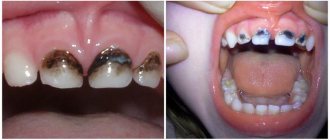
What factors provoke the destruction of tooth enamel?
The integrity of tooth enamel also depends on the quality of hygiene procedures when using the “correct” brush. If food debris and soft plaque are not removed from the surface of the teeth in a timely manner, acid-forming pathogenic bacteria begin to actively multiply in them. In this case, the acidity of the environment increases sharply, and under the influence of acid, demineralization of the enamel coating occurs. The loss of calcium leads to the destruction of enamel and the looser and softer dentin located underneath.
It has been noted that a wedge-shaped defect is often formed:
- abuse of carbonated drinks and sour juices;
- in patients with malocclusion - due to non-physiological movement of the jaws chewing food and increased chewing load on individual teeth;
- when correcting the bite with braces;
- in patients with periodontal diseases, accompanied by subsidence of inflamed gums and, accordingly, exposure of the necks of the teeth.
Even properly selected treatment helps to avoid undesirable consequences only if the problem is identified at an early stage. Therefore, patients should promptly seek dental care when the very first signs of pathology are detected.