A panoramic photograph of the teeth (orthopantomogram or OTP) is an x-ray of the upper and lower jaw. Allows you to examine most dental problems in detail, conduct high-quality diagnostics and prescribe treatment.
A panoramic dental photograph shows not only the teeth, but also the jaw joints, paranasal sinuses and facial bones. All this makes it indispensable for orthodontic, surgical and reconstructive treatment. You can obtain OPTG using an orthopantomograph.
Orthopantomogram: description of the procedure
An orthopantomogram, or OPTG, is an overview of a circular photograph of the teeth, in which the entire jaw is visible, unfolded in a plane. In addition to the teeth, their roots, canals, the presence of fillings and crowns, the panoramic image shows the condition of the periodontal tissues, maxillary sinuses, and sinuses.
A panoramic photograph of the teeth, or orthopantomogram, allows you to detect hidden carious cavities, inflammation, cysts, abscesses, the presence of periodontal pockets, identify the exact condition of the roots and periodontal tissues, and assess the condition of the maxillary sinus and temporomandibular joint. It is also necessary to take an orthopantomogram when planning orthodontic treatment with braces or aligners.
In the process of preparing for dental implantation, using an orthopantomogram, a specialist can determine whether there is enough bone tissue for implantation or a sinus lift is needed, how many implants will be needed and what size they should be, and also predict the dynamics of their engraftment.
It is very important to take a panoramic photo to determine the position and condition of your wisdom teeth. Only an orthopantomogram can give an adequate idea of the “eights”; no targeted photograph will help here. Therefore, competent dentists decide whether to treat or remove wisdom teeth only after x-raying the entire jaw.
Orthopantomogram or panoramic photograph...
In the daily practice of a dentist of any specialty, for the most correct and complete diagnosis, there is often a need to obtain a panoramic X-ray of the patient’s oral cavity (survey X-ray, orthopantomogram). What kind of research is this, why do it, and what can be seen in such a picture? The answers to these and other questions can be found below in the article, which its author, Moscow colleague Stanislav Vasiliev, kindly shared with me.
What is an orthopantomogram and why is it needed?
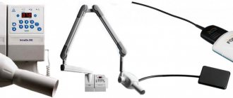
An orthopantomogram (or “OPTG”, “panoramic image of the dental system”) is one of the types of diagnostic radiography. In dentistry, OPTG is of key importance - many types of treatment cannot be started without this diagnostic method. Technically, it is carried out as follows: the beam source (X-ray tube) and its receiver (film or digital sensor" move around the object under study in opposite directions. As a result, a very limited part of the object of study is in focus, everything else is blurred. Depending depending on the settings, we can get clear only a specific layer that interests us, and everything else in the image will be blurred. Panoramic images are taken using orthopantomographs. There are different orthopantomographs - film and digital. Film OPTG are almost history, while “digital” takes more and more place in modern dentistry. A modern orthopantomograph looks something like this:
This photo shows a Planmeca dental tomograph with a cephalostat. The latter is needed for teleradiography, a study that is widely used in orthodontics and maxillofacial surgery.
There is a widespread belief that this type of research is harmful. In fact, the volume of radiation even from a film orthopantomograph is such that it is possible to take panoramic photographs every day for a month without significant harm to health. And the radiation from digital devices is several times less than that of film devices, and the radiation dose received is much less than what you receive, for example, during a two-hour flight.
When is an orthopantomogram needed?
In principle, it is always needed. In dental treatment, prosthetics, orthodontic treatment, in surgery and implantology, even in rhinology when examining the paranasal sinuses, the value of panoramic images cannot be overestimated. However, in some cases it is impossible to navigate only by OPTG - nevertheless, we are transferring a three-dimensional image to a plane, and therefore distortions are possible. But orthopantomography should be considered as a primary x-ray examination, based on the results of which the tactics of both further, more in-depth diagnosis and treatment are built.
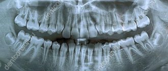
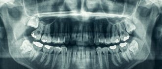
Orthopantomography of an adult.
For example, here's a snapshot:
Pay attention to the shape of the dentition. It turns out that the person in the picture seems to be smiling))). If we don’t see a smile (on the contrary, a “sad form”), then the picture was taken incorrectly and the amount of distortion in it will be very high. Such an image is not suitable for a good diagnosis.
They look at an orthopantomogram not as if in a mirror, but as if they were looking at another person. That is, the left side is on the right, and the right side is on the left. Sometimes, for convenience, the letter L (left) is placed on the left side or the letter R (right) on the right side. Or they sign the picture so that the patient’s data can only be read on one side.
An adult normally has 32 teeth. But you have repeatedly seen on my blog that we are discussing, for example, the treatment of the 44th tooth or the removal of the 48th tooth... How does this happen?
The fact is that each tooth, in addition to its name, has a number. According to the WHO classification accepted throughout the world, the dentition is divided into four segments. The top right segment is tens. Upper left - twenties, Lower left and lower right, respectively - thirty and forty. The numbering comes from the center of the dentition. It turns out that the right upper central incisor is the 11th tooth, the tooth following it (the right upper lateral incisor) is 12, the lower left second premolar is 35, and the lower right wisdom tooth is 48 teeth.
Although my friend, Valera Kolpakov, who now lives and works in the USA, said that they have a different classification: teeth are simply designated by a clockwise serial number. In our country such designations are very rare. Europe is still...
In the picture, yellow numbers indicate the numbers of an adult’s teeth. Now you can surprise your dentist by telling him that your pain is not just some lower molar, but specifically the 36th tooth. He will understand everything and look at you with such respect...
Some doctors name teeth by segment number. For example, “four, two,” lower left lateral incisor. This account is being vigorously imposed in institutes and universities, mainly by teachers of pre-retirement age. This classification is fundamentally incorrect. Why? Because initially this teeth counting system was intended for machine processing, and the machine works with the number “forty two” and not with the number “four two.” Therefore, you need to name the teeth correctly.
Most dental materials are made radiopaque. This is done so that the volume and location of this material on x-rays can be controlled. For example, filling materials for root canals are indicated by the letter A in the picture.
When an orthopantomogram of the dental system reaches a nearby ENT doctor, the latter immediately notices the penetration of the roots of the teeth into the maxillary sinus (letter B in the picture). Moreover, old dental textbooks mention this. In fact, the roots of teeth very rarely penetrate the maxillary sinus. Most often, they go around the edges of it so that the bottom of the sinus is between the roots of the teeth.
Orthopantomography gives a good idea of the location of wisdom teeth (letter C in the picture). Even targeted photographs do not provide a complete picture of the structure and localization of figure eights. Therefore, without performing an OPTG, I highly do not recommend undertaking the removal or treatment of “wise” teeth
In addition to teeth, OPTG also shows other structures that may be of interest to the dentist. For example, the mandibular canal, which runs through the thickness of the lower jaw and contains a neurovascular bundle, is highlighted in green. The latter innervates the teeth, the corresponding half of the lip and chin. This bundle exits through the mental foramen (indicated in dark green), which some “specialists” sometimes confuse with a cyst.
The borders of the maxillary sinuses and nasal passages are indicated in red. The image shows a curvature of the nasal septum and, as a consequence, asymmetry of the nasal passages. This is an indirect sign of chronic rhinosinusitis, including allergic nature.
The temporomandibular joints are indicated in blue. Normally, they should be symmetrical and have a certain shape. A noticeable difference in the shape of the joints, as well as their asymmetry, is one of the signs of chronic arthritis. In this image, the joints are almost symmetrical, therefore, the patient has no problems with the joints.
Radiopaque materials are highlighted in purple. The pictures clearly show all the fillings, the quality of the canal fillings, etc. The thin purple stripe on the front teeth are retainers installed after orthodontic treatment.
There are also structures on the OPTG that are not very interesting, but are still visible:
For example, the earlobes are highlighted in red, and the spinal column is highlighted with a blue dotted line against the background of the lower jaw. On either side of it is the hyoid bone. It comes into focus only in a lateral projection, and therefore is visible as two separate parts.
The horizontal line above the upper jaw is the hard palate, and on the sides of it the zygomatic bones are clearly visible. In the contour of the earlobes, one can distinguish the styloid process, the mastoid process and the opening of the external auditory canal. Thus, a panoramic image, although distorted, can give an almost complete picture of the state of the dental system - due to this, it is simply necessary in the high-quality diagnosis of dental diseases.
Orthopantomogram of a child.
Oddly enough, children have more teeth:
But the principle of their numbering is the same. Unless baby teeth are designated not by tens or twenties, but by fifties, sixties, seventies or eighties - as indicated in the picture. For example, tooth 75 is the left lower primary molar.
Under the milk teeth are the rudiments of permanent teeth (indicated by yellow numbers). At different ages they look different and have different shapes. In this image, for example, you may not see the crowns of the forming teeth 15, 25, but you can see the contours of the rudiment - therefore, there will be teeth.
The blue color in the picture indicates the mandibular canal.
Blue color - temporomandibular joints. Note how the relative position of the joint head, articular tubercle and socket differs from the structure of the adult joint.
The purple line is the contours of the maxillary sinus. In children it is relatively less than in adults. Therefore the voice is quieter and higher.
Green color - nasal septum and nasal turbinates. While relative symmetry is observed, however, due to injury, chronic rhinitis, allergies, or simply due to uneven growth, a curvature of the nasal septum is possible. As I already wrote, this is a risk factor for the development of chronic rhinosinusitis (sinusitis).
Red arrows and the letter A indicate artifacts from earrings. In some cases, jewelry in the form of piercings, chains, etc. can introduce significant distortions and complicate diagnosis. Therefore, before performing orthopantomography, it is better to remove all jewelry from the face and neck.
Of course, experts see much more ordinary people in the photographs. And radiologists are much more than ordinary specialists. It would be great if any radiologist would comment on this post.
In general terms, the diagnostic picture from panoramic images looks exactly like this. Now you can take your panoramic photo, look at it carefully and no longer say: “I don’t understand anything here.” After all, it’s better to see once than to hear a hundred times, right?
Orthopantomogram on film or digital: which one to choose?
Today, electronic devices have almost replaced film devices. Their cost is higher, but they do not require a large amount of consumables, as for film orthopantomographs. To develop film, you need a darkroom, which is prohibited by law from being placed in residential buildings, which can become a problem for small private clinics. In addition, the irradiation exposure of film devices is several times higher than that of electronic devices. An orthopantomogram of the jaws, made on a digital device, is immediately transferred to the doctor’s computer, who can be located anywhere. It is much easier to store such an image: it will simply remain in the patient’s electronic record and can be viewed at any time. If desired or necessary, you will receive the result of the orthopantomogram on disk or printed on transparent film.
Unlike a panoramic photograph on film, a digital orthopantomogram will not be lost, which means that the patient will not have to undergo radiation again, which is present during dental x-rays, albeit in very small doses: only 0.002 - 0.003x mSv, whereas, according to Sanitary Regulations , a person can receive up to 1 mSv per year for research purposes, and from the environment we receive 2 - 3 mSv over the same period. In addition, the image clarity and image resolution on digital cameras is much higher than on film cameras.
How is an orthopantomogram done?
No special preparation is required for the study. You will need to remove glasses, earrings, hair clips, other metal objects, and dentures. An orthopantomogram is usually done in a standing position. A special X-ray protective apron-cape is put on the chest. The position of the head is fixed with special clamps. As directed by the doctor, you will need to bite a special plastic mark (which is located in a disposable cover) - this will allow the image to be correctly centered. The tongue should be kept pressed to the palate. During an orthopantomogram, the module will move around the head, this will take 15-20 seconds, and during this time you must stand strictly still in order for the image to be of the proper quality.
You can get an orthopantomogram in Moscow at Family Doctor JSC.
Sign up for diagnostics Do not self-medicate. Contact our specialists who will correctly diagnose and prescribe treatment.
Orthopantomogram 3D
A three-dimensional panoramic x-ray of the jaw can be performed with a computed tomograph. He takes many sequential photographs in different projections and then completes the image in a computer program. The result is a three-dimensional image of the jaw without distortions, which are inevitably present on a conventional otopantomogram, since it conveys a curved object in a flat form. Only with the help of computed tomography does it become possible to look at an object from a different angle or in a different projection without taking new pictures. Also, this method of examination allows the doctor, in the absence of the patient, to see an image of his jaw and examine it from any side and even at any depth.
Contraindications
Like any medical procedure, an orthopantomograph examination has some contraindications. If a person, due to the nature of his work or due to undergoing radiation therapy for oncology, regularly receives small portions of radiation, then the possibility of research is considered on an individual basis. Compared to other devices, dental equipment is practically harmless.
If you only need to take one photo, then there is no point in worrying. It is important to understand that without it, it is very difficult to carry out full treatment, especially for complex pathologies. Lack of proper preparation can lead to the development of complications and medical errors. This is especially important before installing artificial teeth, which should serve their owner for many years.
Some doctors warn that it is not recommended to conduct such an examination during pregnancy and breastfeeding. However, as you know, the presence of an infection in the mouth, even due to a small carious cavity, can cause much more harm to the body of the mother and baby than its treatment. It is impossible to remove the affected areas without taking a picture; acting blindly can cause even more harm.
Where can I take a panoramic photograph of my teeth or an orthopantomogram?
Today, an orthopantomogram is included in the list of services in almost every dentistry. You can use it in the same place where you decided to undergo treatment, or you can take a panoramic photo at a better price in another place. Some clinics offer regular clients a panoramic dental photo at a significant discount, which means it can be quite inexpensive. Prices for an orthopantomogram in Moscow start from 750 rubles and reach 2500 rubles. On average, dentistry in Moscow offers a high-quality panoramic photograph of teeth for 1000 - 1300 rubles.
Painless procedure
The procedure itself is absolutely painless and quick. Unlike MRI, where a person is practically in a confined space, there is nothing to be afraid of. During one procedure, a person receives a minimum dose of radiation, which is incomparable to what can be obtained with radiation treatment (no more than 0.11 m3v). Fortunately, modern equipment allows us to reduce harm to minimal values, namely up to 0.04 m3v.
To better understand, you can compare a panoramic shot with a flight from America to Europe or vice versa. For 8-9 hours in the air, a person is exposed to radiation in the amount of more than 0.05 m3v, that is, a trip across the ocean and back can be easily compared with more than two studies on an orthopantomograph. The most dangerous device used by doctors is a CT scanner. The dose will be almost 30-40 times higher, so conscientious specialists do not prescribe a CT scan unless absolutely necessary.
If the patient needs to take several pictures in a year, then there is no need to worry either. Ultimately, the benefits of treatment will be much more noticeable than the harm from the device used. After completion of the procedure, there is no discomfort, discomfort, dizziness or pain. Immediately after leaving the clinic, you can return to the normal rhythm of life, drive a car, communicate with colleagues. If for any reason you feel unwell, it is best to immediately seek advice from your dentist. Only he will be able to understand the cause of what is happening and prescribe preventative therapy or medication.
Rehabilitation after panoramic dental imaging (optg)
Unfortunately, many patients are afraid to have x-rays and panoramic photographs because they simply do not have enough information about these procedures. Because of this misconception, fear and mistrust arise, but it's not that bad at all. Some sources claim that photographs are allowed to be taken only once a year, otherwise you can harm your body. This is also not true! Lack of timely treatment or doctor's work without prior examination can be more dangerous.
As with an X-ray, there are no special recommendations after an OPTG; you do not need to make changes to your schedule, go on a diet, or stop exercising. Just a couple of minutes after you leave the room with the device, you can forget about visiting it. It is extremely rare for people to experience pain and discomfort, but this is associated more with the fear of the dentists themselves than with the influence of the equipment.
There are no consequences from radiation either. Doctors give standard recommendations regarding recovery: you need to drink more warm liquids, eat soups, and it is best to avoid eating spicy foods, coffee, strong tea and alcohol. If mild stress occurs, you can take a prescribed sedative.
How is the procedure done?
The procedure is carried out using a special device - an orthopantomograph. The patient is placed in the center of the device and allowed to bite on a special plate. Next, the moving parts of the device rotate around the patient’s head, which takes up to 20 seconds. Designs come in digital and film types; on film orthopantomographs the image is more blurry. In our clinics we use more modern digital equipment, which guarantees a clear result of the final images.
How safe is it to take panoramic X-rays of the jaw? If we resort to calculations, then the maximum permissible radiation dose for a person is a dose not exceeding 1000 μSv per year. Taking into account the radiation from a digital device during the procedure, it is better to take a picture no more than once a month.
Before and after panoramic dental x-ray (optg)
A panoramic photograph allows you to study the structure of all tissues of the oral cavity with maximum accuracy; the roots, canals, joints and other details are clearly visible in the photograph. When visiting a professional medical institution, sheets with explanations are immediately issued for the images. In the text you can familiarize yourself with the details of the study; the ongoing pathological processes and deviations from normal indicators when they are detected will certainly be indicated.
Even an ordinary person can draw certain conclusions when viewing an image, because everything is quite simple. In the picture, wisdom teeth are clearly visible; they can grow in the right direction or at an angle in relation to adjacent units. Also, the number of previously filled canals and composite fillings will definitely catch your eye; quite often inflammation in the pulp chamber, changes in the root apex, etc. are clearly visible.
Without much experience, it will be more difficult to assess changes such as a decrease in bone volume or the degree of caries damage to the tooth. Bone deficiency can occur due to advanced periodontitis and other serious dental problems. Thus, the image provides comprehensive information about all the nuances of the location and structure of individual parts of the oral cavity, which, of course, is very helpful in drawing up a treatment plan.