When is an X-ray prescribed for a child?
If X-rays in adolescents and adults do not cause concern, then X-ray diagnostics of young children worries many parents.
The devices used in modern medicine do not harm the body, but the study is still carried out only if indicated. Dental X-rays are recommended for children for the following indications.
- Deep caries. The procedure is carried out to determine the lesions and affected area. The image allows the specialist to assess whether the roots and rudiments of permanent teeth are damaged.
- Developmental anomalies. X-rays make it possible to determine the causes and characteristics of irregular teething, as well as delays in their growth.
- Control of permanent teeth. Diagnostics allows you to see all the features of the rudiments of permanent teeth, as well as predict the period of their eruption and identify possible pathologies.
- Malocclusion. X-ray is the most accurate diagnostic method for determining abnormalities in tooth growth and jaw position.
- Abscess. The study is aimed at confirming or refuting the development of an abscess if signs are present.
- Jaw injuries. In case of mechanical damage, an x-ray is taken to assess the damage to bone tissue that has occurred.
X-rays are often used to monitor the treatment being carried out. Diagnostics is also used when restoring teeth using temporary prosthetics or installing orthodontic structures.
How is a dental x-ray performed?
An X-ray of a child’s jaw with baby teeth is a painless procedure that takes 3 to 5 minutes.
The course of dental radiography in children consists of:
- the pediatric dentist gives a referral for examination;
- the child, with or without a parent, is sent to a special room;
- a special apron with lead plates is put on him;
- clamps a plastic plate with his teeth;
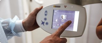
- the specialist fixes the position of the head and leaves the room;
- The device turns on and takes a photo.
If there is a need for a targeted X-ray, the child is seated on a chair, a film is placed in the mouth, and an X-ray tube is placed near the mouth and held for several seconds. During the procedure, the child must sit still, otherwise the images may be distorted.
The results are delivered in 20–30 minutes. With the received images, the patient is sent to the attending physician. You can get an X-ray of a child in Moscow at the Novodent dental clinic. There is advanced and modern equipment with minimal radiation doses, but high information content. There are doctors who will find an approach to every little patient, inspire trust, and listen to their recommendations. More detailed information about the clinic’s capabilities can be found on the website or by phone.
Types of X-rays for examining children
Examinations of the teeth and jaw may require different areas to be examined or results obtained from different types of images. Depending on the indications for x-rays, a certain type of procedure is used.
Types of x-rays of a child's teeth and jaw.
- Sight shot. Obtaining an image that displays a separate area (one or two teeth with closely spaced soft tissues).
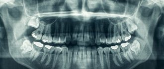
- Panoramic shot. An image showing a large area of the dentition along with the jaw bone and the rudiments of molars.
- 3D pictures. A modern method of obtaining three-dimensional images of the upper and lower jaw.
There are also differences in technique. There are internal or external diagnostic methods. If it is necessary to obtain a targeted image, internal x-rays are used; in other cases, the external method is used.
Popular types of x-rays of baby teeth in children
Modern pediatric dentistry allows us to solve any problems with baby teeth. But in order to consider the disease and make a correct diagnosis, it is not enough just the child’s complaints and his description of pain and discomfort. In order for treatment to be as effective as possible, it is necessary to build a treatment plan based on diagnostic data that can be obtained after one of four popular types of radiography:
- Sighting (periapical). Indicated in cases where clarification is necessary on one or more adjacent teeth (from 1 to 3). Provides data about the units themselves, their roots and surrounding soft tissues.
- Occlusal (palatal). As a result, the attending physician receives a complete projection of one of the child’s jaws. The technique is used to determine possible malocclusions and anomalies of the dentition in general.
- Panoramic (orthopantomogram). Allows scanning of the entire oral cavity. An orthopantomogram for children is indicated for a detailed study of pathologies of the upper and lower jaw, the temporomandibular joint and the maxillary sinuses. The results of such an x-ray are necessary for further dental correction. This type of examination is prescribed for children with a fully formed dental system, that is, from about 9 years old.
- 3D pictures. A three-dimensional X-ray image of children's primary teeth allows us to obtain data on all units (existing and not yet erupted). The entire maxillofacial area is subject to examination. The photographs can be used to evaluate the structure of the entire skeletal system of the jaws, the condition of the root system (including pathologies), inflammation in soft tissues, etc. Such dental x-rays are prescribed for the purpose of making a diagnosis and as a control study after canal filling.
Depending on the existing problems with baby teeth, the child may be prescribed one specific or several different types of examination. The attending physician will definitely explain the advisability of obtaining images and the need for x-rays.
You might be interested in:
Dental diagnostics
Pediatric orthopantomogram
3D image of teeth
What can a dental x-ray show?
Carrying out X-rays of a child’s teeth or jaw is aimed at identifying pathologies and determining the anatomical features of the development of the dental system. Dental x-rays are performed before treatment, during treatment or upon completion for control.
X-rays may show:
- decrease in tooth weight due to inflammation of soft tissues;
- foci of carious lesions in any area;
- changes occurring in dental roots;
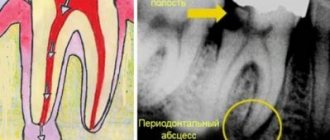
- purulent processes flowing into an abscess;
- rudiments of permanent teeth;
- anomalies in the structure and development of teeth and jaw;
- impacted teeth;
- supernumerary teeth;
- damage to the jaw bones;
- and other.
The result of the image is assessed exclusively by a specialist. Based on the information received, the doctor can determine the diagnosis, prescribe or adjust treatment, or evaluate the effectiveness of the measures taken.
When is a child's dental x-ray prescribed?
It is important for parents to understand in what cases it is necessary to take an x-ray of children’s baby teeth. Detailed diagnostics may be required in a variety of cases (depending on the individual characteristics of the structure and development of the dental system). Most often, radiographic examinations are prescribed in the following cases:
- To identify deep caries. Milk teeth, unlike permanent teeth, have more fragile enamel. Consequently, they are more susceptible to exposure to aggressive substances that enter the mouth with food. Carbonated drinks, fast food, sweets - all this causes the appearance of caries, which is not always noticeable upon visual inspection. At routine visits to the dentist, with x-rays, you can verify the absence or confirm the presence of deep caries.
- To study abnormalities in the development of teeth and jaws. If a child has problems with teething, abnormal growth, or pain when closing the jaws, this may be a reason to take an X-ray of the children’s baby teeth. The results will allow us to study the existing problems in detail and intervene in a timely manner, solving issues of delayed growth of units and many others.
- To monitor the condition of permanent teeth. Not only baby teeth, but also permanent children’s teeth require special attention even before they appear. Studying the rudiments allows not only to determine possible anomalies, but also to make predictions about the timing of eruption.
- Bite study. An incorrect bite can cause premature tooth wear and the appearance of various pathologies. An X-ray of a child’s teeth can provide detailed data on the position of the jaws in order to identify even the slightest deviations from the norm.
- Detection of abscesses. Inflammatory processes in the initial stages may be asymptomatic. An advanced abscess can cause serious operations, including surgical ones. At the same time, x-rays can show foci of inflammation and begin treatment on time.
Some children are so mobile and active that they often get injured, including in the jaw. Serious injuries may be visible during visual inspection, and some cracks and misalignments can only be diagnosed after X-raying the child's teeth.
Are there any risks?
Modern X-ray machines are characterized by a low dose of radiation, but since exposure to X-rays cannot be completely ruled out, it is recommended that X-rays be taken no more than 2 times a year for children with baby teeth and no more than once every 1.5 years for adolescents. The need for diagnosis is assessed by the attending physician. If the risk from x-rays is lower than the risk of complications from dental pathology, then the specialist may prescribe a study with a more frequent frequency.
There are no risks of side effects after radiography if all the rules of the procedure are followed. There are no contraindications to the study. The exception is the patient's serious condition or previous irradiation.
For children under the age of one year, an X-ray of the teeth/jaw is taken if other diagnostic methods have turned out to be uninformative or there are contraindications to their use. Thus, children are examined mainly only in case of mechanical injuries.
Indications for x-rays of primary teeth
X-rays for children are carried out as prescribed by a doctor, at least 2 times a year. The image allows the doctor to recognize the following dental disorders and diseases:
- condition of dental roots and soft tissues, including gums;
- determine the rudiments of the roots of permanent teeth;
- inflammatory processes;
- deep caries;
- nerve damage;
- pathological changes in tooth roots;
- purulent processes;
- anomalies in the structure of the teeth and jaw;
- impacted and supernumerary teeth;
- malocclusion;
- damage to the jaw bones.
Indications for dental X-rays in children may include other conditions - diseases of the teeth, jaw apparatus, but in any case, the image should be taken on the recommendation of a specialist who needs a complete clinical picture.