Diagnostics plays an important role in any field of medicine, and dentistry is no exception. It is very important for the doctor to see the full picture of the patient’s condition in order to make an accurate diagnosis and select the appropriate treatment. In dentistry, diagnosing something is not so easy, which is explained by the difficulty of accessing some parts of the oral cavity. In some cases, a 3D image of the teeth helps clarify the picture. What is it and how is it made?
Methods for diagnosing dental condition
Most methods for diagnosing the condition of the teeth and oral cavity fall into the category of visualization, that is, they can clearly show everything that happens in this part of the body. At a minimum, it is now completely impossible to imagine dentistry without radiography. Now this type of examination is included in the standard set of procedures that a patient undergoes when visiting the clinic with any complaint.
On a note! An examination is necessary not only when diagnosing the condition of a specific tooth, but also if the patient is faced with problems with the functioning of the entire maxillofacial apparatus.
Table. Main types of diagnostics in dentistry.
| View | Description |
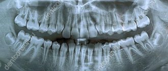
| This is the most common diagnostic option, which can be carried out in any, even the most modest clinic. It provides the opportunity to correctly and accurately assess the condition of the canals in the roots of the tooth and identify developing pathologies in the area of hard tissues. Often used for endotonia. An X-ray image is a small black and white image on a special film or on a CD. | |
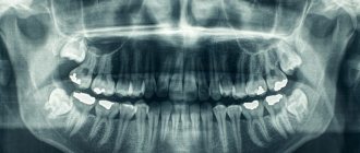
| This is otherwise called a panoramic image of the dental system. Using it, the doctor will be able to identify pathologies in the development of the jaw in general and teeth in particular, detect all carious cavities, understand how much hard tissue is destroyed during periodontal disease, clarify whether endotonic treatment helped the patient, etc. The image will also allow you to see whether there are any or deviations or changes in the condition of the lower sections of the maxillary sinuses. An orthopantomogram is also done in relation to the temporomandibular joints. It is often prescribed before prosthetics. | |
| This is a picture of not just the jaw, but the entire skull in one projection or another. It is necessary for measuring parts of the facial part (cephalometry). Based on this image, you can plan treatment with an orthodontist. | |
| It is this picture that is called 3D, which is discussed in the article. We'll talk about it separately below. |
Panoramic shot or 3D x-ray of the jaw: which is better?
Modern adult and pediatric dentistry is impossible without accurate diagnostics. During a visual examination, the doctor can only obtain about 50-60% of all information, because information about the roots of the teeth and the condition of the jaw is simply not available to him. Naturally, to obtain the necessary data, an X-ray examination of the teeth is prescribed, the results of which allow one to obtain a complete picture of the condition of the patient’s dentition. X-ray examination of the jaws allows the dentist to consider all the nuances. Until recently, the most accurate type of diagnosis was considered to be a panoramic photograph of the teeth, which gives a general understanding of the condition of the teeth, their roots and the bones of both jaws. Using a panoramic image, you can accurately diagnose most diseases of the teeth and periodontal tissues.
But it’s not for nothing that dentistry is called one of the most dynamically developing areas of medicine - most advanced technologies are used both in diagnosing and in treating dental diseases.
An innovative solution was the use of computed tomography when examining patients. Scanning the patient’s oral cavity using a tomograph, in contrast to a panoramic X-ray, makes it possible to obtain information in the smallest detail. Compared to a “flat” panoramic image, a 3D X-ray of the jaw allows you to obtain a three-dimensional model of the area under study, while it is possible to “examine” each layer separately. Computer diagnostics gives a clear picture of bone density, the presence of hidden inflammatory processes, neoplasms, etc.
The table below shows comparative characteristics of the capabilities of different types of diagnostics:
| Panoramic shot | 3d x-ray |
| General picture of the condition of the jaws and periodontal tissues | Accurate image of all tissues of the area under study, the ability to obtain a lateral projection, integration with a facial scanner |
| Static two-dimensional (flat) image | 3D volumetric imaging |
| Low degree of detail - you can distinguish between hard and soft tissues, inflammation and neoplasms are visible | High degree of detail - the smallest structural changes are visible in both hard and soft tissues |
| More powerful radiation during the photo | Minimal radiation exposure during diagnostics (2 times less than with panoramic) |
| There are restrictions on diagnosing diseases | High accuracy of diagnosis of any pathological changes |
What is a 3D photo?
A 3D image or computed tomography of teeth is an x-ray that is performed using a special device - a tomograph (CBCT). The picture is a three-dimensional image that demonstrates the entire dental system in detail. The resolution of the image is high, which means that even the smallest details can be seen in it. The doctor, using a computer program, will be able to thoroughly examine absolutely any part of the patient’s jaw, moreover, at any depth and at any angle. The snapshot is provided on disk, where it is stored. If you need to view the disc, you can insert it into your computer and see everything you need on the monitor. Moreover, sometimes this diagnostic option saves the patient from a number of unpleasant manipulations on the part of the doctor.
On a note! The scanning volumes of such an image vary from 5x5 to 13x15 cm.
An orthodontist, implantologist, maxillofacial surgeon and other doctors can give a referral for such a plan. The technique is very accurate, and often without it, doctors simply will not do most of the manipulations. And the effectiveness of treatment thanks to this image increases significantly.
The advantages of taking a 3D photo are as follows:
- high image accuracy and detail;
- the ability to view all parts of the mouth - the image is three-dimensional;
- errors are excluded when identifying the location of pathology development, since there are no distortions in the image;
- the ability to assess the condition of the jaw from any side;
- the picture is taken quickly - a few seconds are enough to get a detailed image;
- taking a picture is safe for health, since the radiation exposure experienced by the patient’s body is extremely small. Especially if you compare its level with the level with the same x-ray.
3D image of teeth in dentistry
Diagnosis in dentistry is often complicated by the fact that the doctor is not able to see the oral cavity or an individual tooth in the most detailed angles. Therefore, to obtain an accurate picture of the condition of the dentition, hardware examination methods are used, which allow you to see what is inaccessible with a normal visual examination.
Technologies are constantly improving, and a 3D image of the teeth has been added to the traditional X-ray and orthopantomogram. As a rule, it is computed tomography that is prescribed by most plastic and maxillofacial surgeons, orthodontists and implantologists.
This is a modern diagnostic method that allows a specialist to see a detailed image of the jaw in different projections and from any angle. The examination in 3D format visualizes the oral cavity for a thorough examination of the dentition for the presence of pathologies and assessment of the general condition.
Content
- What does a 3D CT scan of teeth show?
- Indications for the procedure
- Main contraindications
- Can this be done for children?
- Main types of examination
- How do they take 3D photographs of teeth in specialized clinics?
- What are the benefits?
- Where can you take a 3D photo in St. Petersburg?
What does a 3D CT scan of teeth show?
This type of research is considered one of the most informative diagnostic methods in dentistry. With its help, it is possible to obtain the most reliable information about the condition of bone tissue, the circulatory system of the face and soft tissues.
Speaking about what a 3D photograph of teeth shows, we can highlight the following:
- Identification of foci of pathologies in the studied areas using a large-scale image.
- Visualization of the condition of the jaw in three-dimensional format.
- Diagnosis of cysts, tumors and other neoplasms.
- Evaluation of the effectiveness of previous treatment.
- Position of teeth, including unerupted ones.
- Separate areas of the oral cavity at the required angle.
3D dental x-ray allows you to visualize the condition of not only bone, but also soft tissue. This greatly simplifies the process of diagnosing many pathologies and inflammatory processes.
In addition, the resulting images will show the beginnings of teeth, the location of nerves and blood vessels. The popularity of this diagnostic method is due to the fact that without its use, correct installation of orthodontic structures, implantation and planned surgical interventions in the maxillofacial area are impossible.
Indications for the procedure
The use of dental computed tomography is necessary for most orthodontic and surgical procedures. This procedure is also indicated in the following cases:
- Pathologies of the temporomandibular joint.
- Anomalies in the structure of the jaw.
- Fractures of the maxillofacial region (to determine the severity of the condition).
- Preparation for upcoming dental prosthetics.
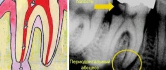
- Periodontitis and periodontal disease.
A 3D photograph of teeth is taken if there are suspicions of inflammatory processes that cannot be diagnosed by other methods.
Main contraindications
3D computed tomography of teeth is based on x-rays, so this procedure cannot be called completely safe. The radiation exposure during this examination ranges from 0.045 to 0.06 mSv. This is not a very high figure, given that the annual exposure limit is 5 mSv (according to the Russian Ministry of Health).
As for contraindications, they are standard for all types of x-ray examinations. The main limitation is the period of pregnancy (especially in the first trimester). In this case, the situation is considered individually, based on the ratio of harm to the child and benefit to the mother.
If a contrast agent is used (it is not used so often in CT scans of the teeth and jaw), then the following can be added to the contraindications:
- Patients suffering from thyroid diseases.
- Kidney failure.
- Allergy to drugs containing iodine.
The lactation period is not a contraindication, but after the procedure at least 48 hours must pass before the next breastfeeding.
Can this be done for children?
Many parents think that the use of 3D computed tomography is impossible in childhood due to the negative impact on the child’s body. This is not entirely true, because special attention to this study is given specifically in pediatrics.
If you follow all safety rules and do not exceed the frequency of procedures (for children – once a year), then the examination will not lead to any pathological changes in the child’s body.
3D CT in pediatric dentistry allows you to assess the condition of the sinuses, gums and see the rudiments of baby teeth. This method also allows you to identify malocclusion pathologies in a child.
Main types of examination
Three-dimensional images are taken using various types of tomographs. It is extremely important to know which device the examination will be used on, because the dose of radioactive radiation received and the degree of information content depend on this.
There are several main methods:
- Step-by-step computed tomography. It was carried out on old tomographs that produced images of low resolution and detail. Now it is no longer used, because the minimum radiation dose was 1500 μSv (with a standard of 1000 μSv).
- Multislice CT (MSCT). Body scanning is carried out using sensors located on a rotating tube. The table moves slowly through the ring, allowing you to take pictures in several planes at once. This method is characterized by the fact that the detectors are arranged in several rows. Such equipment is better suited for examining lymph nodes, muscles and other soft tissues. The radiation dose ranges from 300 to 400 μSv.
- Cone beam CT (CBCT). This technique is considered the most modern and informative in the field of dentistry. The main advantage is the maximum accurate image of teeth and bone tissue, which cannot be achieved using MSCT. CBCT is also considered the safest type of computed tomography (on average, the radiation dose ranges from 40 to 120 μSv).
How do they take 3D photographs of teeth in specialized clinics?
The average time of the procedure is no more than 1 minute. During this short period of time, the device takes about 200 pictures in various projections.
Before the procedure itself, you need to remove all metal objects in close proximity to the area being examined (chains, piercings, etc.). The examination itself looks like this:
- The patient's head is securely fixed using immobilization devices. This is very important, otherwise the pictures will turn out blurry.
- A protective apron is put on the chest.
- The rotating tube of the tomograph rotates around the head, taking a series of images.
- All information is immediately displayed on the computer monitor connected to the device.
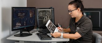
- Further interpretation of the images is carried out by a specialist.
What are the benefits?
Computed tomography is a completely painless procedure that does not cause any discomfort to the patient. The only thing is that he must remain motionless for a short amount of time.
The use of 3D images is due to the following reasons:
- Minimum radiation dose.
- The examination procedure lasts no more than a few minutes.
- Get results quickly.
- Three-dimensional images have a high diagnostic value.
- 3D images eliminate errors in prosthetics and dental surgery.
Where can you take a 3D photo in St. Petersburg?
Diagnostic X-ray - invites you to undergo a 3D CT scan of the jaw if your physician has prescribed this procedure for you. We have no queues, so you can quickly receive images for further delivery to a medical institution.
For the convenience of our clients, we have organized courier delivery within St. Petersburg. You also have access to recording results on digital media, extended consultation and other additional services. You can find out more detailed information on the website.
To make an appointment, you need to call the specified phone number, or leave an online application.
Sign up for a study by phone
+7 (812) 332-52-54
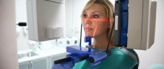
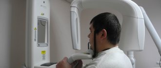
How is a 3D photo taken?
The image is taken using a special tomograph. The procedure is carried out as follows:
- a person takes a sitting or standing static position;
- then a special protective apron is put on his chest - the same as used during radiography;
- there is a special small plate in the mouth that you need to bite with your teeth;
- then you need to rest your forehead against a special support, and grab the handrails with your hands to avoid unnecessary movements;
- then you need to freeze for about 15 seconds at the doctor’s command to get an accurate image;
On a note! The tomograph will make only one full revolution, but will have time to take about 200 mini-images.
- After this, the pictures are processed and compiled into one overall picture - this is how the desired three-dimensional image is obtained. The treatment only takes a few minutes.
At the exit, the patient receives a disk with an image recorded on it, which he brings to the doctor to assess the condition of the jaw system.
Dentistry "Eurodent" - the best 3D dental x-ray in Almaty
The Eurodent dental clinic is equipped with the most modern diagnostic equipment, which makes it possible to accurately and timely diagnose a wide variety of dental diseases. Our clinic has the latest generation computed tomography device, GALILEOS Comfort PLUS, from the German company SIRONA. The improved characteristics of the device allow you to obtain a huge amount of information that is not available with other types of diagnostics (including a panoramic image).
The data provided by 3D dental examination using GALILEOS Comfort PLUS can be used:
- In endodontics - for preoperative diagnostics, to determine deep root damage, when diagnosing injuries to the dental system, to determine the degree of external and internal resorption of root canals, when performing apictoectomy;
- In implantology - to obtain a complete picture when assessing the implantological and orthopedic parameters of prosthetics, to install implants in the least traumatic way;
- In surgery - in the diagnosis of injuries, fractures, displacements of teeth, their roots and jaw bones, in the diagnosis of neoplasms, diagnosis of sinuses, orthodontic operations;
- In orthodontics - when diagnosing pathologies of the palate, jaw, diagnosis of impacted teeth, root resorption, during orthodontic analysis;
- In therapeutic dentistry - for complex diagnosis of clinical conditions, when planning conservative treatment of periodontal diseases, to clarify the results of X-ray studies, etc.
If you have a question: “Where can I get a dental x-ray done?” and “How much does it cost?”, then feel free to contact our clinic. Eurodent dentistry guarantees its patients the European level of diagnosis and treatment, while the cost of our services is quite affordable for all categories of the population.
We are waiting for you for a free initial consultation!
Based on: 9 votes
Indications for this type of examination
3D tomography has a lot of possibilities, for which it is valued by doctors. Thus, it will allow you to assess the condition of not only the jaw, but also the gums, see the quality of previously installed fillings, and reflect all pathologies and anomalies. Indications for this may include:
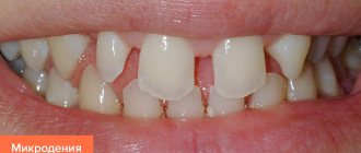
- abnormalities in the structure of the jaw and the shape of the teeth;
- suspicions of pathological changes in dental tissues;
- identification of the rudiments of permanent teeth in children, assessment of the condition of milk teeth;
- the presence of cysts, inflammation, cracks in the roots of the tooth;
- suspicion of traumatic injuries to the jaw;
- preparation for implant installation;
- the presence of tumors in the jaw area;
- preparation for plastic surgery;
- preparation for operations in the jaw area.
Thus, a detailed three-dimensional image is necessary in all complex cases that require detail and a thorough assessment of the patient's current condition. It is especially important in assessing abnormally growing teeth, as well as in case of complex fractures and dental pathologies.
On a note! A CT scan is also often performed during preparation for implant placement. Moreover, its efficiency and effectiveness are much higher than that of orthopantomography. It is often necessary when performing a so-called sinus lift.
Table. Areas of application of CT.
| Sphere | Explanation |
| Orthodontics | The ability to assess how distorted the bite is. |
| Surgery | Assessment of the possibility of certain manipulations by the surgeon, as well as the possibility of installing teeth. |
| Maxillofacial Surgery | Assessment of the condition of the jaw and surrounding areas of the skull. |
| Therapy | The ability to detect deep-lying caries and choose the right treatment option. |
Computer X-ray of teeth: indications
Among the indications for computed tomography of the jaw, or, as it is also called, 3D dental imaging, are anomalies in the structure of the jaw, the temporomandibular joint, the presence of impacted (not erupted or not fully erupted) and dystopic (rotated in the wrong direction or grown in the wrong direction) in its place) teeth; complex fractures of the jaws, damage to the dentition; preparation for maxillofacial operations. CT is also significantly superior to orthopantomography in preparing and performing implantation, in diagnosing inflammation in the area of tooth roots, determining the number of roots and root canals of a tooth, diagnosing cracks in the roots and detecting tumors at the initial stage; when assessing the severity of periodontal and periodontal disease. When planning a sinus lift or bone grafting, you will also have to take a 3D photo of your teeth. If bone tissue is clearly visible on a regular x-ray, then CT also allows you to see soft tissues, blood vessels, canals and changes in the mucous membrane. It is believed that a 3D image of teeth is more informative than one panoramic image and a set of targeted images of all teeth together.
Are there any contraindications?
This procedure also has contraindications. Whatever one may say, there is an effect on the body from x-rays. In general, the load is small, especially if you remember the SanPiN standards (1 mSv/year), and is only 0.045-0.06 mSv. However, there is still a load with CT. So the indications for a CT scan must be justified - it is not advisable to just take a picture.
A 3D photo cannot be taken or is permitted with caution under the following conditions:
- pregnancy, especially the first months when the fetus is actively developing;
- breastfeeding period;
- problems related to the thyroid gland;
- diabetes;
- certain forms of allergies;
- chronic renal failure.
Attention! In some cases, a doctor, despite contraindications, may prescribe a 3D image, but only if the risk of the study is completely justified, and doctors have no other choice.
There is also a so-called tomography with contrast agent. It makes it possible to obtain a more detailed picture of soft tissues, so it is used extremely rarely to take photographs of the jaw. But it has many more contraindications than conventional CT. Substances that may cause undesirable effects should not be used as contrast. It is important to inform your doctor or specialist before the examination if you have any allergic reactions.
How to prepare for a 3D photo?
Step 1. The first thing you need to do is visit a dentist to understand whether it really makes sense to spend money on such an expensive examination. In some cases, a dental CT scan may not be strictly necessary.
Step 2. Next, if the doctor has confirmed the need for an examination, it is important to evaluate the market for this service in your city and choose a clinic based on the price-quality ratio.
Step 3. Then it is important to sign up for a convenient day and arrive at the clinic at the appointed time.
Step 4. Before taking the photo, it is recommended to clean your mouth by chewing gum or brushing your teeth.
Step 5. After the picture has been taken, you need to receive the results and take them to an appointment with your doctor.