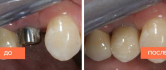
Where is the dentistry located?
Dentists on Rimskaya
How does a targeted radiovisiograph image differ from an x-ray?
Why do you take a targeted photo?
Types of targeted dental imaging
Advantages and Disadvantages of Sight Shot
Technology
How much does it cost to take a targeted photograph of a tooth on Rimskaya
A targeted dental image is a planar image of one or more teeth made using an X-ray machine. A targeted image allows you to see the internal structure of the coronal part of the tooth, assess the condition of the tooth canals and adjacent bone tissue. Even minor changes in the tooth can be seen on it, which simplifies the diagnosis of diseases in the early stages.
There are film radiography, which is done on X-ray film, and digital, which requires a special device - a radiovisiograph.
Are you looking for where to take a targeted photo of your teeth on Rimskaya in Moscow? Do you need a clinic with positive experience in solving dental problems in the Central District of the city? Do you live or work in the Tagansky district or Ploshchad Ilyich metro station? We invite you to the AB Clinic on Sergius of Radonezh 23-25с1. Make your dream smile a reality!
Dentists on Rimskaya
Gneusheva Elena Viktorovna
Dentist-orthodontist, pediatric orthodontist
Experience: 20 years
★ For more than 13 years she worked at the Moscow State Medical University at the Department of Orthodontics and Children's Prosthetics, Ph.D.
Gorokhova Inga Vitalievna
Dentist-therapist
Experience: 14 years of total experience
★ Expertise in restorative dentistry to make your teeth shine
Konstantinov Oleg Nikolaevich
Dental surgeon
Experience: 25 years
★ Virtuoso of painless treatment. Patients fall asleep in the doctor's chair
Nikolaychuk Anna Vladimirovna
Chief physician, dentist-therapist
Experience: 13 years
★ Chief specialist in the treatment of toothache. Masterfully deals with problems quickly and permanently
Frolova Alexandra Ivanovna
Dentist-therapist
Experience: 25 years
★ A brilliant specialist with many years of experience as a manager in public dentistry
Radiology and computed tomography
The role of correct diagnosis in dental treatment is difficult to overestimate. Both the success of treatment and the ability to save the patient from unnecessary inconveniences that accompany the dental treatment process fully depend on the accuracy and completeness of the diagnostic picture of the disease.
Diagnosis of the oral cavity
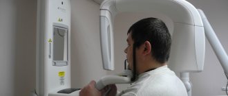
In our clinic, diagnostics are carried out using an ultra-modern panoramic X-ray machine Sirona, manufactured in Germany. When examining patients using a new orthopantomograph, the dose of ionizing radiation is 80% (!) less than when examining using traditional devices. It achieves excellent image quality through the use of panoramic dental X-ray equipment, which has a number of excellent features. This device has the ability to scan jaws layer-by-layer for the purpose of performing dental implantation operations.
Our doctors can instantly receive an accurate image of the patient’s teeth (panoramic image, etc.) on a computer monitor and can quickly process it - increase its size, enhance the contrast. The use of this device allows you to obtain high-quality and informative images of the upper and lower jaw in real time, and also allows you to perform 3D reconstruction of the image. This allows the doctor to detect the presence of a pathological focus at the earliest stages of the disease, monitor the quality of treatment, providing the patient with the most comfortable and effective treatment.
The equipment of the RuDenta clinic allows you to obtain complete diagnostic information of the highest quality, however, for successful treatment and further monitoring of patients, the quality of storage, recording and cataloging of the obtained diagnostic data is equally important. In the special computer patient registration system of the RuDenta clinic, a personal folder is created for each of them, where the entire history of treatment and diagnosis is stored, including all x-rays taken. Any of these images is available for viewing by specialists directly at their workplace.
We carefully collect anamnesis, which allows us to trace the history of the development of the disease, stages of development, and identify factors contributing to the progression of the process. Many systemic diseases are both triggering and supporting factors in the development and course of oral pathology. Timely identification of aggravating factors greatly contributes to the correct choice of treatment method.
How does a targeted radiovisiograph image differ from an x-ray?
These methods are based on one principle: the tooth is illuminated with X-rays. They differ in different ways of fixing the resulting image. Having taken an X-ray of a tooth, you get a targeted photograph on film in a single copy, while a visiograph gives a digital image that can be duplicated as many times as you like. Instead of film, it uses a sensor that is more sensitive to X-rays, so the patient receives 10 times less radiation than required for film radiography.
The data is either immediately transferred to the computer if a wired visiograph sensor is used, or it is read by a scanner if a wireless analogue is used. Digital radiography is more accurate than film radiography, so Moscow clinics are increasingly choosing a method that is more informative and less harmful to the patient.
Photo of teeth: types
Dental X-ray is a study of the soft and bone tissues of the oral cavity using an X-ray image. The image informs the doctor about what he does not see during a visual examination. X-rays are prescribed to identify the degree and depth of inflammation, detect pathologies, defects and diseases.
There are many types of dental x-rays. Our clinic uses targeted radiography. A targeted photograph of teeth is a simple and accessible technique in dental research. This is a 2D image that shows the doctor 1-2 teeth located nearby. Dental images are taken using a digital radiovisiograph.
Sight radiography allows the doctor to:
- Identify inflammation in the early stages in the area of the apexes of the roots of the teeth.
- Check the quality of tooth root filling and bone tissue restoration before prosthetics.
- Identify carious lesions, pulpitis, periodontitis.
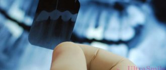
A targeted photograph of teeth, depending on the equipment used, can be of two types:
- X-ray on film.
- Electronic image (the image is transferred to a digital medium, which allows you to enlarge the required area for detailed diagnostics).
Why do you take a targeted photo?
This procedure may be prescribed to you at any stage of treatment. It is necessary for the initial diagnosis, for monitoring treatment and for checking results. It is prescribed when it comes to treatment and filling of canals. Your doctor will need to know their length before starting work. After treatment and filling, you can evaluate how well the filling was installed. In addition, a targeted photograph can show the development of secondary caries in an already filled tooth, without compromising its integrity.
In general, indications for the procedure may include:
- caries,
- pulpitis and periodontitis,
- cysts or granulomas,
- adjustment of treatment,
- removal of a tooth.
The only contraindication for this procedure is pregnancy. X-rays negatively affect the development of the fetus, so it is better to refrain from X-rays during this period.
Contraindications for the study
Sight radiography has such a low radiation dose that there are practically no contraindications to its implementation. It is not recommended to conduct radiographic examinations in the first trimester of pregnancy, therefore preparation for bearing a child should begin with sanitation of the oral cavity. Also, the procedure may not be comfortable for patients with an increased gag reflex in cases where examination of large molars is necessary. However, specialists carry out it very carefully, which most often does not cause any discomfort.
Advantages and Disadvantages of Sight Shot
Since film radiography is being phased out, we will talk about the advantages of digital over it. Among them:
- rapidity,
- image clarity,
- the ability to store and search images in databases,
- less radiation exposure for the patient,
- the ability to take more pictures.
Among the disadvantages are:
- the image is obtained in one projection,
- covers a small area of the dentition.
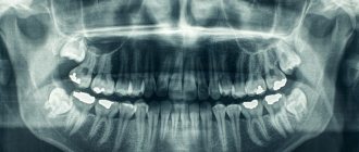
This method cannot obtain a panoramic image of the entire jaw; a maximum of 4 teeth can be captured. Suitable for diagnosing local problems only.
General overview
From a technical point of view, the procedure is an X-ray analysis of the state of one or more adjacent units, characterized by the speed of obtaining complex data and ease of execution. The resulting image allows the dentist to:
- To formulate a correct diagnosis, the formulation of which on the basis of visual examination data alone may be impossible.
- Determine and prescribe an effective course of treatment, as well as control its pace.
- Monitor the intensity of the spread of pathology formed in the oral cavity.
- To study the condition of the structure of bone tissue and root canals, dentin and blood vessels.
- Identify hidden anomalies and pathological processes, determining their localization.
To obtain targeted dental images, digital equipment is used - a radiovisiograph, one of the functional advantages of which is the minimum radiation dose to the patient during the shooting process. The directed beam flow maintains focus, affecting only a selected area of the jaw row, which makes the diagnostic procedure safe for the patient’s health.
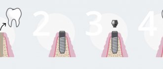
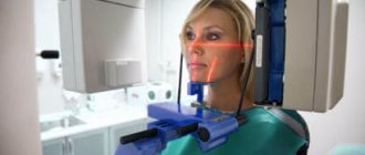
Technology
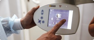
The procedure takes place in an x-ray room. The patient wears a protective lead apron to reduce exposure. Next, install the sensor or film to the desired tooth. After this, a targeted photo is taken. It lasts no more than a couple of seconds. The sensor is taken out and the information is displayed on the screen. If the image is taken on film, it is first developed in a dark room in a special solution, after which it is given to the patient. Film radiography is slower than digital radiography; image clarity is affected by the quality of the film and the technique of working with it, so it is becoming a thing of the past.
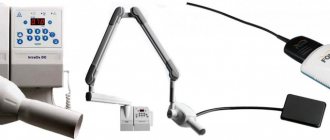
X-ray equipment
To obtain a targeted image, two types of equipment are used:
- X-ray machine of an outdated model;
- digital radiovisiograph (modern version).
The first allows you to get a film image that distorts the image quality. Using an innovative device, you can obtain an electronic image of high quality and resolution. For convenience, it is possible to enlarge the image with detail of the required areas.
The main advantage of the radiation machine is considered to be the minimization of radiation exposure (three times lower compared to the outdated version).
Using a digital camera you can take two types of photographs:
- Interproximal. It is used to assess the condition of the crown when treating caries with fillings, as well as to assess the correctness of prosthetics.
- Intraoral. It is performed to identify caries of nearby teeth and assess the quality of fit of the installed filling. This image is the most informative; it allows you to diagnose caries in the initial stages and identify periodontal pathologies.
How is the procedure performed?
X-rays are taken in a special room, and the procedure consists of several stages:
- The patient sits in a chair. The patient is wearing a protective apron to minimize the negative effects of x-rays.
- The doctor fixes the head in such a way as to obtain an image in the required area and asks the patient to sit still.
- The beam is directed onto the inner or outer surface. The procedure itself takes a few minutes, and it takes 10-15 minutes to prepare an electronic or printed image.
Advantages and disadvantages
Advantages of a targeted photograph performed using modern equipment:
- Safe diagnostic procedure.
- Clarity and image quality.
- The ability to zoom in on the desired area to see the details.
- Print the photo.
- Improving conditions for clinical research.
- Reducing the duration of the procedure.
Dentists consider this diagnostic method to be one of the most accurate and informative, since with the help of such an image it is possible to identify latent pathologies, as well as diseases in the initial stages, when they do not cause discomfort and are not visible to the naked eye.
The method under consideration also has disadvantages:
- limited scope of the study;
- photographs are taken from only one angle;
- a small viewing area, which allows you to evaluate no more than three teeth;
- inability to identify cracks in the buds and determine perforation.
Varieties
Targeted radiography can be carried out in several ways: using an X-ray machine, a visiograph or a computed tomograph (but this will already be tomography - layer-by-layer scanning of the object of study with the reconstruction of its three-dimensional model). There are two types of equipment.
- Analog X-ray machine. The image is recorded on X-ray film. The disadvantages of the technique include the need to install a film in the oral cavity, a long waiting period for the conclusion, as well as a limited shelf life of the results - up to 2 years. After a few years the film simply fades.
- Digital radiovisiograph. A device that makes it possible to obtain images in electronic form with a high degree of resolution. The dentist gets access to an online image that can be enlarged and detailed. Images are stored indefinitely in an electronic database. The examination is carried out using a sensor. Radiation exposure is reduced by approximately 3 times compared to traditional X-rays.
According to the direction and area of examination, there are pictures:
- interproximal. Informative when it is necessary to identify hidden carious cavities. Used to diagnose caries under fillings and crowns. Makes it possible to assess the distance between the filling and the nerve;
- periapical. Used to assess the length and shape of roots, as well as their number. Makes it possible to diagnose inflammatory processes in bone tissue and assess the condition of periodontal structures. Detects various deformations, determines the degree of obstruction and can help detect foreign bodies, in particular, fragments of instruments.
During root canal treatment, the dentist takes a series of photographs for precise control. The following studies are performed:
- diagnostic. Helps to identify the presence of old filling material and determine the condition of the roots;
- instrumental. The exact parameters for carrying out therapeutic actions are established;
- verifier. It is carried out with a master pin at the final stage of trying on materials before installing a filling.
After filling the dental canals, the doctor takes a final image, which is entered into the patient’s electronic or regular medical record.
Indications for examination
X-rays in dental practice are prescribed when it is necessary to diagnose the condition of a tooth (for example, to identify an inflammatory process) or to control the quality of manipulations - after canal cleaning, filling canals, restoring the structure of bone tissue, treating caries, pulpitis, periodontitis and other pathologies. The procedure must be completed to confirm such diseases.
- Caries. In addition to obvious cavities, the image makes it possible to identify carious processes under crowns or fillings, to detect superficial and medium caries, which is not accompanied by acute pain.
- Periodontitis or pulpitis. X-rays are performed to prescribe the correct endodontic treatment. Makes it possible to obtain information about the condition of the roots and their characteristics.
- Cystic neoplasms or granulomas. The examination is carried out to identify the size of the formation and determine its location.
- Pericoronitis. Photographs of wisdom teeth are relevant if the development of inflammatory processes in the “eights” is suspected, and are also necessary to obtain information about how the tooth germs are located in the tissues.
- Anomalies of tooth growth.
- Gum diseases – periodontal disease, periodontitis and others.
X-rays are also prescribed if there is pain in the area of a certain tooth and severe inflammatory processes. The dentist may refer you for this study if it is necessary to establish a deficiency of bone tissue in the patient’s jaw or its destruction.
This method is often used in surgical practice when choosing a treatment method after dental trauma. This examination is indicated as a control examination after therapeutic treatment.
X-rays are taken without fail before prosthetics, implantation, installation of braces or the use of other methods of correcting the bite, as well as during surgery. But performing targeted X-rays in such cases is impractical, since, as a rule, it is necessary to evaluate the condition of several teeth, their relative position, and the characteristics of periodontal tissues. To solve these problems, an orthopantomogram - a panoramic photograph of the dentition - will be more effective.
Radiography is often powerless in diagnosing perforation and tooth root cracks.
Carrying out x-rays in children
X-ray examinations in pediatric dental practice are indicated from the age of two. If there are injuries or life-threatening conditions, the doctor may decide to perform x-rays at an earlier age.
Dental radiography is a common procedure in pediatric dentistry. Baby teeth are especially susceptible to caries, and often carious cavities are located in places inaccessible to visual assessment.
Digital visiographs are recommended as equipment for pediatric diagnostics, since they have a low level of radiation and do not require placing a film in the oral cavity. Parents can remain near the child during the procedure, using radiation protection equipment.
Restrictions and contraindications
Standard contraindications for dental x-rays using the targeted image method are the first and third trimesters of pregnancy. This type of examination is also not prescribed when acute viral diseases are detected.
A relative contraindication is the period of lactation. It is usually recommended not to breastfeed your baby immediately after having an x-ray. Milk should be expressed, and feeding should begin no earlier than three hours after the end of the procedure.
Age and condition restrictions are waived in life-threatening situations.
Further detailed analysis of the image
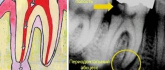
A detailed analysis of the image is carried out by a doctor who determines a treatment regimen or prepares for surgery, or by a radiologist. The dentist examines the image and evaluates the color of various areas. He makes a conclusion about the rigidity, density, homogeneity of the bone structures of both jaws, and the placement of parts of the dentition. The analysis makes it possible to identify carious cavities, granulomas, cysts, periodontitis and the consequences of injuries. The image shows areas of inflammation, blood vessels and nerves.
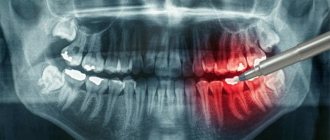
The most common dental diseases on a dental photograph are manifested by the following signs.
- Caries. This is the process of destruction of dental tissue. The picture shows areas of transparency of the enamel and the hard part of the tooth. The clearing zones have uneven, unclear contours.
- Pulpitis. Damage to the bone structure is characterized by heterogeneity of color in the interroot zone against the background of hypertrophy.
- Periodontitis. The progression of granulomatous periodontitis is accompanied by destruction of the hard part of the tooth and cement. Granulomas are observed in the area of tooth root formation, the interdental gap is enlarged, its edges are blurred.
- Periodontitis. The image shows signs of osteoporosis - the density of bone structures decreases, the height of the partitions in the dentition decreases, and pockets form.
To read the photo correctly, the image must be unblurred and in focus.