Rogatskin D. V. Radiologist, Ortos LLC (Smolensk)
Until recently, radiation diagnostics in dentistry was considered as an additional examination method, that is, optional, without which, in principle, full treatment could be carried out. However, in the 21st century the situation has changed dramatically, new technologies, new specialties and new requirements for the examination and treatment of patients have appeared. Currently, not a single civilized dental appointment is complete without a detailed radiodiagnostic examination of the patient, and it can be argued that radiodiagnostics in dentistry is now one of the main and most popular research methods.
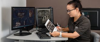
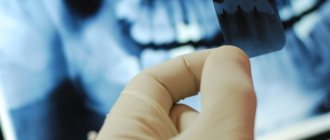
The main difference between digital radiography (radiovisiography) and traditional one is that in this case, instead of film, the image receiver is a sensor that perceives radiation and transmits information to a computer. The equipment required for radiovisiography consists in sequence of a radiation source, a device for reading information, a device for digitizing information, and a device for reproducing and processing images.
Modern low-dose generators with a minimum timer value, designed to work as part of a visualization complex, are used as a radiation source. The visual imager itself consists of a sensor, which is a sensor based on a CCD or CIMOS matrix, an analog-to-digital converter and a computer program designed to optimize and store images.
At first glance, the original digital photographs may differ slightly from the usual film ones, and therefore require processing using software options. The highest quality image is the one that is closest in visual perception to analogue, therefore, even despite the highest technical characteristics of the visiograph, the quality of the final image largely depends on the capabilities of the program and the ability of a specialist to work with it.
What is a targeted dental photograph?
Another name for the procedure is targeted intraoral contact radiography - this is a simple and fast method of X-ray diagnostics in dental practice. Research is carried out in clinics using analog X-ray machines or digital radiovisiographs. The doctor gets the opportunity to examine both the condition of the tooth being examined and those located nearby.
- Carry out diagnostics. For example, detect the development of an inflammatory process.
- Evaluate the results of the therapy. This is necessary not only in the treatment of pulpitis, caries or other dental diseases, but also in preparation for prosthetics.
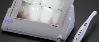
When using analog X-ray equipment, an image is created on film and then transferred to special paper. A digital device allows you to obtain an electronic photograph. If necessary, any area of the image can be enlarged on the monitor for a more thorough examination.
- Interproximal. Allows you to diagnose pathologies of the crown part of the tooth, detect the presence of carious cavities, as well as defects that can form under fillings and crowns.
- Periapical, allowing to assess the condition of bone tissue. This type of imaging helps monitor the quality of therapy provided.
The radiation dose for a dental x-ray is 2-3 μSv, which is very small. For comparison, with fluorography we receive a radiation dose of 500-800 μSv.
Sight radiograph
Sighting dental radiography
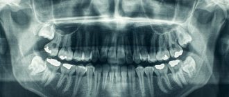
A targeted radiograph (or dental radiography) is a detailed image that produces an image of a selected area of the jaw with a possible pathological process. X-ray allows you to make a diagnosis or identify hidden pathology, monitor the quality of sealed canals and the process of bone tissue restoration.
Main indications for use
- abscesses and carious cavities,
- malocclusion,
- jaw fracture,
- periodontitis,
- focus of infection in the root canal,
- cyst, granuloma,
- tumor, abscess,
- unerupted teeth
- deviations in the process of wisdom tooth growth.
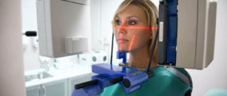
Radiovisiograph and its features
To create a targeted image, a digital device is used - a radiovisiograph, which has a low level of radiation and a precise, directed beam. Before the procedure, the patient is always wearing a special apron that protects from reflected radiation. This device allows you to take the most accurate image of the tooth; the image is stored electronically or in natural form. Having several images makes it possible to track the smallest changes in the jaw area. A clear picture allows the specialist to determine the nature of the disease and the degree of damage to the tooth.
Advantages of Maestro Dent
- The X-ray room is equipped with a modern Italian Evolution device, which provides maximum accuracy of images of the entire jaw area and its joints;
- attentive attention to the patient’s condition and well-being; a personal treatment plan and recommendations for oral care are drawn up for each patient;
- individual terms of payment for services are considered;
- The clinic provides treatment under the VHI policy. The center cooperates with all insurance companies represented in the region.
in Rostov conducts dental x-rays for various age groups. This study is safe even for children, since modern X-ray devices have low radiation exposure. Using X-rays, the doctor evaluates the condition of the jaw tissue and also diagnoses possible problems with the oral cavity. Therefore, you should put aside your fears; if the doctor insists on the procedure, then you must undergo it.
You can sign up for an x-ray online at any time on our website or by calling our number.
You can view the cost of the service in the adjacent tab or in the “prices” section on our website.
Convenience and safety
When an x-ray is prescribed, a number of factors are taken into account, starting from the feasibility of the study and choosing the most effective technique, ending with ensuring maximum safety for the patient. Therefore, the clinic’s X-ray room is equipped with a modern orthopantomograph, which provides minimal radiation exposure. For targeted photography, the Italian digital camera Evolution is used, which is distinguished by high beam accuracy and excellent image detail. You can sign up for a study at the Maestro Dent center on the website, or simply by calling us.
Pros and cons of the diagnostic method
The use of a dental visiograph has a number of advantages compared to analog X-ray machines. First of all, it is an opportunity to get a clear picture of the tooth and the tissues around it. It is convenient to store the resulting images on a computer or other electronic media, printing them if necessary. A digital image allows a more detailed assessment of the clinical picture, since the image can be enlarged several times. In addition, digital dental radiography is a safe procedure. If necessary, multiple procedures may be performed. For example, during implantation, control images are taken before and after each implant is installed. And also after complete completion of prosthetics. The equipment used is characterized by a reduced level of radiation exposure. The dose of radiation that the body of the person under study receives is so small that it does not cause any harm to health. Most often, it does not exceed the natural background recorded in some megacities. Despite the large number of advantages, this method has disadvantages. For example, the image can be performed only in 1 plane covering a small area of tissue. Therefore, the maximum effectiveness of the method is achieved at the stage of early diagnosis or to control the quality of therapy already carried out.
Execution steps
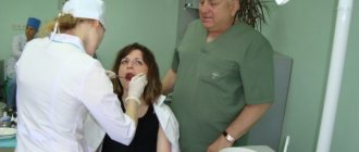
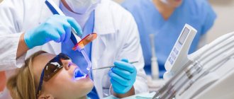
Interproximal radiography is one of the types of intraoral examination that allows you to obtain a picture of 1 section of the oral cavity with an image of the upper and lower dentition. To obtain the image, a special holder is secured between the closed teeth. It allows you to identify interdental caries and various changes in bone tissue due to gum disease. And also check the correct installation of crowns, dentures or fillings. An X-ray of the tooth is taken by a doctor in an office specially equipped for this. Before taking an X-ray of a tooth, the doctor gets acquainted with the problem and studies its location.
To obtain a clear image of the problem area, the patient's head is fixed in the required position. The process of visiography takes a matter of seconds, but during this time the patient is required to remain completely still. The video in this article shows how to take targeted photographs of teeth so that the image is as informative as possible. Using a digital sensor, the radiologist directs a beam of rays to the required area. The photograph is taken either from the inside of the mouth or from the face. During the manipulation, the patient does not feel any pain or discomfort.
The original digital image differs from the traditional film image because it is processed using a special program in a few seconds and transferred to the monitor screen.
General information
An X-ray examination that allows you to get a real clinical picture of one or more teeth located nearby. A simple and reliable method for accurate diagnosis, used in most dental clinics.
Problems that can be solved by analyzing a targeted X-ray image:
- making an accurate diagnosis;
- drawing up a treatment plan with monitoring the dynamics of the condition;
- assessment of the degree of development of pathology;
- assessment of the restoration of hard tissues and the condition of the dental canals;
- assessment of the actual condition of blood vessels, soft tissues, rudiments;
- identification of pathologies in a latent course;
- identifying the area affected by caries, as well as identifying the source of inflammation.
Targeted X-rays are done using a digital radiovisiograph, which minimizes the radiation dose. The equipment produces a direct beam beam that directly affects the area being examined. The technique is absolutely safe for health.
Features of the procedure in children
Contact intraoral radiography can be prescribed for children in cases where tooth damage cannot be examined in any other way. The technique allows early detection of disturbances in the process of teething, bone tissue diseases, and prescribing effective treatment. In addition, this method allows you to control the implementation of orthodontic manipulations if the child has problems with the formation of the jaw.
The study is carried out in the same way as in adult patients. Children under 2 years of age are recommended to undergo x-rays only in case of urgent need. For example, in case of injury during childbirth, to monitor the development of the jaw, or after a fall from a height, to assess the integrity of the teeth.
Indications for use
Factors that determine recommendations for radiovisiography include:
- Pain in the dental area.
- Routine diagnostics prescribed to detect pathologies.
- The need to identify the degree of bone loss before implantation.
- Identified diseases of gum tissue, including periodontal disease.
- Determination of the severity of mechanical damage received.
- The need to control the growth of “eights”, or wisdom teeth.
- Checking the quality of root canal cleaning.
The procedure is mandatory before installing prosthetic structures or orthodontic devices, as well as when planning surgical intervention.
Restrictions and contraindications
How often a dental x-ray can be taken can only be determined by a doctor, guided by the severity of the problem and the patient’s condition.
It is forbidden to take dental x-rays during pregnancy, especially in the early stages. A woman needs to take care of the condition of her teeth in advance in order to eliminate the need for treatment while carrying a child. X-rays are contraindicated for children under 2 years of age.
In case of urgent need, an x-ray can be performed, but not earlier than the second trimester. The only exception is when emergency assistance is needed. Manipulation must be carried out using all possible means of protection. The period of breastfeeding is not a limitation for the study.
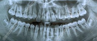
Interpretation of a dental radiograph
Only a dentist or radiologist can decipher and describe a dental image. The image shows the tooth: its root, internal canals, shape, anatomical features.
- Examines the rigidity, density and uniformity of the bone structure. Evaluates the location of each element of the dentition.
- Determines the presence of signs of clearing or darkening, indicating the development of an inflammatory process, cysts, granulomas, neoplasms.
- Depending on what the dental x-ray shows, a diagnosis is made.
Carious formations in the picture look like light areas of various shapes with unclear boundaries. The development of pulpitis is characterized by bone damage. The image shows a violation of its homogeneity in the interroot space. With the development of periodontitis, a granuloma appears in the area of the tooth root in the form of a darkened round shape with clear contours. With periodontitis, the image shows a decrease in the density of the bone structure, a decrease in the height of the partitions between the elements of the dentition, and the formation of “pockets.”
Indications
Radiophysiography is prescribed to patients with the following symptoms:
- toothache;
- carious lesions;
- cystic formations in the root of the tooth;
- pulpitis;
- periodontitis;
- bone tissue deficiency;
- atrophy and tooth destruction;
- pathologies of soft tissues of the oral cavity;
- tooth damage due to trauma;
- malocclusion.
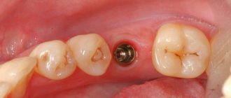
The image is prescribed before the installation of removable and fixed dentures, braces, plates, implants, as well as before surgery.
Where can I take a photo and how much does it cost?
Targeted radiography can be performed in any specialized medical institution. The average cost of the procedure is about 400-450 rubles. Some clinics include in the cost of conducting several studies at once, which involve monitoring the treatment being carried out.
All categories of patients, both adults and children, can take a targeted photograph of a tooth. The procedure is prescribed with caution to pregnant women and infants under 2 years of age.
A referral for a radiovisiographic image is given by a dentist. The resulting image makes it possible to determine the presence of a problem, confirm or refute the diagnosis, and prescribe treatment. To monitor the patient’s condition during treatment, the doctor may recommend taking a second photo of the tooth.