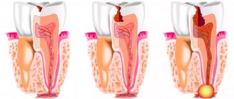
When a root canal infection occurs, a formation called a cyst may develop at the top of the tooth root, which appears as a pus-filled cavity with a fibrous lining on the inside.
A cyst, also often called a periodontal abscess, can gradually enlarge over time, causing serious discomfort to a person. The cyst located on the teeth of the upper jaw has the highest growth rate, which is facilitated by more porous bone.
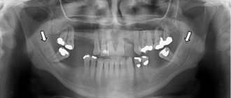
If we look at the x-ray, then a strong darkening in the upper region of the root is a cyst.
Diagnosis of dental cysts
Since at an early stage the disease has practically no symptoms, it can be very difficult to determine. Nevertheless, there are certain diagnostic methods that allow you to identify a cyst:
- external examination, the task of which is to determine whether there is growth of the capsule in the gingival tissue;
- checking the integrity of existing fillings (if there are any violations in the root part, they begin to collapse, which is noticeable to the naked eye);
- X-ray;
- tomography of the jaw.
The surest way to make a diagnosis is an x-ray: the capsule filled with cystic fluid will appear as a bright white area (its shape can be round or oblong) located under the gum pocket.
Based on the location of the formation and the current stage of development of the disease, the doctor will be able to prescribe the most effective treatment.
Diagnostics
Often a cyst is found when neighboring teeth begin to be treated. A radicular cystic formation is clearly visible on an x-ray - it is a round-shaped shadow with a clear border, adjacent to the apex or wall of the root.
To clarify the diagnosis, electroodontometry is done. To clarify the nature of the neoplasm, a puncture with a thick needle is prescribed. To check the extent and distribution, you will additionally need radiography of the paranasal sinuses, sometimes contrast radiography and computed tomography of the upper jaw bone.
Types of cysts
To prescribe the most effective treatment, the dentist needs to determine the type of cyst and identify why it formed.
Cystic formations are:
- radicular (occur at the root of the tooth or in close proximity to it);
- residual (formed after tooth extraction);
- retromolar (formed as a result of problematic eruption of wisdom teeth).
Based on their origin, cysts are divided into:
- odontogenic (they are caused by various dental diseases);
- non-odontogenic (their formation is not associated with dental diseases).
Separately, it is worth highlighting follicular cysts, which contain the tooth germ: they are formed as a result of improper care of baby teeth.
What is a radicular cyst?
Radicular cysts of the jaw are round-shaped defects in the bone adjacent to the root of the tooth, which appear in the presence of chronic inflammation that affects the root of the tooth.
Education of this type most often appears between the ages of twenty and forty-five years. It is important to note that radicular cysts usually appear on the upper jaw.
In most cases, the disease is caused by teeth that are affected by granulomatous periodontitis in an advanced chronic stage.
Main symptoms of an abscess
A dental cyst may not reveal itself for a long time: a person may not realize that there is something wrong with his teeth and gums. Sometimes an abscess causes mild pain when biting or pressing on the gum. But barely noticeable and far from constant pain can force few people to go to the doctor: a cyst at this stage of development is discovered by chance when a person goes to the dentist about completely different teeth.
An infection in the abscess cavity can worsen if immunity decreases: the process of pus formation accelerates, severe pain appears, the cheek swells, the person feels weak and lacks strength, and the temperature rises.
Why does a cyst occur?
The main reason for the development of an abscess is the appearance of infection in the root canals. But there can be several reasons for infection:
- Advanced caries that has developed into pulpitis due to ignoring the need for its treatment. In the tissues on which the carious formation has appeared, pathogenic microorganisms settle, first entering the pulp, as a result of which its inflammation begins, then, if the pulpitis is not treated, penetrating beyond the boundaries of the tooth, resulting in a periodontal abscess.
- Unsatisfactorily filled root canals. Canal filling is a fairly common procedure that dentists have to perform during treatment. However, despite the routine nature of the operation for a dentist, not all specialists, due to lack of experience or due to inattention, carelessness and the desire to complete the work as quickly as possible, do it efficiently. The canal must be sealed along its entire length, since in the unsealed part there is a high probability of infection, which, just as in the case described above, will sooner or later go beyond the boundaries of the tooth and lead to the appearance of an abscess. Official statistics on poorly filled canals are depressing: in more than half of the cases, dentists make mistakes during the procedure, which subsequently result in the appearance of a cyst. In the picture, the unsealed part of the canal is always clearly visible. Sometimes it happens that the root canal is completely filled, but the material used for filling is loose, which is also not good.
Symptoms of a radicular cyst
Unlike some other oral diseases, a radicular cyst can go unnoticed by the patient for a long time. Do not cause discomfort at all or cause minor inconvenience, which in most cases is ignored. However, with a dental examination performed by an experienced specialist, such a disease can be easily identified.
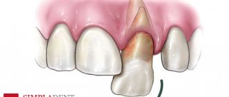
One of the main signs of the appearance of a radicular cyst is considered to be darkening of the tooth or advanced caries. In addition, in the presence of a cyst, displacement of adjacent teeth often occurs. In about a third of cases, patients experience facial deformation, which, if the tumor grows further, can lead to a jaw fracture.
Tooth cyst: treatment
Treatment can be divided into therapeutic (use of drugs) and surgical (gum incision and root resection).
Conservative treatment is justified if:
- no unsealing required;
- if the canal is unsatisfactorily sealed along its entire length;
- if the diameter of the cyst is large (over 1 cm), which is accompanied by pain and swelling noticeable even to a non-specialist.
Surgery is recommended when:
- there is a pin in the channel;
- there is a crown on the causative tooth;
- the root canals are unsealed at the upper part of the root for 1/3 of their length;
- The gums swell, which is accompanied by pain.
Description of therapeutic method of treatment
This method takes a lot of time, requiring a lot of patience from the patient (you will have to visit the dentist more than once), a lot of free time and significant financial expenses.
The process consists of a number of sequential stages:
- First, the doctor works with the canals. The dentist looks to see if they have been filled: if not, the pulp is removed, and the canals are processed using the instrumental method; if they were, then they need to be unsealed before proceeding with subsequent actions.
- The cyst contains pus, which is why after unsealing and getting rid of the pulp, repeated thorough rinsing of the canal with an antiseptic solution will be required.
- A potent medicine is removed from the apex of the root.
- The canal is temporarily filled with a paste that has an antiseptic effect.
- Since the medicine needs to be changed once every certain period of time, the patient will have to come to the dentist every time for this over the next couple of months. After the next change of medication, the canal is again filled with medicinal paste.
- From time to time, an x-ray is taken to monitor the progress of treatment and evaluate its progress. If the cyst in the picture becomes smaller over time, then this indicates positive dynamics.
- When the cyst shrinks, the doctor fills the canal with gutta-percha - this filling is permanent and periodic refilling is not required.
- Installation of a seal.
But even after placing a permanent filling, the patient still must visit the doctor for an image for another two to three months. It should show a reduction in the size of the existing abscess and restoration of natural tissue.
Treatment at Dr. Korenchenko’s clinic: a modern and professional approach
See also Treatment of ENT diseases Removal of a cyst in the nose Cyst in the maxillary sinus Treatment of a maxillary sinus cyst
Treatment of maxillary sinus cysts in Dr. Korenchenko’s clinic is carried out in accordance with international standards and using modern techniques. With this disease, thermal and physiotherapy procedures are unacceptable, and the use of any drugs is inappropriate. After examination, if indicated, endoscopic removal of the cyst is performed. The doctor carries out all the necessary manipulations intranasally - through the natural anastomosis of the maxillary sinus and nasal cavity. An endoscope with a camera and micro-instruments are used.
This operation does not lead to disruption of the natural ventilation of the maxillary sinus and gross scar tissue changes. It can be performed even on patients with various concomitant somatic diseases and contraindications for general anesthesia.
Drug treatment of dental cysts
If the size of the mass is less than 1 centimeter, your doctor may recommend trying medication.
The range of prescribed drugs usually includes:
- antibiotics (for example, azithromycin, amoxicillin, etc.);
- immunomodulatory agents (arbidol, dibazon);
- antihistamines (ketotifen, acrivastine, etc.).
Depending on the individual characteristics of the development of the disease, multivitamin complexes and antifungal drugs may be prescribed. On average, treatment takes about 2 weeks minimum; every 4 days it is advisable to visit a dentist, who, after an examination, will adjust the medication intake.
Removal of a tooth cyst
The operation is called root resection. It takes no more than an hour: the procedure time depends on the location of the damaged tooth (back teeth are easier and faster to treat, front teeth take much longer).
The operation consists of the following steps:
- A couple of days before the procedure, the canals are sealed (days, not weeks, otherwise early filling can cause purulent inflammation).
- Immediately before the operation, the doctor gives the patient a pain-relieving injection - local anesthesia (it does not hurt, the only thing is that it may hurt a little when the injection begins to “go away”).
- The gum is cut, exposing the bone. A hole is made in the bone tissue near the tooth in which the cyst has formed, for which a drill is used (due to the anesthesia, the patient will not feel anything).
- The hole will allow the doctor to see part of the root and the cyst attached to it. This area of the root is cut off with a drill and removed from the resulting wound using tweezers.
- Since the cyst occupied a certain space in the tissue, after removal a cavity appears in its place. If the cyst is large, the doctor fills it with synthetic bone tissue, which stimulates the growth of normal bone.
- The dissected mucosa is sutured, and for a more effective outflow of the ichor, a drain is installed, which must be removed after a few days.
Resection in no way affects the lifespan of the tooth. The doctor’s task is to completely get rid of the cyst, since even leaving the slightest remnant of it can lead to its re-formation.
Removal or treatment of a dental cyst?
The question is of great importance for people who are afraid of dental operations.
Before deciding on a treatment method, you need to consider a number of nuances:
- if you treat the cyst with medication, it will take at least 3 months; in case of surgical intervention, you will have to visit the doctor only 3-4 times;
- Nowadays, in most surgical operations, a laser has replaced the scalpel, but in some cases it is still impossible to do without traditional incisions;
- if there are deep cracks on the tooth, you cannot do without surgical intervention - no modern medications will help in this case;
- Regardless of the chosen treatment method, the probability of cyst recurrence is about 10%.
In any case, the final decision regarding the method of treatment remains with the doctor, who, based on the analysis of the x-ray and other measures, can make the right choice about the development of the disease.
Disease detection
Most often, this disease is diagnosed using x-rays. In the picture, the cyst appears as a round or oval shadow with clearly defined boundaries, and the roots of the adjacent tooth are displaced.
But in some cases it is not possible to detect such a disease using x-rays. This is due to the fact that the root of the tooth is not completely within the visibility range of the image.
If the dentist suspects that the patient has a cyst, then a special method is used to confirm the diagnosis, which involves passing an electric current through the tooth in order to detect a reaction to such an irritant. If the sensitivity threshold corresponds to a certain range, then the dentist can accurately determine the presence of a cyst.
It is extremely important to determine whether the identified formation is festered. To do this, the dentist must perform pulpation with a special needle. If the neoplasm has not festered, the discharge will be a yellow liquid with some cholesterol grains. To prevent further spread of the tumor, it is recommended to take an x-ray of the paranasal sinuses.
Complications of a cyst on the root of a tooth
If the cyst was detected too late, complications cannot be ruled out. The following scenarios are possible:
- deformation of the dentition, which is fraught with the installation of removable orthodontic appliances to correct the situation;
- purulent inflammation that develops into abscesses and fistulas;
- pulp destruction;
- damage to healthy teeth;
- blood poisoning and further transfusion.
The only way to avoid complications is timely detection of the disease, so it is extremely important to visit the dentist at least once a year, even if there are no obvious reasons for this.
Is it possible to get rid of a cyst without visiting a doctor?
Dental treatment has long ceased to cause severe pain, which has become possible thanks to the development of medical technology, but many people still do not like the hassle of going to the dentist, delaying the visit until the last minute.
A person who notices a cyst often tries to get rid of it with the help of strong antibiotics, hoping that the drugs will eliminate pathogens and stop the inflammatory process. However, none of the most modern medications will change the situation for the better if you do not get rid of the infected tissue, treat the root canals with an antiseptic and do not seal them thoroughly. Yes, antibiotics are also needed, but they play the role of an auxiliary, not a primary remedy.
All other traditional methods of treatment (applying lotions, rinsing with various solutions, using homeopathy) bring temporary relief and do not solve the problem. The pain may subside, and the cyst may even decrease slightly for a while, but the inflammatory process will continue to progress.
An abscess is quite insidious because it can develop over a long period of time without any symptoms at all. Dentistry knows many examples when a cyst grew to enormous sizes, turning into a pus-filled sac 6 cm in diameter. In such advanced cases, saving the tooth becomes extremely problematic.
Symptoms
The disease has no pronounced symptoms. Minor signs that are sometimes observed are attributed by patients to other pathologies and ignored. When examining the oral cavity, a dentist may suspect a cyst when he discovers a darkened tooth or advanced caries. If you probe the root canals, a yellowish liquid discharge and absence of pain during manipulation will indicate a cyst.
The formations can reach 5 cm in diameter. With significant dimensions of the radicular cyst, displacement of the teeth occurs; upon palpation in the affected area, pliability and a “parchment crunch” are felt.
The cyst develops asymptomatically until it suppurates. It is accompanied by pain localized around one tooth, weakness, and fever.
Sometimes the infection spreads to the inner ear and sinuses, causing an acute inflammatory process. A large cyst can deform the walls of the maxillary sinus, cause atrophy of the spongy bone and sinusitis.
Prevention measures
To avoid cyst formation, you need to:
- brush your teeth twice a day, or better after every meal. If this is not possible, then it is worth purchasing an irrigator that will allow you to rinse the mouth;
- once a year take a photo of the jaws and maxillary sinuses;
- correct any dental disorders at an early stage, preventing their development;
- try not to injure the jaw;
- Visit the dentist twice a year to examine the condition of your teeth and gums.
Causes
In most cases, granulomatous periodontitis in an advanced stage leads to the disease.
The cause of this disease can be factors such as:
- Trauma or mechanical impact;
- A root-filled tooth that was treated incorrectly;
- Individual tendency to develop cysts;
- Insufficient oral hygiene.
In most cases, the cyst is a consequence of other diseases of the oral cavity, and first of all, chronic periodontitis.
Choosing a clinic for cyst treatment
A large number of hospitals and dental offices have now opened, but not all of them provide truly high-quality services: situations where people, having parted with money for treatment, not only did not get rid of the disease, but ended up with an even bigger problem, are, unfortunately, not uncommon .
To avoid mistakes, you should try to find out the following information before your visit:
- how long has the dentist been practicing, does he have enough experience to perform surgical interventions;
- The clinic also has dental microscopes in its arsenal, which allow us to examine the affected area in as much detail as possible and efficiently fill the canal.
Choosing a professional doctor is the key to quality treatment the first time.