S. M. Kochanov Ph.D., CEREC-trainer, dentist
Today, CEREC debunks the myth that increasing the height of the lower third of the face and, accordingly, the bite is a labor-intensive task that can only be accomplished in collaboration with the laboratory. With CEREC equipment, total dental reconstruction with increased bite height can be performed in one visit.
This is possible thanks to the latest software. Options such as smile design, a virtual articulator and the ability to virtually mark tooth contacts make total bite reconstruction an easy and fun task. The presented clinical case describes a technique for increasing the height of the bite in a patient with occlusal wear facets in one visit. The technique described below, I am sure, is not new, and although not described in the literature, it is used by many clinics equipped with CEREC technology. In particular, in the author’s clinic of Tamara Prilutskaya, this technique has been successfully used for several years.
It should be understood that dental reconstruction should be carried out in the absence or subsidence of clinical manifestations of temporomandibular joint dysfunction. And after reinstalling the lower jaw into a new correct position, if necessary, relative to the initial one using, for example, an orthotic, in the future, using CEREC Omnicam, you can simulate a new bite in one visit.
How is the bite formed?
The process of bite formation is long - from birth to adolescence. There are 5 periods.
- Initial (up to 6 months): no teeth, the upper jaw is larger in size than the lower jaw. At this time, an active feeding process is extremely important for the proper development of the jaw apparatus.
- Emerging temporary bite (6 months - 3 years): baby teeth and a bite appear, but it is still temporary. This stage is accompanied by inflammation of the gums, a rise in temperature, often grinding of teeth (bruxism), and emerging teeth may be crooked. It is important to maintain hygiene without taking any orthodontic measures.
- Formed temporary occlusion (3 - 6 years): baby teeth have erupted and can close in different ways, jaw growth continues. This is the time of active use of teeth. Characteristic wear of temporary teeth is considered normal before the upcoming change.
- Changeable dentition (6-12 years): jaw growth, loss of baby teeth and appearance of permanent teeth. Often new teeth, especially the lower ones, erupt unevenly. During this period, it is necessary to decide on a plan for further correction of the bite.
- Permanent dentition (12 - 15 years): replacement of milk teeth with permanent ones, all 28 teeth are visible, no wisdom teeth. The period is most favorable for orthodontic treatment.
In order for the bite to form correctly, there must be careful attention to teething, oral hygiene, and a timely plan to correct emerging problems.
Causes
Reasons why the distance between the jaws becomes smaller:
- changes in the surface of the teeth;
- teeth grinding, which changes the condition of the enamel;
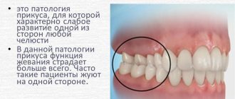
- habit of chewing on one side;
- removal of molars;
- lack of substances important for the normal functioning of the body;
- unqualified installation of prostheses.
To professionally make dentures, many measurements will be required, including the patient’s jaw at rest.
Correct teeth bite
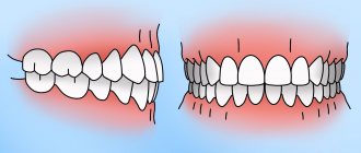
An ideal orthognathic bite implies the presence of the following features:
- The upper dental arch is slightly inclined forward and has the shape of a semi-ellipse, the lower, in the shape of a parabola, is slightly backward.
- When the jaws close, each upper tooth contacts the opposite tooth below.
- There are no obvious gaps between the teeth.
- The upper teeth overlap the lower teeth by about a third of their height.
Despite the fact that there are the above parameters for a perfect smile, the question “What should be the bite of the teeth?” cannot be answered unambiguously. In addition to the orthognathic bite, modern orthodontics allows for several options for the normal structure of the dentition and the location of the jaws.
Orthognathic
The upper row of teeth overlaps the lower one by 30% of the height, there are no gaps between the teeth or spaces between the rows.
Biprognathic
The dentition is slightly directed towards the vestibule of the mouth.
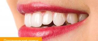
Progenic
The lower jaw moves forward slightly.
Straight
When the jaws close, the upper teeth come into contact with the lower cutting edges.
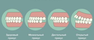
Malocclusion of teeth
Look at your reflection in the mirror. If you notice an excessively protruding upper or lower lip, teeth that “overlap” each other, or gaps between the dentition when the jaws are closed, this is a clear reason to consult a specialist. Descriptions of dental anomalies and photographs of teeth with malocclusion pathologies are presented in the table below.
Distal (prognathic)
In a distal bite, the upper jaw is more developed than the lower jaw.
Mesial (medial)
With a mesial bite, the lower jaw is pushed forward.
Cross
In a crossbite, the rows of teeth intersect in the manner of scissors when the jaws close.
Deep
In a deep bite, the upper teeth significantly overlap the lower teeth.
Open
The presence of pronounced gaps between the dentition when the jaws are closed indicates an open bite.
Initial consultation with an orthodontist
During the consultation, the following problems were identified: the absence of the 35th tooth (there is no place for its prosthetics), pathologically deep bite, bruxism, severely worn teeth in the lateral regions, minor chips on the front teeth, displacement of the jaw when opening the mouth, clicking in the TMJ on the left.
Bruxism, deep bite, increased tooth wear, TMJ dysfunction - all these problems, which are of the same nature, will only get worse without proper treatment. Treatment should be carried out comprehensively with the participation of several specialists: an orthodontist, an orthopedist, a neuromuscular dentist, an osteopath.
Bite in the absence of teeth
A person without teeth has problems with the functioning of the temporomandibular joints, facial aesthetics deteriorate, and wrinkles appear due to loss of skin tone. To avoid unpleasant consequences, it is necessary to restore the bite using a complete denture made using a special technique.
Bite restoration with complete dentures
- The central ratio of the jaws is determined - the position of the lower jaw in relation to the upper in three planes: vertical, sagittal and transversal. The role of the dentition is played by wax rollers.
- Measurements are taken using a device consisting of an external facebow-ruler and an intraoral plate with a flat frontal part and curved distal parts.
- The plaster model marks the boundaries of the future structure, the line of the middle of the alveolar process, the tubercles of the upper and lower jaw, and the midline.
- Artificial teeth are placed in such a way that when you smile, the part of the prosthesis that imitates the gum is not visible.
Mock up
A complete mock up is very useful for checking the functional and aesthetic result at the same time.
In addition, the precision and aesthetics of bisacrylic resin (luxatemp star, DMG) help to win over the patient.
Fig.8 Following the concept of a full mock up, the posterior part of the mock up is used as a reference for subsequent preparation and creates the necessary space for the occlusal veneer.
Fig. 9 Almost no preparation is required to install an occlusal veneer. All you need is a mark so that the technician can accurately recreate the boundaries of the restoration. It is necessary to make a thick edge of the veneer to avoid fracture or crack.
Fig. 10 Each veneer is fixed one after the other with a flowable dentin body shade composite (enamel HRi flowable UD1) to reduce the appearance of the transition. In addition, some subtle “waves” were created to enhance the aesthetics of the restoration.
Fig. 11 Final view of the restoration performed with emax LT A1. If the patient is not satisfied with the aesthetics, cheek veneers may be offered.
Fig. 12 View of a one-piece lithium disilicate veneer from the palatal side. Thanks to the translucency of the material, good aesthetics are created along with functionality.
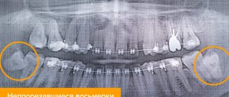
Bite after wisdom tooth removal
Some doctors are convinced that wisdom teeth are an atavism and must be gotten rid of. In ancient times, they were used to chew tough foods, such as raw meat. With the development of civilization, the human diet changed, his jaw began to gradually decrease - and the “eights” turned out to be superfluous. Most often, before starting to correct the bite, wisdom teeth are removed. This frees up space for the correct formation of rows, and the result of teeth alignment will be much better.
What affects the bite of teeth?
The negative impact of defects in the dental system on the condition of the entire organism has been repeatedly proven by scientists. The list of identified health risks is impressive even for ardent optimists.
- Due to improper distribution of the chewing load, tooth enamel wears off faster, teeth begin to react painfully to cold and hot, their necks become exposed, and noticeable gaps form between the teeth.
- Diseases of the temporomandibular joints occur, accompanied by headaches and a characteristic clicking sound when the jaws move.
- The aesthetics of the face suffers: its proportions change, wrinkles form, teeth become crooked, and the smile becomes unattractive.
- It becomes difficult to bite food, chewing functions are disrupted, causing diseases of the gastrointestinal tract.
- Speech defects appear, facial expressions are distorted, and self-esteem decreases.
- Crowding and crooked teeth cause accelerated tooth decay due to frequent chipping and carious lesions.
- The oral mucosa is injured - cheeks, gums, tongue, and the tissues of the oral cavity become inflamed.
- The quality of breathing deteriorates, which leads to diseases of the nasopharynx, trachea, and hearing aid.
Wax up
Wax up - Wax up of teeth is performed by a technician (Hilal Kudey, Bodrum, Turkey) and closely follows clinical observations and recreates lost volume.
Fig.5 To perfectly move the wax up in the mouth and increase the accuracy of the mock up restoration, wax up is 3D printed from .stl files to obtain the required hardness. This template is lined with silicone material to increase adhesion to the existing template and to prevent swelling during insertion into the mouth.
Fig.6 This 3D printed template is made of bisacrylic plastic (luxatemp star, DMG) and placed in the oral cavity to create a complete mock up. A new smile line is visible in the anterior and posterior regions, as expected during the registration of the bite height.
Fig.7
Treatment of dental malocclusion
How to change the bite of teeth? Pathologies of the dental system can only be eliminated through orthodontic treatment. An exception is the correction of a low bite, when the height of the teeth decreases with age due to their wear. In such cases, the bite is corrected by dental prosthetics with crowns or veneers, as well as by implantation. Other anomalies of teeth and bite can be removed at any age with special orthodontic structures or through surgical intervention.
However, maxillofacial operations are performed only in case of serious deviations from the norm. Usually, to correct dental malocclusion, doctors suggest correction with braces or aligners.
Now that you know about all the health risks that can cause pathologies of the dental system, and how to correct your dental bite, all that remains is to take specific actions. Even if at first glance it seems that your teeth are straight, make an appointment with an orthodontist. Diagnosing the condition of the dentition will help maintain the health of other organs and systems. And timely orthodontic treatment will make your smile more attractive and significantly reduce the number of future visits to the dentist.
Measurement methods
Anatomical method
The use of this method of measuring bite height is aimed at identifying the physiological transformation of the lower part of the facial zone.
Calculation of central occlusion using this method is based on identifying features of facial anatomy.
A decrease in bite height is expressed in the following points:
- sunken lips;
- increasing the depth of nasolabial folds;
- moving the chin forward;
- reduction in the height of the lower part of the face.
When using the anatomical method, you need to pay attention to the following points:
- the lips should be mobile and in contact with each other along the entire length without effort;
- the functionality of the orbicularis oris muscle should be high;
- The elevation of the corners of the mouth and the severity of the nasolabial folds should be determined.
Experts in the field of dentistry note that this research method is quite subjective , so it is practically not used at present.
Information content of teleroentgenograms in orthodontics in lateral and direct projection.
This material is devoted to modern diagnostic methods in dentistry.
Here https://orto-info.ru/ortodonticheskoe-lechenie/podgotovitelnyiy-period/miogimnastika.html we’ll talk about the effectiveness of myogymnastics in orthodontics.
Anatomical and physiological
The anatomical and physiological method for determining the height of the bite is based on identifying the height of physiological rest.
The technique for performing the procedure is as follows:
- The dentist places two marks on the patient's skin - at the base of the nasal septum and in the central part of the chin.
- Next, the patient is asked to make a couple of swallowing movements or pronounce several phrases, the pronunciation of which involves the lips.
After completing these actions, the lower jaw row comes to a state of rest. At the same time, the lips touch each other without tension, without stretching or sinking, and the nasolabial folds are moderately visible. - The dentist, using a special ruler with divisions, measures the distance between two points that were previously applied to the patient’s skin.
- Special templates with occlusal ridges are placed in the patient’s oral cavity, which must be lightly bitten.
- The specialist re-measures the distance between the points located on the nose and chin. The resulting indicator determines the occlusal height, which normally should be less than the previously determined height of physiological rest by 2-3 mm.
If, when determining the height of the bite, the occlusal height is equal to the indicator determined at rest, then the specialist concludes that the bite is elevated. Conversely, when the occlusal distance exceeds the resting height by more than 3-4 mm, this indicates an underbite.
After determining the bite index, the specialist removes or increases the height of the lower bite ridge until the occlusal height reaches the norm.
In this case, the dentist evaluates the condition of the tissues around the mouth. When the physiological occlusion is restored, the contours of the lower part of the face are normalized, the corners of the mouth are raised, and the nasolabial folds become less pronounced.