MAKE AN APPOINTMENT
- Removal cost
- Indications
- Preparation
- Methods and steps
- Complications
- What to do after the procedure
- Our doctors
- Reviews
- To make an appointment with a doctor
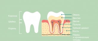
In practice, specialists at Plomba dentistry periodically encounter cases of complete destruction of the crown of a tooth and infection of the root system, in which a purulent-inflammatory process occurs. Most often, this problem can be solved only by removing the root and remains of the tooth. But sometimes the root system can be preserved for subsequent prosthetics. How the removal will be carried out, what measures and tools will be used, the doctor will be able to decide after familiarizing himself with the clinical picture.
Indications for tooth root removal
- Complete destruction of the coronal (supra-gingival) part, affecting the root system;
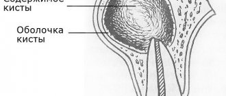
- extensive purulent-inflammatory process at the root: cyst, abscess;
- longitudinal axial fracture;
- previous incorrect extraction - during removal, fragments remained in the hole, causing an inflammatory process that affected nearby tissues.
The damaged area of the tooth is easily identified visually. Additional symptoms of the need for urgent medical intervention and even possible removal of a diseased tooth are:
- twitching, throbbing pain;
- acute pain due to mechanical action - pressing, biting, chewing food;
- unpleasant odor;
- gum hyperemia;
- a purulent process is a direct indication of the need to remove the root of a diseased tooth;
- elevated body temperature.
If one or more symptoms are present, the destroyed units are removed. In some cases, incomplete removal is performed - resection of the tooth roots. This usually occurs when the root apex is affected by periodontitis, a small cyst, or granuloma. Often in such diseases the coronal part is preserved. In this case, the damaged part is removed through an incision in the gum. Subsequently, installing a crown solves the problem of restoring the chewing unit.
How is a decayed tooth removed?
The process of tooth extraction is a simple dental operation. The difficulty increases when you need to remove the root of a tooth that is completely destroyed. Factors requiring the intervention of an experienced specialist:
- small size of the remaining crown;
- condition of surrounding tissues;
- the location of the remaining hard tooth tissues under the upper edge of the gums;
- defects of gums, roots.
Affects the complexity of the operation and whether the position of the upper or lower jaw belongs. In the upper jaw, the walls of the sockets are longer and thicker; accordingly, teeth are removed from them with great difficulty - a highly qualified dentist is required to perform the manipulation.
Wisdom teeth, from which only the root remains, are removed in the same way as ordinary molars, but in some patients, healing after such an intervention is very painful.
Examination and preparation
The procedure begins with a thorough examination and preparation of the patient. When you first visit the clinic, the doctor will take an x-ray and examine the oral cavity.
Doctor's tasks:
- determine the condition of the tooth, assess the extent of destruction;
- clarify the presence of allergies, contraindications, inflammation;
- choose a method of pain relief;
- draw up an operation plan;
- prepare tools.
The tools used to remove a rotten tooth root are a drill, forceps, and a set of elevators (photo).
A prerequisite is hygienic treatment of adjacent tissues. Extraction is possible only after removing stones and plaque from the molars, incisors or canines surrounding the surgical field. Immediately before extraction, the oral cavity is treated with a Chlorhexidine solution.
Anesthesia
There are cases when dentin is destroyed gradually, without inflammation, without the appearance of rot. In such situations, painless root removal is possible without the use of painkillers, however, anesthesia is more often necessary.
The choice of drug is carried out taking into account:
- age;
- allergic status;
- presence of somatic diseases;
- individual intolerance to drugs;
- presence of chronic diseases: epilepsy, diabetes;
- complexity of the upcoming operation.
The patient must notify the dentist about any deviations before the procedure begins. In most cases, the tooth root is removed under local anesthesia - one or two injections for incisors, 2 to 4 injections into the gums for molars. But if two teeth are destroyed, or the jaw is to be opened, the patient receives general anesthesia - he will sleep until the doctor finishes pulling out the tooth.
Features of pain relief for a tooth with a rotten root
An anesthetic injection is given at the site of the projection of the tooth roots. But if the medicine is injected into the rotten area, it may not work and the person will be hurt during the extraction process.
Treatment of patients with rotten roots is carried out in two stages. On the first visit, the dentist numbs the gum, prepares it and cleans it of pus. During the second visit, anesthesia is repeated, and the doctor removes the root that has rotted inside the gum.
Removal
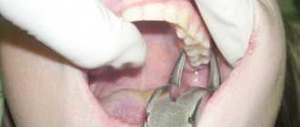
The doctor decides how to remove a tooth if only the root remains. Usually they start with the use of forceps. Even if the destroyed roots remain under the gum, the holes do not completely heal - the dentist can carefully pick up the remnants of hard tissue and easily pull them out.
If the tooth has crumbled to the very base, it is pulled out with an elevator. Having inserted the instrument between the gum and dentin, the doctor presses on the handle and makes rotational movements of small amplitude. As a result, the periodontal fibers shift and the root is squeezed out of the socket.
A drill is used when it is necessary to crush the hard tissues of a molar before removal. With proper anesthesia, this procedure is painless; discomfort is possible only when the gums are already healing.
Relieving inflammation
When removing a rotten root, an inflammatory process is often detected. To ensure that the wound heals safely and does not fester, it is customary to treat it with an antiseptic. But one treatment will not provide adequate prevention, so an anti-inflammatory drug is placed in a fresh hole. With it, the hole will heal faster, and the patient will have less chance of developing alveolitis.
Stitching
To extract the root system, the doctor separates and lifts flaps of soft tissue; they can only be attached back by suturing. It is customary to tighten the edges of the holes with threads during double or triple removal, when a significant part of the gum has been subjected to preparation. This is done so that the affected area heals faster and does not cause discomfort to the patient.
Preparing for tooth extraction
Removing a tooth or its roots is a rather complex surgical dental procedure, but it will not be difficult for the patient to prepare for it. If local anesthesia is planned to be used for pain relief, the patient should eat a large meal before visiting the dentist because:
- After removing a tooth or root, it is forbidden to eat for several hours;
- salivation after eating is significantly reduced, which will make the dentist’s work easier;
- after eating, blood glucose levels are normalized and the risk of loss of consciousness under the influence of local anesthesia is reduced
In the case of general anesthesia, on the contrary, it is necessary to abstain from eating for several hours before the removal procedure begins. Drinking alcohol before visiting the dentist is prohibited. Alcohol affects the structure of the blood and does not combine well with anesthetics, not to mention the negative impact on the human psyche and behavior.
Inflammatory and infectious diseases of any nature must be cured before surgery to remove a tooth or its roots. The dentist must be warned about the presence of allergies to certain medications, in particular to anesthesia drugs.
A normal pregnancy in general is not a contraindication to dental procedures. However, during this period the use of a number of drugs used in dentistry is prohibited, so information about pregnancy is entered into the patient’s dental record. Also, detailed information about the patient’s chronic diseases, especially heart pathologies, is recorded in the dental record.
In what cases should teeth not be removed?
Contraindications to the operation are:
- acute infectious diseases;
- menstruation in women;
- hypertensive crisis, severe arrhythmia and other cardiac disorders;
- I and III trimesters of pregnancy;
- taking medications that reduce blood clotting;
- hemophilia and other blood diseases;
- epileptic seizures;
- schizophrenia and other mental illnesses.
If the listed pathologies and conditions are identified, the issue of tooth extraction is considered on an individual basis.
Methods and stages of removing teeth or their roots
Most often in modern dentistry, only two methods of removing teeth or their roots are practiced:
- removing a tooth from the gum using forceps;
- rocking of the tooth and its rotation around its axis by elevators.
In cases where the roots are deep, the gum tissue can be cut with a scalpel. In general, the process of removing teeth or their roots is divided into the following stages:
- separation of the round ligament from the neck of the tooth (ligamentotomy);
- applying (installing) forceps to the tooth;
- advancing the fixing elements of the forceps under the gum;
- final fixation of the forceps;
- rotation (rotation) or luxation (swaying) of the tooth;
- extracting a tooth or its roots from the socket.
Main stages of the operation
- Syndestomy;
- Forceps delivery;
- Advancing the forceps to the area of the tooth neck under the gingival margin;
- Fixation;
- Luxation (rotation);
- Traction (extraction).
When removing a tooth with forceps, you must follow the following basic rules:
- Apply the cheeks of the forceps only to the vestibular and internal surfaces.
- The longitudinal axis of the cheeks of the forceps must coincide with the longitudinal axis of the tooth being removed.
- Avoid placing the tip of the cheeks of the forceps on the edge of the alveolar process and avoid subperiosteal resection of the edges of the socket.
- When dislocating a tooth, you must remember the possibility of a fracture in the neck or at the apex of the root.
When removing the upper teeth, the patient sits in a dental chair with the back slightly reclined, resting the back of his head against the headrest. The chair is raised to a position in which the tooth to be removed is at the level of the doctor’s shoulder joint. The doctor stands to the right and in front of the patient. When removing lower teeth, the chair is lowered so that the tooth being removed is at the level of the elbow joint of the doctor’s lowered arm. When removing lower incisors, canines, premolars and left molars, the doctor stands to the right and in front of the patient; when removing right molars, the doctor stands behind and slightly to the right.
Common complications after tooth extraction
Tooth extraction is essentially a full-fledged surgical intervention. Symptoms such as pain and inflammation in the surgical area are considered normal for the rehabilitation period, unless they are too severe and are not eliminated 3-4 days after tooth (root) extraction. The rehabilitation period may also be characterized by increased body temperature and enlarged lymph nodes.
More serious clinical complications include:
- renewed bleeding from the socket after tooth (root) removal - methods for eliminating minor bleeding can be discussed by the dentist; in case of intense bleeding, it is necessary to urgently consult a specialist;
- incomplete removal of the tooth root - the presence of residues is diagnosed by x-ray and follow-up and quickly eliminated;
- alveolitis is a dangerous, but easily eliminated by antibiotics, inflammatory process in bone tissue, characterized by a significant increase in body temperature, swelling, severe pain, and requires immediate treatment, as it can lead to sepsis.
In the first hours after removal, the patient should not eat. Cotton swabs from the surgical area can be removed 30 minutes after completion of all manipulations. In the first days after surgery, it is not recommended to eat sour, sweet, salty, very chilled or hot foods. Short-term cold compresses can relieve pain in the first days after tooth extraction.
Tooth extraction is a last resort in modern dentistry. A qualified specialist must strive by all means to save the tooth even in the most difficult cases.
The final stage of restoring oral health is not the removal, but the replacement of teeth (except for wisdom teeth).
Your feedback
I had my wisdom teeth removed at the RUTT clinic.
After six years of torment, during which he constantly made himself known at the most inopportune moments, she finally decided to break up with him. Removal was difficult (or so they said), with sawing and pulling out in parts. In terms of time, the doctor worked for at least 20-30 minutes. But it doesn’t feel like it hurts at all. Apart from picking at the jaw and pressing, I actually felt nothing. Unpleasant - yes, but not painful at all. Victoria |
10/31/2020 Leave a review Other reviews
What to do after tooth extraction
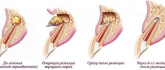
The operation cannot be prescribed without an X-ray examination, which makes it possible to determine the amount and degree of root destruction and the extent of the inflammatory process. Before root removal, the patient is given anesthesia, after which the gum is separated from the neck of the tooth. The surgeon's further actions are determined by the clinical picture. Dentists call the most difficult operation the removal of the deep and often twisted roots of the “figure eight” – the eighth tooth in the dentition. But the specialists of the Plomba clinic successfully cope with this too.
How to remove a rotten tooth root while preserving the crown
A tooth in which only the root remains is not always pulled out entirely. For example, if an inflammatory process develops at the root apex, but the tooth itself can still be saved, resection of the root apex is performed - partial removal.
The procedure is carried out after filling the canals, under local anesthesia. The operation is simple and lasts no more than half an hour. Its main stages:
- Anamnesis collection.
- Preparation of the surgical field.
- Anesthesia.
- Cutting the gum to access the root.
- Delamination of soft tissues.
- Sawing out a “window” in the bone.
- Cutting off the inflamed area of the root with a granuloma or cyst.
- Placing drugs into the cavity that stimulate bone growth.
- Stitching.
Tooth eruption problems. Wisdom tooth removal in advance
Many patients think that if the wisdom tooth is not visible on the gum, then there is no need to worry. However, over time, this molar increases in size and begins to interfere with the others. Waiting until the wisdom molar appears can lead to the following problems:
- pain in the tooth and gum;
- caries between molars;
- curvature of the roots of a healthy chewing tooth;
- infection in the place where the molar erupts because it grows incorrectly;
- curvature of the bite.
It is easier for a doctor to pick up and remove a tooth that has not yet come out. So while there is no pain, it is better to have surgery in advance.
Tooth extraction involves forcibly tearing the tissues connecting the tooth root with the walls of the socket and gum, and removing it from the alveolus. When diverging and curved roots are removed from the hole, its walls shift during the intervention, and the entrance to it expands.
The tooth is removed with special forceps and elevators. In some cases, they cannot remove the tooth. Then a drill is used to remove the bone that is preventing the root from being extracted (root cutting operation). When using a drill, it is necessary to cool it with an isotonic sodium chloride solution or Ringer's solution to prevent overheating of the bone.
When removing teeth, the lever principle is used. The pliers for removing teeth and roots have cheeks, handles and a lock. Some pliers have a transition part between the cheeks and the lock. The cheeks are designed to grip the crown or root of a tooth. The handles are the part of the forceps by which they are held and to which force is applied during surgery. The lock is located between the cheeks and handles and serves for their movable connection. To better hold a tooth or root, the cheeks have a groove on the inside with a fine longitudinal groove. The outer surface of the handles is corrugated over a considerable extent, while the inner surface is smooth.
The design and shape of the forceps are not the same. Their design depends on the anatomical structure of the tooth and its place in the dentition.
To successfully perform the operation, you should use forceps, the design of which corresponds to the anatomical features of the tooth being removed.
The operation begins with the separation of the circular ligament from the neck of the tooth and the gum from the edge of the alveoli. It is best to do this with a smoothing iron or a narrow flat rasp. Careful separation of the circular ligament and gums facilitates the advancement of the cheeks of the forceps under the gums and prevents rupture of the mucous membrane during the intervention.
Tooth extraction consists of a number of techniques carried out in a certain sequence: 1) application of forceps; 2) moving the cheeks of the forceps under the gum; 3) closing the forceps (fixation); 4) tooth dislocation (luxation or rotation); 5) extraction of the tooth from the socket (traction). The success of surgical intervention depends on the precise and consistent implementation of these techniques.
The tooth extraction operation is performed in a dental chair. The outcome of the operation largely depends on the correct position of the patient and the doctor during this intervention.
Removal of individual groups of teeth in the upper jaw. The method of removing each tooth has its own characteristics. It depends on the shape, number and location of the roots, the thickness and density of the bone around the root of the tooth, as well as the type of instrument.
Removal of incisors. The central and lateral incisors have one root that is cone-shaped and rounded; the lateral one is thinner and shorter than the central incisor. The root of the lateral incisor is slightly compressed from the sides, so its cross section has the shape of an oval. The apex of the root is sometimes curved towards the palatine side. The outer wall of the socket in the area of these teeth is thinner than the inner one.
To remove incisors, the doctor must stand to the right and in front of the patient. When removing a lateral incisor on the left side, the patient should turn his head slightly to the right, and when removing the right lateral incisor, he should turn his head slightly to the left. For a good overview of the surgical field and fixation of the alveolar process during the operation, the doctor with the second finger of his left hand moves the patient’s upper lip and places it on the outside in the area of the alveolus of the tooth being removed, with the first finger covering the alveolus from the palatal side. The central incisor is removed with straight forceps with wide cheeks, the lateral incisor is removed with the same forceps, but with narrower cheeks.
Due to the cone-shaped and rounded outline of the roots of the central and lateral incisors, they are removed by rotation (rotation). Sometimes rotational movements fail to dislodge these teeth from their sockets. Then they resort to rocking the tooth in the labial and palatal sides, then rotate it again. After this, the tooth becomes mobile and is easily removed down and out, where the wall of the socket is thinner.
Fang removal. The fang has one long, massive and laterally compressed root; its cross section resembles the outline of a triangle. The upper part of the root is curved in 30% of cases. The bone on the outside of the root is thinner than on the inside. However, both walls of the alveoli are much thicker than those of the incisors. All this creates certain difficulties when removing a fang.
The position of the doctor and the arrangement of the fingers of the left hand are the same as when removing incisors. When removing the right canine, the patient must turn his head slightly to the left, and when removing the left one - to the right. This head position is more convenient for surgery.
The canine is removed using straight, wide-browed forceps. When removing, combine rocking to the labial and palatal sides with rotation around the longitudinal axis of the tooth. The first dislocation movement is made to the outer wall of the alveolus, since it is thinner than the palatine wall, then in the opposite direction. After this, rotation is carried out.
When removing a fang, significant force is often required due to anatomical features. By successively rocking and rotating, they break the periodontal fibers that hold the root and push the walls of the socket apart. After this, the tooth is brought down and out.
Removal of small molars. The roots of these teeth are compressed in the anteroposterior direction. The root of the first small molar in 50% of cases is completely split into two thin roots (buccal and palatal), rarely - into three (two buccal and one palatal). The root of the second small molar is flattened, has longitudinal grooves on the lateral surfaces, and its apical section is split. If the root of the first small molar is split, then the palatal root is located deep in the bone. The outer wall of the alveoli of these teeth is thinner than the inner.
During removal, the patient's torso is tilted posteriorly and the head is thrown back. It is more convenient to remove the right small molar when the patient’s head is slightly turned to the left, and when removing the left one, to the right. When removing these teeth, the doctor stands to the right and in front of the patient, with the first finger of his left hand (for removal on the right) or the second finger of the same hand (for removal on the left), he pulls the upper lip and the corner of the mouth outward. Accordingly, he places the second or first finger on the side of the palate and fixes the alveolar process from the vestibular and palatal sides in the area of the tooth to be removed.
The upper small molars are located in the middle part of the dentition, so they are removed with special forceps that have an S-shaped bend. This shape of the forceps allows you to correctly apply them to the tooth and carry out lateral dislocation movements without encountering obstacles from the lower jaw.
Small molars are removed by rocking to the vestibular and palatal sides. The first dislocation movement is made outward, towards the thinner and more pliable wall of the alveoli. Movements should be smooth, especially when removing the first small molar, since sudden movements can cause a fracture of its thin roots. These teeth are removed from the hole downwards and outwards.
Removal of large molars. The first and second molars each have two buccal and one palatal roots. The buccal roots are compressed laterally, shorter and thinner than the palatine. The palatine root is massive, cone-shaped. Sometimes (especially in the second large molar) fusion of the buccal roots occurs with each other or the buccal with the palatal, less often - all three roots. The roots of the first large molar are longer than those of the second and diverge more to the sides (especially the palatal root). The top of the roots of these teeth may be slightly crooked.
The outer wall of the alveolar process in the first large molar is thickened due to the zygomaticalveolar ridge, in the second it is thinner than the palatine one. Due to the divergence of the roots, these teeth have powerful bony interradicular septa. All this complicates their removal.
The position of the patient, the doctor and the fingers of the left hand is the same as when removing small molars. The first and second large molars are removed using S-shaped curved forceps, which have different cheek structures for the teeth on the left and right sides. One of the cheeks has a spike at the end; it is placed on the outside of the tooth. The spine enters the groove between the buccal roots. The other cheek with a semicircular or flat end is located on the palatal side.
Large molars are removed by rocking to the buccal and palatal sides. The dislocation of the first large molar begins in the palatal direction, the second - in the buccal side. The tooth is removed from the socket downwards and outwards.
Removal of the third major molar. This tooth has several, often fused roots, forming a conglomerate of a cone shape. The crown of the tooth is smaller and the roots are shorter and more curved than those of the first and second molars. To remove this tooth, special forceps are used, which have short and wide cheeks with rounded ends and pits (to cover the crown of the tooth) on the inside.
The tooth is dislocated by rocking first to the buccal side, then to the palatal side. Removing a tooth with fused roots is usually not difficult. Removing a tooth with diverging, curved, or bent roots can be more difficult.
Removal of individual groups of lower jaw teeth. Removal of incisors. These teeth have one straight, thin and significantly compressed root from the sides, having the shape of an elongated oval in cross section. The root of the lateral incisor may be slightly bent. The bone of the alveoli in the area of these teeth is thinner on the outside than on the inside.
When removing the lower incisors, the patient sits in a chair in an upright position, the head is tilted slightly forward, the chin is down. The doctor stands on the right and slightly in front of the patient, with the first finger of his left hand he pushes back the lower lip and rests it on the outside against the alveoli of the tooth being removed, with the second finger he presses the alveoli from the inside, with the third finger he places it on the chin and holds the lower jaw with it.
The lower incisors are removed using rib-curved forceps with narrow cheeks. Wide jawed forceps can cause damage to the adjacent tooth. The forceps are placed on the tooth so that one of the cheeks is located on the lingual side, the other on the labial side, and the handles are on the outside of the jaw. The tooth is dislocated from the socket by rocking. First, it is shifted to the labial side, where the bone is thinner and more pliable, then to the lingual side. The tooth is removed from the socket upwards and outwards.
Fang removal. The root of this tooth is wider and longer than that of the incisors. It is compressed from the sides and has a cone-shaped shape. There are well-defined longitudinal grooves on the lateral surfaces. The apex of the root is curved and very rarely bifurcates into lingual and labial parts. The outer wall of the alveoli is thinner than the inner.
The position of the patient when removing the lower canine is the same as when removing the lower incisors. The doctor stands to the right and in front of the patient. When removing the left fang, the patient turns his head slightly to the right, and the right one - to the left. The placement of the fingers of the doctor's left hand is similar to their position when removing the lower incisors.
To remove a fang, forceps are used that are designed to remove lower molars and have wider cheeks. The tooth is dislocated by swinging it first to the labial, then to the lingual side. To finally free the tooth root from the tissues holding it, you can make light rotational movements. The tooth is removed from the socket upwards and outwards.
Removal of small molars. These teeth have one rounded root, somewhat compressed from the sides (especially in the upper part). Sometimes it is crooked. The root of the second molar is more massive and long; its bifurcation at the apex is very rare. The buccal wall of the alveoli in these teeth is somewhat thinner than the lingual, or both walls are almost the same thickness.
When removing right small molars, the doctor stands to the right and slightly behind the patient. Grasping his head with his left hand, he inserts fingers I and II into the oral cavity and grasps the alveolar process on both sides. At the same time, the second finger pulls back the corner of the mouth and pushes back the cheek, the first finger - the tongue. The remaining fingers of the left hand support the lower jaw by the chin.
When removing small molars on the left side, the doctor stands to the right and in front of the patient, turns his head towards him, with the second finger of his left hand he pushes back the cheek, with the third finger he moves the tongue, with the first finger he supports the lower jaw by the chin.
Forceps for removing small molars are the same in shape and design as for removing lower incisors, only with wider cheeks. Due to the thick walls of the alveoli, it is not possible to push the cheeks of the forceps deeply. This causes certain difficulties during removal, especially if the tooth crown is insufficiently strong.
The small molars are dislocated by rocking, first to the buccal, then to the lingual side. The shape of the tooth roots allows these movements to be combined with light rotational movements. The dislocated tooth is removed from the socket up and towards the cheek.
Removal of large molars. The first and second lower molars have two roots: anterior and posterior. The roots are compressed in the anteroposterior direction, flat. The anterior root is longer and thicker, often having a slight arched bend forward. The posterior root is straight, deviated posteriorly. In some cases, significant divergence and curvature of the roots are possible. Complete fusion of roots or only their apical sections rarely occurs. The sockets of these teeth have thick and durable walls. In the first large molar, the thickness of the buccal and lingual walls of the socket is the same, in the second, the buccal wall, due to the oblique line passing here, is thicker and more powerful than the lingual.
The position of the patient, the doctor and the fingers of the left hand is the same as when removing small molars. Remove with beak-shaped or plane-curved forceps, which have wide cheeks with triangular protrusions (spikes) at the ends. The forceps are applied and advanced so that the triangular protrusions (spikes) of the cheeks enter the space between the roots.
These teeth are dislocated with rocking movements. The first large molar is dislocated first in the buccal, then in the lingual side, the second - in the lingual, then in the buccal side. The tooth dislocated from the socket is removed upward and to the buccal side. The presence of two diverging roots and the significant thickness of the bone walls of the alveoli sometimes create great difficulties when removing large molars.
Removal of the third major molar. This tooth has anterior and posterior roots, which can grow together into one cone-shaped root. Often the roots are significantly curved and bent back. In some cases, this tooth has three diverging or fused roots or more. On the outside the alveolus has a very thick compact layer of bone (due to the oblique line), on the inside it is thin.
Anomalies of the tooth (size, shape, eruption), and structural features of the bone often create great difficulties when removing it. The position of the patient, the doctor and the fingers of his left hand is the same as when removing small and large molars. Removal is carried out using beak-shaped forceps or forceps curved along a plane, having triangular protrusions at the end of the cheeks. Dislocate the tooth with smooth movements, shifting it first to the lingual, then to the buccal side. Sometimes it is not possible to remove a tooth with forceps, then elevators are used. The tooth is removed from the socket up and towards the cheek.
"Surgical Dentistry" edited by Robustova T.G.
Fourth edition. Moscow "Medicine" 2010
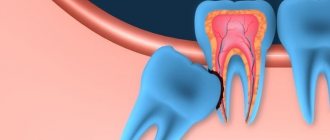
When is it necessary to remove the lower wisdom tooth?
Eighth teeth are removed in two cases: when they have not yet sprouted or are already protruding from the gums. In the first case, the operation is performed if the patient feels discomfort, and during an external examination the doctor does not see diseased teeth. The appearance of pain means that the molar in the gum is pressing on its neighbor and disturbing him.
If the wisdom tooth has erupted incorrectly - that is, not straight up, but with a deviation, at an angle to the jaw and neighboring teeth, then removal is also a mandatory procedure. The slope of the figure eight is:
- distal - when the “eight” is tilted back, away from the “seven”. In this case, there is a risk of injury to the gums and damage to the roots of the adjacent tooth;
- medial - when the “eight”, on the contrary, is inclined towards the neighboring tooth. This increases the likelihood of damage to the crown part of the “seven” and the development of caries on both teeth;
- buccal – in this case, the wisdom tooth is turned towards the cheek and constantly touches the mucous membrane, causing irritation and hardening of the tissues. This condition can eventually lead to the formation of a tumor on the mucosa.
The removal of a wisdom tooth in the lower jaw is definitely necessary in the following cases:
- Caries on a wisdom tooth.
- Lack of space in the row for teeth. The last molars put pressure and cause discomfort.
- Pain in the gum where the wisdom tooth is located, at any time of the day, even after sleep.
- Incorrect growth sideways towards the tongue or larynx. This phenomenon is called dystopia.
- It interferes with implantation.
A tooth can erupt without causing pain once it appears; it will still be removed because it is not growing properly. The dentist makes an accurate diagnosis. To make an appointment, leave a request on the website or call one of the numbers: +7-924-444-05-45 - administrator
Removing a wisdom tooth in the lower jaw is a more complex procedure than removing an upper tooth. The bottom eights have a more powerful root system, and the jaw bones are denser and thicker. To remove a lower molar, it is necessary to perform a complex operation with dissection of the gum. In some cases, the surgeon has to remove part of the bone, divide the root into several parts and extract them separately.
Sign up for a free consultation
Removal of a wisdom tooth on the lower jaw
Most people of conscious age go to the dentist because of problems with their lower wisdom teeth.
They often interfere, put pressure on neighboring molars, and cause discomfort or pain after eating. Most often, wisdom teeth in the lower jaw appear between the ages of 20 and 25, although the timing varies from person to person; for some, they may appear at the age of 16, for others closer to 40 years. In most cases, the appearance of figure eights is accompanied by various complications that require their removal. Malocclusion, difficulty in teething, tumors and trauma to the mucous membrane and much more.
Make an appointment for lower wisdom tooth removal
Byshlyaga Dmitry Yurievich
Byshlyaga Dmitry Yurievich Orthopedic dentist
Extensive practical experience. Regularly undergoes internships and advanced training courses.
Working hours: from 9.00 to 20.00, daily, seven days a week, by appointment
Make an appointment
More details
Asatryan Alexander Aramovich
Asatryan Alexander Aramovich Dentist therapist - orthopedist - surgeon
Extensive practical experience. Regularly undergoes internships and advanced training courses.
Working hours: from 9.00 to 20.00, daily, seven days a week, by appointment
Make an appointment
More details
×
Tools used for complex removal
When extirpating figure eights, a number of instruments are used:
- Pliers consisting of cheeks, a handle, and a lock are used if the crown is not destroyed or is slightly damaged (there is something to grab onto). They differ in shape, structure, and size. Different forceps are used to extract the upper or lower tooth.
- The elevator acts like a wedge - it loosens the dental unit, tearing the ligaments and pushing it out of the socket. The tip is immersed in the periodontal fissure, then rotated around the axis using a handle. It removes fragments well from a damaged crown, a shallow fracture, and is suitable for the extraction of dystopic, impacted teeth.
- An excavator is used to extract softened parts and root fragments. It has an angular shape with pointed edges. Easily inserted between the alveolus and the remaining tooth.
- The chisel allows you to destroy the interroot connection, actually gouges out the tooth, but is rarely used for the figure eight.
- The drill is used to divide a multi-root system into parts, especially in cases of crown destruction, atypical location, impacted teeth. The gradual extraction of dental elements causes less trauma to surrounding tissues.
- An osteotome is used to cut and remove part of the bone that is blocking the removal of a molar.
Modern techniques include the use of laser or ultrasound equipment. In this case, fewer complications arise, and the recovery period takes less time.
Removal methods
Today, there are 4 techniques for extracting molars: simple, surgical or complex, atraumatic and according to the indications of an orthodontist. A simple method is usually understood as extracting a tooth that has completely erupted above the surface of the gum. In this case, removal occurs using special forceps. First, the tooth is slightly loosened from side to side and then removed.
The surgical method is intended for more complex cases, for example, to remove impacted or hard-to-reach, decayed teeth. In this case, the specialist cannot limit himself to using only forceps; a scalpel and other devices necessary for dissecting soft tissues will also come to the rescue. After such an operation, the resulting wound can take quite a long time to heal, which will cause a lot of inconvenience in everyday life.
Atraumatic removal is the most progressive method, which is painless and fast. First, the doctor administers the required dose of local anesthetic and then removes the tooth without using forceps. The cost of this procedure is quite high, but the risks of complications and infection in the wound are minimal.