Osteotomy is a surgical operation aimed at eliminating deformity or eliminating anatomical and functional disorders by artificially breaking the bone and correct fusion. This operation is performed to treat both congenital and acquired pathologies, for example, in case of improperly healed fractures.
Indications for osteotomy:
- improperly healed bone fractures;
- ankylosed joints;
- change, shortening of the limb;
- rachitic curvatures;
- formation of a false joint;
- osteomyelitis;
- osteoarthritis, spondioarthrosis;
Depending on the surgeon’s goal and the patient’s clinical picture, the following types of osteotomy can be performed:
- wedge-shaped;
- linear (oblique and transverse);
- hinged (can be arc-shaped, that is, made in one plane, or angular, spherical - in several planes);
- staircase;
- Z-shaped;
- derotational.
By purpose they are distinguished:
- Corrective osteotomy - to correct deformity after complications that led to malunion.
- Hinge osteotomy is an operation to lengthen the limbs.
Osteoplasty techniques are also divided into closed and open. The first is performed with minimal access, through a 2-3 cm incision. The open method involves wide access - an 8-12 cm incision is made exposing the bone. In some cases, when there is a risk of damage to nerves and large vessels, preference is given to open osteotomy, despite its low invasiveness, over closed one.
The choice of method depends on the type of pathology, volume and area for surgical intervention - pelvis, jaw, hip, etc. Most often, the operation is aimed at restoring the function of the musculoskeletal system and removing bone deformations.
To equalize the lengths of the lower extremities, osteotomy is performed according to the principle of oblique cutting of the bone in mandatory combination with extrafocal compression-distraction osteosynthesis. The design for osteosynthesis is selected individually for each patient.
Progress of the operation
The instruments used are osteotomes (chisels) with a grooved or flat section, Gigli files, ultrasonic devices for cutting bones, and electric medical saws. After fragmentation of bones during osteotomy, their fixation is required, for which knitting needles, plates, screws or special devices are used. In some cases, plaster casts are additionally applied, but they try to avoid this so as not to cause discomfort to patients and eliminate the risk of developing joint contractures. Anesthesia - general anesthesia or local anesthesia, it all depends on the general health of the patient and the scope of the intervention.
Open osteotomy
A wide surgical field is created, the bone is exposed, the periosteum is separated and a dissection is made in the desired area. The fragments are connected with metal structures or a plaster cast is applied.
Closed method
The external incision on the skin is minimal - 2-3 cm. The muscle tissue is separated above the site of the intended bone dissection. The chisel is positioned longitudinally to cut the periosteum. After this, the instrument is turned perpendicular to the bone and cut through it with several blows with a hammer on the chisel. Nearby soft tissues, nerves and blood vessels are protected with special instruments to prevent damage.
Osteotomies are also classified according to their intended purpose: corrective (for example, for immobility of the knee joint), aimed at improving the supporting function, shortening the bone or lengthening it, derotational, etc.
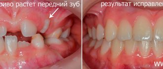
If the patient has a clear bow of the big toe, a proximal corrective osteotomy is indicated. It is performed to correct the first metatarsal bone and is needed for the following symptoms:
- inability to put on shoes;
- pain syndrome;
- incorrect gait;
- bursitis.
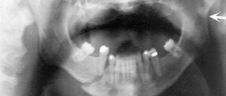
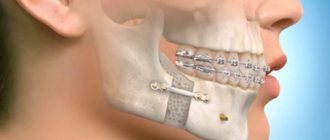
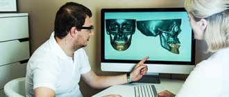
Osteotomy of the upper and lower (planar) jaws can be performed on only one of the jaws or on both at the same time. It is indicated for developmental anomalies, serious problems with bite, existing negative consequences from previous operations, displacements, and fractures. Surgical intervention can be complete or fragmentary. As a result, after the operation, the process of biting and chewing food is easier for patients, the period of tooth wear is reduced, the risk of tooth decay due to poor bite is eliminated, the relationship between the jaws is normalized, which has a good effect on the appearance in general, and the risk of developing diseases of the temporomandibular joint is reduced.
Osteosynthesis of the lower jaw
In order to immobilize fragments of the lower jaw, open focal osteosynthesis is used (bone suture, mini-bone plates with screws, staples with predetermined properties, osteoplast glue, quick-hardening plastic, surrounding suture without a supragingival splint, internal bone rods and sometimes Kirschner wires), closed focal osteosynthesis (Kirschner wires, surrounding suture), closed extrafocal osteosynthesis (surrounding suture with a splint, S-shaped and unified hooks, extraoral clamp and pin devices) and open extrafocal osteosynthesis (surrounding suture with a splint, extraoral clamp and pin devices).
Typically, osteosynthesis is used in cases where conservative methods have not given the desired result or when, after examining the patient, it has become clear that conservative methods will not provide adequate reposition and effective fixation of fragments.
Indications for osteosynthesis are fresh, old, improperly healing linear or comminuted fractures of the lower jaw of any location with or without a bone tissue defect:
- fractures within the dentition with an insufficient number of stable teeth on both jaws;
- fractures within the dentition with the formation of a large toothless fragment;
- fractures within the dentition with significant displacement of fragments and the impossibility of their reposition in any other way;
- fractures behind the dentition with displacement of fragments;
- pathological fracture resulting from tumor growth or chronic osteomyelitis;
- large- and small-comminuted fractures of the body and branches of the lower jaw;
- defects of the body and ramus of the jaw with preservation of the condylar process;
- osteoplasty of the lower jaw;
- reconstructive operations for congenital or acquired deformation of the lower jaw.
When describing a specific type of osteosynthesis, additional recommendations for each of them will be given.
The operation of osteosynthesis of the lower jaw is carried out under conduction, including stem, and infiltration anesthesia or under anesthesia with intubation through the lower nasal passage. With open focal osteosynthesis in the area of the condylar process, angle and lateral parts of the body of the lower jaw, the skin and underlying soft tissues are dissected 1.5-2 cm below the base of the jaw so as not to damage the marginal branch of the mandible (R.marginalis mandibulae) of the facial nerve. In the anterior parts of the body of the lower jaw, a tissue incision is made in the submental region. The base of the jaw is exposed layer by layer, the periosteum is dissected, and the bone is skeletonized with a rasp for at least 2 cm on both sides of the fracture gap. Access can also be performed using intraoral access, when all incisions are made from the side of the oral cavity. After examining the fracture gap and adjacent areas of the bone, removing loose bone fragments, extracting blood clots and soft tissue embedded between the fragments, clarifying the nature of the displacement of the fragments, the method of their reposition and temporary retention for applying a fastening device, they choose the type of osteosynthesis planned before the operation, or decide to use another method (or methods) of osteosynthesis. Closed focal osteosynthesis can be carried out only if the fragments are easily reduced by hand. The same condition will allow the use of extraoral clamp and pin devices - closed extrafocal osteosynthesis. Some types of closed extrafocal osteosynthesis (surrounding suture, S-shaped and unified hooks) do not require such a condition, since they themselves perform reposition and fastening of fragments of the lower jaw.
Rehabilitation after osteotomy
Let's consider the recovery period after surgery using the example of corrective osteotomy.
The patient is in the hospital under the supervision of doctors for 3 to 7 days. At this time, regular treatment of the surgical wound is carried out, painkillers, antibiotics and anti-thromboembolism drugs are given. It is important to start getting up as early as possible and not to lie around.
After discharge, the patient must continue taking the prescribed medications at home. On the 10-14th day, the sutures are removed. Next, rehabilitation is indicated - the use of crutches for up to 6-12 weeks, regular visits to the doctor for examinations. Heavy physical activity will be allowed only 6-8 months after the operation. Sometimes installed metal structures are removed after 1-2 years.
Hospital After the operation, you will remain in the hospital for 7 to 15 days, depending on your general condition and the characteristics of the surgical procedure. After the swelling goes away, the pain decreases and a more or less normal rhythm of eating and activity is restored, you will be discharged for home rehabilitation. You will be provided with all the necessary documents and recommendations.
Nutrition. For 4 weeks after surgery, you will need to eat high-protein foods in the form of purees or broth (for example, ground chicken, yogurt, fruit puree, etc.). Food should be cold or warm, not hot. Subsequently, you can gradually return to your usual diet.
Medicines. You will be prescribed a set of necessary medical support (antibacterial and anti-inflammatory therapy, pain relief). Upon returning home, you must take prescribed medications and procedures. Remember, it is not recommended to take aspirin or aspirin-containing medications. You should not take any non-prescribed medications and avoid drinking alcoholic beverages for 4 weeks after surgery. Do not take medications on an empty stomach.
Temperature. You may experience an increase in body temperature up to 38.0 degrees in the first 3-7 days after surgery. Do not worry. This is the body's reaction to the intervention. If the temperature is significantly higher or lasts more than 5 days, please report.
Edema. You will experience swelling, which peaks on the 3rd day after surgery. The swelling may be quite massive and spread to the neck. Do not worry. This is fine. To prevent swelling from increasing, it is strongly recommended to apply ice to the cheeks and neck on both sides of the face for 3 days after surgery. As the swelling resolves, the skin color will change places several times (maroon, blue, green and yellow). This picture can be observed up to the décolleté level and last up to 2-3 weeks. Swelling and discoloration are never the same on both sides of the face and neck. Remember that each area heals independently and independently. It is also important to understand that swelling will not affect the results, most of which can be seen after 5-6 weeks. The final result is assessed after 1 year.
Sensitivity. In the postoperative period, a decrease or even loss of sensitivity (numbness) may be observed in the area of the lips, chin, lower jaw on both sides and in some places of the oral cavity. This occurs as a result of the surgical protocol in the vicinity of nerve fibers. On average, restoration of sensitivity takes from 2 to 6 months. In some cases, full recovery may take up to 12 months. You will be recommended a set of exercises to speed up the recovery of sensitivity.
Lips. Immediately after surgery, you will “lose control of your lips,” which may result in slight drooling, difficulty swallowing liquids, and difficulty speaking. Difficulties in controlling lip movement arise for a number of reasons: surgical trauma to the soft tissues of the lips, movement of the jaws and teeth into a new position, an expected decrease in sensitivity, the presence of orthodontic structures with rubber traction, and the possible presence of a splint. It is necessary to actively work with your lips - try to close them. By doing such exercises you will speed up the restoration of coordinated movements, sensitivity and reduce swelling of the area. All this will help to quickly normalize speech and the act of swallowing. Always use Vaseline or similar products to moisturize your lips. You will be recommended a set of exercises to restore lip function.
Nose. As a rule, nasal congestion occurs immediately after surgery and can last up to 7 days. It is caused by swelling of the nasal mucosa and hardening blood clots. You may also have a bloody nose for the first 2 to 5 days after surgery. This is normal, don't worry. Keep the visible part of your nose clean. You can use petroleum jelly or similar products to moisturize the area. If you sneeze, allow the air to escape through your mouth.
Hygiene. You will need an antibacterial mouth rinse to prevent inflammatory complications. You should brush your teeth with a soft toothbrush starting the next day after surgery. Care must be taken not to injure areas where there are postoperative wounds. You can start washing your hair one day after surgery (if there are no previously agreed restrictions). It is important that someone is present during this process, as you may feel weak and dizzy. For the first week after surgery, you must wash your hair strictly while sitting.
Bandage. You may have a bandage applied to your face and/or neck. Do not attempt to remove or replace it yourself. It is important to keep the dressing dry.
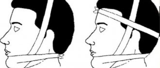
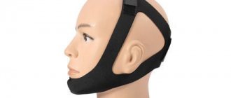
Splint. In some cases, you may have a splint (plastic positioner) installed, which will be clamped between the jaws to hold them in the required position using rubber rods. The duration of its stay can be up to 3 weeks. Don't try to get it.
Dream. For the first week after surgery, I recommend sleeping with your head elevated (for example, with two pillows). This position promotes the natural drainage of fluid from the facial area and helps reduce swelling. You need to understand that your sleep cycle may be disrupted in the first 1-2 weeks after surgery. This is a possible normal reaction. Drive with caution for the first 2 weeks after surgery.
Physical activity. Stay quiet for the first day or two after surgery. Avoid stressful situations. For the next 4 weeks, it is necessary to limit physical activity (for example: weight lifting, long walks, fitness training, active sports). During this time, exercise at a quarter of your capacity. Also, after 2 weeks after surgery, you can begin to perform a set of exercises to develop the lower jaw.
Cosmetics. If possible, refrain from using cosmetics for the first two weeks after surgery.
Possible complications
Some complications arise already during osteotomy, others during the rehabilitation period.
- Incorrect fusion of bones. Occurs due to improper fixation of bone tissue fragments during surgery. With such a complication, repeated intervention is necessary.
- Non-union of bones. It may occur due to severe concomitant pathologies, smoking, impaired blood supply to the operated area, or osteoporosis. For treatment, a repeat operation is performed and special rehabilitation is prescribed.
- Compartment syndrome. Occurs if, during surgical manipulation, the muscles were strongly compressed by a tourniquet. For treatment, certain drugs are prescribed; if the case is severe, a fasciotomy is performed.
- Improper functioning of the joints located next to the surgical field. This complication is typical for the absence or violation of rehabilitation rules. Exercise therapy is prescribed.
- Infections. They can be introduced during surgery or due to improper wound care. An antibiotic is prescribed for treatment; in severe cases, revision surgery will be required.
- Nerve damage is a surgeon’s mistake or a peculiarity in the location of the patient’s nerve endings. The function of a damaged nerve cannot be restored.
- Thromboembolism. Occurs when anticoagulants are prescribed incorrectly, getting out of bed late, or the inability to wear compression stockings. To eliminate this complication, high doses of antiplatelet agents and anticoagulants are needed.