Osteomyelitis of the jaw is an inflammatory and infectious process with purulent contents that covers the structural elements of the jaw bone. Gradually, the disease provokes bone decomposition, that is, osteonecrosis gradually progresses. The pathology requires urgent professional help to avoid the spread of infection to other organs and systems of the human body. Osteomyelitis of the lower jaw is diagnosed twice as often as the same disease, but affecting the upper jaw system.
Osteomyelitis of bone: what is it?
Bone osteomyelitis in the concept of general surgery is an inflammation of bone tissue, which has a rather complex pathogenesis. In modern medicine there are many theories of its origin. However, it is impossible to determine the most reliable one, since each of the theories does not exclude the others, but complements them. Thus, osteomyelitis is a multifactorial disease, in the development of which not only the penetration of any infectious agent into the bone tissue plays a major role, but also the state of the human body’s immune system, local circulation disorders with deterioration of trophism.
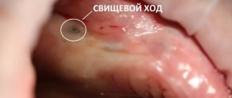
When an infectious agent enters bone tissue, a violent reaction of the body develops, manifested by purulent inflammation. To destroy the infection, leukocytes begin to actively migrate to the site of the lesion, which produce a huge amount of enzymes. They gradually destroy bone structures and form cavities filled with liquid pus, in which pieces of bone or sequestra can be found. Sometimes the inflammation spreads to the surrounding soft tissue, which leads to the formation of fistula tracts that open on the skin.
If the immune system of a sick person is active enough, then inflammation can be limited on its own and become chronic. But if there is an immunodeficiency in the body, then the infection spreads further with the development of severe purulent complications, such as sepsis, which often leads to disability or even death.
Why does pain occur in the upper jaw?
Injuries
Damage occurs as a result of household, street, sports, automobile, and industrial injuries.
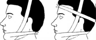
The bruise is characterized by moderate pain that goes away after a few days. Fractures of the upper jaw are accompanied by extremely intense acute pain, rapidly increasing swelling, facial asymmetry, and stepped dentition. In case of fractures of the alveolar process, lacerations are visible on the mucosa, and sometimes the end of the displaced bone fragment is determined. The occlusal contact is sharply impaired, the teeth are mobile. With an isolated fracture of the walls of the maxillary sinus, severe aching pain in the upper jaw, infraorbital area, significant swelling, and hemorrhages is observed. Nasal breathing is difficult. With combined damage to the bone walls of the sinuses, a clinical picture of a concussion and profuse nosebleeds are revealed. Perforation of the maxillary sinus occurs during dental procedures. If the damage was not detected, swelling of the cheeks, a nasal tone of speech, pressing or bursting pain in the jaw, and projections of the sinuses subsequently appear.
In some cases, radiating pain in the jaws is detected in victims with subluxation of the cervical vertebra. Irradiation to the back and shoulders is also possible. The clinical picture includes a forced position of the head, neck pain, muscle tension, and sometimes dizziness, weakness, convulsions, and paresthesia in the arms.
Dental reasons
Discomfort and mild pain may be associated with the use of removable dentures and orthodontic structures. Pulling, pressing, aching pains occur in children due to malocclusion, including those caused by deformation of the upper jaw with a cleft lip and cleft palate. Some soreness is normal after tooth extraction, especially molars and wisdom teeth.
With the development of alveolitis, the pain disappears, and then reappears 3-5 days after tooth extraction. Intense pulsating sensations are noted in the projection of the socket, intensify as inflammation progresses, and sometimes cover the upper jaw and half of the face. Attacks of severe pain spreading along the trigeminal nerve are characteristic of acute diffuse pulpitis. More local pain is observed in acute periodontitis.
Upper jaw pain
Purulent processes
Intense tugging, tearing, bursting pain occurs with purulent inflammation of the upper jaw and nearby soft tissues. Combined with hyperthermia, deterioration of general condition, intoxication syndrome. The most striking clinical picture occurs in acute osteomyelitis. The disease begins suddenly, the symptom progresses quickly, and the temperature rises to high levels. A foul odor emanates from the mouth, and pus accumulates in the gum pockets.
Periostitis has less severe symptoms. With a high intensity of pain, the general condition is slightly disturbed, the temperature is subfebrile. In patients with a perimandibular abscess, the abscess is limited, located in the soft tissues, the condition is moderate or closer to satisfactory. With perimaxillary phlegmon, the infection spreads quickly, twitching, shooting pains intensify with the slightest movements of the jaw, the condition is serious.
With abscesses of the salivary glands, the first symptoms are dry mucous membranes and an unpleasant taste in the mouth. Hyperthermia up to 40°C is noted. Maximum pain is determined in the projection of the affected salivary gland, complemented by pronounced swelling. Irradiation is noted in the upper jaw, neck, and ear.
Neuralgia
With ganglionitis of the pterygopalatine ganglion, a clinical picture of neuralgia of the trigeminal nerve is observed in the zone of innervation of its 2nd branch – n.maxillaris. An attack of intense shooting pain develops spontaneously, often occurring at night. Pain sensations predominate in the upper jaw, eye, hard palate, and at the base of the nose, spreading to nearby anatomical zones. The episode lasts from several minutes to several hours, complemented by autonomic disorders: lacrimation, profuse salivation, hyperemia of half the face.
Atypical facial neuralgia, which is more often detected in middle-aged women, is considered as another possible cause of the symptom. Pathology is provoked by dental procedures. The pain is dull, sometimes burning. They do not reach the intensity typical of other neuralgia. They quickly transform from paroxysmal to permanent.
Diseases of the ENT organs
In otolaryngology, the manifestation is more often provoked by odontogenic sinusitis against the background of injuries, dental diseases, and endodontic treatment. The acute form is characterized by heaviness, bursting unilateral pain in the upper jaw, intensifying when lowering the head, and throbbing headache. There is a sharp pain when chewing food, a subjective feeling of lengthening of the teeth. For chronic sinusitis, the clinic unfolds gradually. The symptom is also combined with a headache, radiating to the forehead, temple, and orbit.
Radiating pain in the upper jaw, orbit, and temporal region can be observed in acute purulent otitis and is caused by irritation of the trigeminal nerve during infiltration of the mucous membrane of the tympanic cavity. Supplemented by severe pain in the ear, intoxication syndrome. A similar irradiation is found in mastoiditis, which develops simultaneously with otitis media or a few days later, manifested by profuse suppuration from the ear, throbbing pain behind the ear.
Tumors of the upper jaw
Against the background of benign neoplasia of the upper jaw (fibromas, cementums, osteomas, osteoblastoclasts), the pain is usually mild, dull, and aching. They do not occur in all patients. They grow slowly over a long time in parallel with the growth of the tumor. Sometimes they are complemented by progressive facial asymmetry. An exception is osteoid osteoma, which is characterized by intense pain that worsens when eating and at night.
With malignant tumors of the upper jaw (cancer, sarcomas), pain appears in the early stages. At first periodic, dull, aching or pressing. They quickly intensify, become permanent, acute, painful, unbearable. They radiate to adjacent anatomical zones. They are supplemented by tooth loss, infiltration of nearby tissues, decay with the formation of ulcers, and enlargement of regional lymph nodes.
Other reasons
Aching, initially paroxysmal, then constant pain in the upper and lower jaw is observed with bruxism and myofascial syndrome. In both cases, the cause is constant excessive load on the masticatory muscles. In patients with Horton's disease, the symptom is caused by irradiation and is combined with a dull headache that gradually increases over several weeks, more pronounced in the temporal region.
Osteomyelitis in dentistry
Osteomyelitis of the jaw bones accounts for approximately a third of all identified cases of this disease. This statistical feature is not accidental and is determined by the presence of teeth, which are often a source of infection of bone tissue. In addition, there are a number of features in the jaw that predispose to the development of such a disease:
- a very abundant network of arterial and venous vessels in the maxillofacial region;
- active growth of the jaw and rapid changes in its structure during the period of replacement of milk teeth with permanent ones;
- the presence of relatively wide Haversian canals;
- very thin and delicate bone trabeculae;
- high sensitivity of myeloid bone marrow to infection.
All this leads to the fact that the penetration of almost any microorganism deep into the bone tissue provokes the development of osteomyelitis.
Causes
The main reason for the development of osteomyelitis of the jaw is the penetration of highly pathogenic microorganisms into the bone tissue. Infection can occur in several ways:
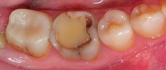
- Odontogenic way, when the source of the infectious agent is a tooth affected by caries. In this case, microorganisms first enter the pulp tissue, after which they spread through small lymphatic vessels or dental tubules to the bone tissue.
- Hematogenously, when pathogenic microorganisms spread to the maxillofacial area through blood vessels from the primary source of infection. This role can be played by any infectious focus present in the body: acute or chronic tonsillitis, erysipelas or furunculosis. In addition, some specific infections can provoke osteomyelitis: typhus, scarlet fever or even the common flu.
- Traumatically, when osteomyelitis occurs against the background of infection after a fracture or surgery on the jaw. It is most rarely seen in dentistry.
With the odontogenic route, the lower jaw is most often affected, and with the hematogenous route, the upper jaw is most often affected. If the infection occurs hematogenously, then the localization of the purulent focus will be deep in the bone tissue, and the phenomenon of periostitis will be minimal.
Classification
Osteomyelitis of the jaws (photo) is classified according to several parameters.
Osteomyelitis may be caused by:
- Infectious
a) Single-gene. It is a consequence of dental disease - caries, stomatitis. The most common type of pathology accounts for 75% of all cases of osteomyelitis.
c) Non-odontogenic (hematogenous). The pathogen enters the bloodstream from other foci of infection. May be a consequence of chronic tonsillitis, diphtheria, scarlet fever.
- Non-infectious (traumatic). The result of mechanical damage to the jaw or a wound through which infection enters. Osteomyelitis can occur under the influence of a growing malignant tumor or become a complication after dental surgery. For example, if the nerve tissue is not carefully removed from the tooth socket.
According to the course of the disease, osteomyelitis of the jaw occurs:
- Spicy. It lasts 7–14 days, then passes into the subacute stage - when a fistula forms at the site of inflammation and exudate flows out from the site of inflammation.
- Chronic. Lasts from a week to several months. It ends with the rejection of dead bone areas through the fistulous tract. Treatment at this stage is mandatory.
- Subacute. The transitional form between acute and chronic disease lasts 4–8 days. The painful signs subside, but the infection is actively spreading.
According to the location of the pathology:
- Osteomyelitis of the upper jaw.
- Osteomyelitis of the lower jaw.
By coverage:
- Limited. The inflammation covers the alveolar process or the area of 2–4 teeth of the jaw.
- Diffuse. The pathology covers most or all of the jaw.
According to clinical and radiological forms, chronic odontogenic osteomyelitis is:
- productive. During the healing process, it does not form sequesters (dead bone areas);
- destructive. Upon recovery, it forms sequesters.
- destructive-productive.
Symptoms
The clinical picture of osteomyelitis depends on whether the disease occurs in acute or chronic form.
Spicy
Typically, the symptoms of this pathology occur suddenly and manifest as local and general manifestations.
General symptoms are nonspecific and reflect only the presence of a severe inflammatory focus in the body:
- A significant increase in body temperature to 39 degrees and above.
- General severe weakness, malaise, headaches and aches in the joints.
- Paleness of the skin and mucous membranes, increased sweating.
Against the background of such general manifestations, local signs of the disease also arise:
- Constant unbearable pain in the area of the tooth that has become a source of infection. As the inflammatory process spreads, the pain syndrome intensifies, loses its clear localization and sometimes spreads to the entire jaw or half of the skull, radiating to the ear area or eye.
- Often inflammation seizes the jaw joint, and arthritis develops, which leads to the fact that a person cannot close his jaw and keeps his mouth open all the time.
- The tooth that caused the disease begins to loosen. With diffuse inflammation, neighboring teeth may also become loose.
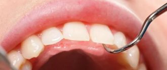
- The mucous membrane of the gums and oral cavity becomes sharply swollen, hyperemic and painful.
- Increasing swelling of soft tissues leads to facial asymmetry and spasm of the masticatory muscles.
- Significant increase in the size of regional lymph nodes.
Hematogenous osteomyelitis usually has the most severe course, since it is characterized by a combination with damage to other skull bones and internal organs, which significantly worsens the further prognosis.
A feature of the course of the traumatic variant of the disease is that the clinical picture in the early stages can be erased due to manifestations of injury. However, when, 3-5 days after a jaw fracture, complaints of increased pain appear, and the patient’s condition worsens, body temperature rises, severe swelling of the oral mucosa and purulent discharge from the wound occurs, then the diagnosis becomes clear.
Chronic
As the disease becomes chronic, the patient's condition improves. However, over a fairly long period of time, such people experience severe pallor of the skin, lethargy, sleep disturbances and lack of appetite.
During examination for chronic osteomyelitis, fistulas are revealed that open both on the surface of the face and in the oral cavity. A small amount of purulent contents is released from the fistula tracts. You can also identify swelling of the mucous membranes, pathological mobility of one or more teeth, and enlargement of regional lymph nodes.
During the remission stage, pain may be absent or insignificant. But during an exacerbation, the pain syndrome may intensify, and the patient cannot always indicate the exact location of the pain.
Treatment of osteomyelitis of the jaw
Treatment of osteomyelitis of the jaw is complex and consists of surgery, drug treatment and restorative therapy, which promotes tissue regeneration.
During surgery:
- the surgeon removes the affected tooth;
- treats fistulas and boils in the oral cavity;
- drains pus;
- removes dead (sequestered) areas of bone;
- fills hollow areas with bone materials;
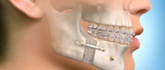
- strengthens mobile teeth with splints.
At the same time, drug treatment is carried out:
- antibiotics;
- immunostimulants;
- vitamin complexes.
To consolidate the result, you must undergo a course of physical procedures:
- UHF;
- magnetic therapy;
- ultrasound therapy.
During the treatment process, the patient is prescribed plenty of fluids and a gentle diet of light, crushed and nutritious foods. It is necessary to carefully monitor oral hygiene.
Diagnostics
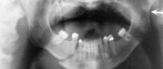
Based on the patient’s complaints and objective data from a general examination, the doctor may suspect osteomyelitis of the jaw bone. Confirmation of such a disease and a full formulation of the diagnosis are possible only after radiological diagnostics (x-ray diagnostics).
There are early and late radiological signs that indicate the presence of such severe pathology.
Early X-ray signs include:
- the presence in the images of areas of rarefaction of bone tissue, which alternate with its compaction;
- blurriness and extreme indistinctness of the bone pattern in the jaw;
- a slight increase in the thickness of the periosteum as a consequence of periostitis.
Late signs of osteomyelitis on a radiograph are:
- the formation of foci of destruction with the formation of sequesters by 7-12 days from the onset of the disease;
- thickening and moderate compaction of bone tissue around the inflamed lesion.
In difficult cases, patients are advised to undergo MRI, which allows them to more clearly see the extent of bone tissue damage, as well as visualize small purulent foci.
In addition to the X-ray examination, general clinical tests are carried out, which reflect the activity of the inflammatory process:
- a general blood test, which can reveal an increase in the number of leukocytes, changes in the leukocyte formula of an inflammatory nature, a decrease in the number of red blood cells and hemoglobin;
- biochemical blood test to identify electrolyte disturbances and the appearance of inflammatory markers.
In order to determine the causative agent of osteomyelitis and identify its sensitivity to antibacterial drugs, a bacteriological study of the discharge of the fistula tracts is carried out with the inoculation of pus on special nutrient media, followed by microscopy of the obtained samples.
Differential diagnosis
Differential diagnosis of osteomyelitis with other diseases that have similar symptoms is important, since an incorrect diagnosis can lead to an incorrect choice of treatment tactics and ineffective therapy. All this increases the risk of an unfavorable outcome of the disease and a poor prognosis for future health.
The differential diagnosis of osteomyelitis should include such diseases as:
- acute form of periodontitis,
- acute pulpitis,
- periostitis,
- suppuration of a tooth cyst,
- acute sinusitis of odontogenic origin,
- abscess of soft tissues in the maxillofacial area (MFA).
Complications
An incorrect diagnosis or untimely initiation of therapy for osteomyelitis leads to the development of severe complications, which have a high mortality rate and often cause disability.
The most common complications of osteomyelitis of the jaw are:
- Abscesses of soft tissues, perimaxillary phlegmons and purulent leaks, which tend to quickly spread to the cervical region and the mediastinum. This pathology is extremely dangerous, since the sepsis associated with it (in non-medical vocabulary the term “blood poisoning” is used) quickly leads to damage to vital organs with the development of septic shock and death.
- Thrombophlebitis of the facial veins, mediastinitis, pericarditis or severe pneumonia.
- Purulent damage to the membranes of the brain with the development of meningitis.
- If the purulent focus is localized in the upper jaw, the infection may spread to the orbital region with damage to the eyeball, atrophy of the optic nerve, which leads to irreversible loss of vision.
Types of periostitis
Based on localization, periostitis of the upper jaw and lower jaw is distinguished. The location of the development of the process depends on the location of the focus that caused the inflammation.
There may be several causes of the disease. Most often, the pathology is odontogenic, that is, the source of infection is a diseased tooth. Advanced caries, pulpitis, periodontitis are risk factors. The infection can spread to the periosteum and cause inflammation. The pathogen can enter the oral cavity through blood or lymphatic vessels from a source located at a distance from the oral cavity. Boils and carbuncles in the face and neck area are especially dangerous. There are also periostitis of a traumatic nature. In this case, the infection penetrates from the external environment through damaged tissue.
According to the nature of the course, acute and chronic forms are distinguished. In most cases, clinical manifestations are pronounced, occur suddenly, and progress rapidly. In a chronic course, the disease is sluggish.
According to the nature of the discharge, periostitis can be serous and purulent. The serous form is possible in the first days of the disease. The process may end with recovery or transition to a purulent form.
Treatment
Treatment of osteomyelitis of the jaw bones consists of simultaneously solving two important problems:
- The fastest elimination of the focus of purulent inflammation in the bones and surrounding soft tissues.
- Correction of functional disorders that were provoked by the presence of a severe infectious process.
All patients, without exception, are subject to hospitalization in a surgical department specializing in maxillofacial surgery. If there is no such hospital, then treatment is carried out in a department with experience in dental surgery.
The complex of treatment measures includes:
- Surgical intervention with opening of the purulent focus, cleaning it of necrotic masses and complete drainage.
- The use of antibacterial drugs with a wide spectrum of activity.
- Detoxification and anti-inflammatory treatment, strengthening the immune system.
General care is also important, with strict bed rest, nutritious but gentle nutrition (hypoallergenic diet including all necessary nutrients, vitamins and minerals).
Treatment of periostitis of the jaw
Therapy includes the elimination of not only the source of inflammation itself, but also the reason why it arose. If periostitis is odontogenic in nature, treatment of the affected teeth is mandatory; in most cases, they have to be removed.
Serous periostitis is treated conservatively. The cause that caused the inflammation is eliminated, physiotherapy is carried out and mouth rinsing with antiseptics is prescribed. In most cases, these simple measures are enough for a quick recovery.
The purulent process requires surgical treatment. The lesion must be opened, cleaned of pus and dead tissue, and thoroughly sanitized. Drainage is installed in the wound, and regular rinsing with antiseptic solutions is carried out. To more effectively fight the infection and prevent its spread, a course of antibiotic therapy is prescribed. Anti-edema and painkillers are used as symptomatic therapy. This allows you to improve the general condition of the patient. After surgical treatment, a diet is indicated; solid foods, as well as spicy and salty foods, should be excluded.
The issue of tooth preservation in acute periostitis is decided individually. Baby teeth in children, as well as permanent teeth that are severely damaged, are always removed. If it is possible to save a tooth and at the same time successfully stop the inflammatory process, a professional dentist will cope with this task. In case of chronic periostitis, the tooth must be removed in any case.
Treatment of periostitis of the jaw lasts for 5-10 days, depending on the stage and extent of the infectious process. With timely measures taken, the prognosis is favorable. If the disease is not treated, the purulent process spreads and can transform into phlegmon, osteomyelitis, and sepsis. An advanced disease causes irreversible consequences and can be life-threatening. At the first symptoms, you should contact your dentist. In this case, the cost of treatment will be lower, and negative consequences will be avoided.
Consequences and rehabilitation after osteomyelitis of the jaw
The consequences of acute or chronic osteomyelitis of the jaw bone can be quite serious and significantly worsen a person’s quality of life.
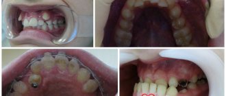
- Often, during the surgical treatment of such a pathology, it becomes necessary to remove not only the causative tooth, but also several others. This leads to the fact that in the future the person will need orthodontic treatment and prosthetics.
- Extensive bone tissue defects can lead to jaw deformities, which is not only a cosmetic defect, but also significantly disrupts the normal functioning of the maxillofacial apparatus.
- Damage to soft tissues often leads to scar deformation, which is also a serious cosmetic problem that needs to be solved with plastic surgery.
- The spread of infection to the joint can provoke inflammation (arthritis) or arthrosis, which subsequently causes the development of ankylosis and a sharp limitation of jaw mobility.
- The consequences of septic conditions against the background of osteomyelitis may also include disruption of the functioning of internal organs, hematopoietic processes and the functioning of the immune system.
- Osteomyelitis affecting the upper jaw can spread to the zygomatic bone and even the orbit with the development of an abscess or phlegmon of the eyeball. This leads to complete loss of vision without the possibility of recovery.
Rehabilitation after suffering purulent inflammation of the oblique jaw sometimes continues for several years. All patients are subject to dispensary registration, from which they are removed only after correction of all violations that have arisen.
Rehabilitation measures include:
- use of physiotherapeutic techniques;
- if necessary, prosthetics for lost teeth;
- repeated surgery for cosmetic or medical reasons;
- prevention of recurrence of such pathology.
Forecast and prevention of osteomyelitis of the jaw
If the patient sought medical help on time, followed the doctor’s recommendations and received the correct treatment in full, osteomyelitis will be successfully cured. Incorrect treatment in combination with a weakened immune system can lead to the following complications:
- sepsis;
- acute purulent inflammation;
- general intoxication;
- the formation of multiple abscesses;
- meningitis;
- inflammation of the facial nerves;
- bone deformities;
- purulent sinusitis and destruction of the walls of the maxillary sinuses;
- infection of the orbit and formation of phlegmon;
- jaw fractures.
To prevent osteomyelitis of the jaw, it is important to:
- Have dental checkups every year.
- Properly brush your teeth, maintain oral hygiene, and use interdental floss and rinses.
- Treat teeth and gums in a timely manner.
- Protect your jaw from injury.
- Strengthen immunity.
- Do not start treatment for infectious diseases, including tonsillitis and ARVI.
Prevention
Preventive measures are not only the key to preventing the development of osteomyelitis, but also a factor that reduces the risk of complications and shortens the recovery period if the disease cannot be avoided:
- Timely treatment of caries, even if it has no clinical manifestations.
- Maintaining normal immune status through regular physical activity, rational and nutritious nutrition.
- Sanitation of all chronic foci of infection in the body.
- In case of injury, in the postoperative period or after tooth extraction, compliance with all preventive medical prescriptions.
In conclusion, it should be noted that, despite all the achievements of modern medicine, osteomyelitis of the jaw in adults and children does not lose its relevance. Timely detection of its signs and adequate treatment increase the patient’s chances of a full recovery and maintaining a high quality of life.
»
Classification of the disease, depending on the location of inflammation. process
- Jaw osteomyelitis after tooth extraction. The nerve endings of the periodontium and gums remain irritated, pain can be felt immediately or after several days.
- Odontogenic osteomyelitis of the lower jaw is the most common form of the disease at an advanced stage of caries. Quite common in young children.
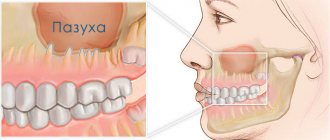
- Odontogenic osteomyelitis of the upper jaw is caused by the anatomical structure of the bone. The infection can provoke inflammation of the maxillary sinuses. If the fang becomes inflamed, it swells, including the infraorbital area.
- Osteomyelitis of the socket is an inflammation of the bed where the extracted tooth was located. A more correct name for the disease is alveolitis, which is accompanied by destructive changes in the bone.
- Hematogenous osteomyelitis of the jaw - affects mainly the upper jaw, after which it affects the cheekbones and nasal bone. The clinical picture is characterized by sepsis, which is accompanied by inflammation in more than half of patients with this diagnosis.