Causes
In addition to mechanical trauma from an impact, the cause of a fracture of the alveolar bone may be some disease that disrupts the normal state of bone tissue:
- osteomyelitis - inflammation of bone tissue;
- fibrous osteitis, characterized by bone dystrophy, thinning of the bone structure;
- various cysts and neoplasms that cause bone degeneration.
If the patient has the above diseases, even a slight impact on the bone can lead to a fracture of the alveolar process.
In children aged 5 to 7 years, that is, at the stage of growth of permanent teeth, trauma to the alveolar process can also be diagnosed due to the presence of follicles of permanent teeth in the bone, which subsequently disappear.
Treatment methods for fractures
Treatment of fractures of the alveolar process of the upper jaw includes several stages:
- pain relief,
- antiseptic treatment,
- repositioning of all fragments,
- immobilization with special structures.
Displaced fractures require complex treatment. In case of serious injuries, the wound is inspected, the sharp edges of bone fragments are smoothed and the mucous membrane is sutured. The bone fragments are returned to the correct position and securely fixed with staples or splints. To prevent complications in the first days after injury, antibacterial therapy and rinsing the mouth with herbal decoctions are prescribed.
With timely provision of qualified assistance, bone callus is formed within 8 weeks. The speed of recovery depends on the individual characteristics of the body.
Symptoms of alveolar bone fracture
An alveolar ridge fracture can be diagnosed if:
- sharp, spasm-like pain in the jaw when chewing or swallowing saliva,
- swelling of the mucous membrane,
- lacerations of the gums (with or without bleeding),
- painful closing of the jaws,
During palpation, the damaged part may move or a separated area may be palpated. Externally, the fracture is characterized by contusions and bruises on the face in the area of the fracture. External examination and x-ray show a crack in the alveolar process or complete separation of the bone from the main cranial bone.
Depending on the angle at which the blow occurred, the broken part may move in different directions, inward or deep into the mouth. If the blow falls on the lower jaw, in this case the impact occurs with the help of the lower jaw on the upper jaw, and the broken part moves upward.
Symptoms
A fracture of the alveolar process of the upper jaw has several characteristic features. The most common symptoms:
- sharp pain
- inability to completely close your mouth,
- occurrence of malocclusion,
- dysfunction of chewing and swallowing,
- hematomas,
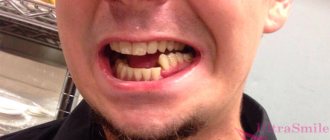
- dislocations and mobility of one or more teeth,
- severe swelling of soft tissues,
- bleeding.
If you suspect an alveolar bone fracture, you should seek medical help as soon as possible.
Diagnostics
The fracture is diagnosed by an oral and maxillofacial surgeon. An x-ray may show different degrees of damage:
- partial damage - damage to part of the bone, not complete detachment;
- non-displaced fracture – damage to all parts of the bone;
- complete fracture - the x-ray shows a gap between the separated part and the skull;
- fracture in different places - damage to the alveolar process in different places, bone fragmentation;
- fracture with deformation - a completely torn off part, displaced at a different angle.
Clinical manifestations
The first sign of a fracture of the alveolar process is a sharp, severe pain. Usually its occurrence is associated with mechanical trauma. The patient cannot completely close his mouth, swallow and chew. The bite is broken.
Upon examination, a hematoma or rupture of soft tissue at the site of injury is usually detected. If the fracture is partial, the deformity may be mild. In complete displaced fractures, bone fragments can come out, breaking through the soft tissue.
The teeth in the area of injury are usually mobile and dislocated. In most cases, severe bleeding is observed.
If a child is injured, the consequences may be adverse. When the alveolar process, which contained the rudiments of permanent teeth, is fractured, they die, so the only solution in the future will be prosthetics.
Treatment
The treatment method is selected depending on the established severity of the fracture. Before any manipulation, the doctor performs anesthesia. Torn tissues are treated with antiseptics to prevent infection. Then reconstruction is carried out (if there are fragments) and fixation of the jaw.
If there is displacement, the doctor opens the gums in the area of the fracture (revision) to eliminate the sharp edges of the fragments, after which he manually puts the displaced part in place. This must be done in such a way that it corresponds to the correct bite. After this, the gum is sutured and an iodoform dressing is applied to the wound.
To fix the alveolar process, a smooth splint-bracket is used, which is attached to three healthy teeth on both sides of the chipped part. If this is not possible, that is, if the teeth on one side are not stable or are completely absent, then I use five additional teeth for fixation. For greater immobilization of the dentition, wearing a chin sling may be indicated.
If the injury occurs in the anterior part of the upper molars, then fixation occurs through the installation of a single-jaw bracket, which is fixed on the damaged area (attached with ligatures to healthy teeth).
In case of complete absence of teeth, a splint made of quickly hardening plastic is installed.
After all surgical procedures, the patient is prescribed antibacterial therapy and medications to accelerate healing and prevent the development of inflammation in the injured area.
Fracture of the upper jaw - symptoms and treatment
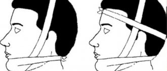
When providing first aid to the patient, it is necessary to stop the bleeding and prevent aspiration (penetration into the respiratory tract) of blood and vomit. If the lower jaw is not damaged and there are a sufficient number of teeth on both jaws, it is necessary to apply a sling-like bandage, pressing the lower jaw to the upper jaw, or perform immobilization (immobilization) with a rigid chin sling [4].
If there is a risk of respiratory failure, immediate insertion of an airway is required to maintain the conductivity of the airways [1]. In addition, it is necessary to provide pain relief and quickly transport the patient to specialized medical institutions. The most important thing at this stage is to preserve the life and health of the patient.
There are many methods of non-surgical treatment of fractures of the upper jaw, for example, various types of bandages and external fixations, which are currently practically not used.
The most common method of orthopedic treatment of fractures is bimaxillary splinting - the application of splints and brackets to the dentition with reposition of fragments and fixation of the bite in the patient’s usual position. This method is conservative and low-traumatic, but in some cases it does not allow obtaining good fixation of fragments of the upper jaw, especially in high and complex fractures. On average, fractures of the upper jaw require immobilization and limitation of chewing load for a period of 4-5 weeks.
The most modern and adequate treatment method at the moment is osteosynthesis (fixation with titanium bone structures) of fractures of the upper jaw. This is a surgical procedure performed through intraoral incisions. With this treatment option, it is possible to accurately compare and fix the fragments to create conditions for their fusion [7].
In the treatment of high fractures, a coronal approach is also used, which allows for cosmetic and wide access to the bones of the entire midface and orbits [5]. Timely implementation of osteosynthesis allows you to prevent late postoperative complications, facilitate the patient’s rehabilitation and speed up recovery.
Fractures with gross violations of the integrity of the upper jaw and significant displacement of fragments towards the pharynx are recommended to be treated surgically. There is no clear opinion regarding other types of fractures - tactics are dictated by the patient’s condition and the specific clinical situation.
It is worth noting that it is very important to constantly wear intermaxillary fixation for tight contact of fragments and to prevent their mobility, especially under the influence of chewing load [9]. High-quality oral hygiene and patient observation by an oral and maxillofacial surgeon are also necessary.
Recovery from fractures takes from four to six weeks, depending on the nature of the fracture, the characteristics of the patient’s body and the method of treatment.
Patients with fractures of the upper jaw should eat liquid food in the early stages, and soft food in the later stages. Eating hard foods and active chewing should be limited. Other recommendations are given based on general somatic and neurological disorders (bed rest, etc.).
Rehabilitation period
If the trauma of the alveolar process did not damage the roots of the teeth, then with proper reconstruction and fixation, after about 8 weeks, a bone callus will form, and the process will grow back to the jaw. The treatment prognosis is favorable.
If the roots of the teeth are damaged, the broken parts may no longer take root. As a rule, it is not possible to achieve consolidation even with successful treatment.
Rehabilitation after a fracture is aimed at full or maximum restoration of the functions of the injured part of the body. It may consist of a set of procedures, which, depending on the characteristics of the injury, may include:
- physical therapy,
- physiotherapy,
- massage.
In order for further rehabilitation to have a positive effect, each of the components of the complex must be selected by a qualified specialist, taking into account all the individual characteristics of the patient.
Fracture of the lower jaw
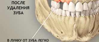
A fracture of the lower jaw most often occurs during the removal of molars with an elevator or chisel. The use of a chisel and hammer to “gouge out” a tooth or root, which was previously recommended, makes the danger of such a complication real. Therefore, a chisel should not be used to remove teeth. It is less traumatic and more effective to use an electric drill with rotating cutting tools (burs, cutters) for this purpose.
A fracture can be considered pathological if a history shows the presence of an inflammatory disease - cysts, tumors, tooth retention, osteomyelitis. Such pathological conditions lead to a decrease in strength, which in turn is a risk factor for fracture.
If a pathological fracture occurs during tooth extraction, it is necessary to carry out transport immobilization of the lower jaw with a chin-parietal bandage and refer the patient to a maxillofacial hospital.
A jaw fracture that occurs during tooth extraction cannot always be recognized immediately. After surgery, the patient may complain of pain in the jaw, difficulty opening the mouth and chewing. A thorough clinical examination and x-ray can determine the presence of a fracture.
CHAPTER V NON-GUNGEOT FRACTURES OF THE MAXILLA.
1. Anatomical structure of the upper jaw and border bones.
The anatomical features of the upper jaw influence the clinical signs when it is damaged and determine the nature of the fracture.
Thus, the upper jaw, being a paired bone and located in the center of the face, is connected with other bones of the facial and cerebral skull: zygomatic, frontal, nasal bones, ethmoid, sphenoid, lacrimal.
There are four surfaces of the body of the upper jaw: anterior, infratemporal, orbital, nasal (Fig. 21).
Front surface
(facies anterior) is bounded above by the inferior orbital margin, laterally by the zygomatic alveolar crest and zygomatic process, below by the alveolar process and medially by the nasal notch. Under the infraorbital margin there is an infraorbital foramen (for. infraorbitale), through which the terminal branch of the nerve and vessels of the same name emerges.
Infratemporal surface
(facies infratemporalis), represented by the tubercle of the upper jaw. The oblique head of the lateral pterygoid muscle is attached to it. In the tubercle of the upper jaw there are 3-4 openings through which the posterior superior alveolar branches enter the thickness of the bone tissue, taking part in the formation of the posterior section of the upper dental plexus.
Rice. 21. Schematic representation of the upper jaw: 1-inferior orbital margin; 2-zygomaticalveolar ridge; 3-alveolar process; 4-nasal cavity; 5-infraorbital foramen; 6-tubercle of the upper jaw; 7- nasal spine; 8-zygomatic bone.
Orbital surface
(facies orbitalis) takes part in the formation of the lower wall of the orbit and forms the lower orbital margin. In the posterior section, together with the orbital edge of the large wings of the sphenoid bone, it limits the lower orbital fissure (fissura orbitalis inferior). Through it, the infraorbital nerve (n. infraorbitalis), a branch of the maxillary nerve, enters the orbit. The latter runs in the infraorbital groove and in the infraorbital canal, which are located on the orbital surface of the body of the upper jaw. The lower wall of the canal is pierced by small anterior and middle superior alveolar openings (foramina alveolaria superiora anteriora et media). They lead into small bone canals that extend to the roots of the incisors, canines and small molars. They carry blood vessels and nerves to these teeth. The medial edge of the orbital surface borders the lacrimal bone, the orbital plate of the ethmoid bone, and the orbital process of the palatine bone. Sometimes it forms cells that are directly adjacent to the cells of the ethmoid bone labyrinth.