15.12.2020
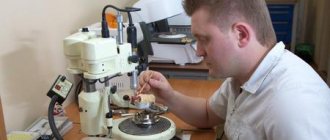
Modeling the base of the prosthesis - creating a wax prototype of the base of the prosthetic structure. This is an important stage on which the aesthetics and functionality of the prosthesis depend. Classic technologies involve carrying out all clinical and laboratory stages manually. With the help of increasingly popular CAM/CAD systems, modeling of the base and the entire prosthesis is carried out on a computer.
Classic basis modeling
The process consists of two stages - preliminary and final basis modeling. The preliminary is aimed at preparing the accluder and articulator for testing the design, the final is aimed at giving the wax base and teeth a look that best matches the finished prosthesis.
Pre-modeling stages:
- Adding wax to areas lacking volume.
- Fastening artificial teeth, and for partial structures and clasps.
- Cleaning the occlusal and vestibular surfaces of teeth from hardened wax.
- Smoothing the wax base, giving it uniform thickness.
- Rounding the edge of a composition.
- Smoothing out irregularities.
After this, the clinic checks the design of the prosthesis, tries it on in the patient’s mouth, returns it to the laboratory and begins the final modeling of the prosthesis base. Its stages:
- Fixation of teeth separated during fitting with wax.
- Removing wax from all surfaces of teeth.
- Design of the edge of the artificial gum - leveling or, on the contrary, creating contours that repeat the anatomical ones. The latter technique contributes to more reliable fixation of the prosthesis. To give a natural look, a lower roller is formed in older patients, and a high one in young patients.
- Modeling the base of the upper jaw. The wire arch is removed, ensuring uniform thickness of the artificial gum. Heated wax is applied to the palatal surface to form a relief, the distal edge is reduced to nothing, and connected to the gum.
- Excess wax is removed from the surfaces of the teeth.
- The wax base is glued to the model.
CAM/CAD modeling of prostheses
Prosthetics for toothless jaws is the most difficult problem in orthopedic dentistry. This is due not only to the complete loss of teeth, but also to atrophic processes due to the lack of chewing load. As a result, the landmarks that determine the height and shape of the alveolar processes are lost, new relationships between the arches of the jaws arise, which significantly complicates the determination of the interalveolar line common to the upper and lower jaws.
CAM/CAD technologies allow you to accurately calculate divergence angles, automate the modeling and production of the prosthesis, and create the most comfortable and aesthetic prosthetic design for the patient. Modeling of the frame, base, teeth is performed on a computer in specialized programs for three-dimensional graphics. First, the jaw is scanned using a tomograph or silicone impressions are made, followed by scanning; further steps are performed in the program. You can construct the model manually or use the library of blanks.
Typically, digital technologies are used in modeling non-removable prosthetic structures, but they are increasingly being used in the creation of clasp and complete removable dentures. Automated systems make it possible to create high-precision structures in a short time with minimal technical costs.
Modeling of a removable denture supported by implants
Joseph J. Massad, DDS David M. Bohnenkamp, DDS, MS Lily T. Garcia, DDS, MS
Introduction
The use of dental implants for complete edentia is, as a rule, considered as an additional means of increasing the retention of the prosthesis. In some clinical cases, the orthopedic dentist, taking into account the financial capabilities of the patients, decides to use an old removable denture for installation on dental implants. When choosing this scenario for implant prosthetics, the patient’s attention is focused only on the possibility of improving the retention of a poorly fitting removable denture, while assessing the quality of the denture itself comes second. Continuing orthopedic treatment with unsatisfactory quality of an existing prosthesis, without taking into account the violation of physiological functions, can lead to less optimal installation of implants and the inability to obtain both functional and aesthetic results.
Edentulism is a form of disability that includes problems with self-esteem and speech, ineffective chewing, and persistent discomfort due to pressure placed on soft and hard tissues. After making a diagnosis of complete edentulism, the prosthodontist determines the prosthetic diagnostic index (PDI) to assess the degree of complexity of the patient's upcoming orthopedic rehabilitation.
Clinical case
An 80-year-old man came to the clinic for a thorough examination of the situation in the oral cavity. At that time, the patient had complete removable dentures installed on the upper and lower jaws. Due to the instability of the mandibular denture, food particles accumulated around and under the base of the denture. Before appearing in our clinic, the patient had already had consultations with several other specialists, who informed him that there was insufficient bone tissue to install implants.
When assessing the clinical situation (view before treatment), an inadequate ratio of the size of the buccal corridor and the base of the upper complete removable denture was revealed.
Front view of the patient before (left) and after treatment (right)
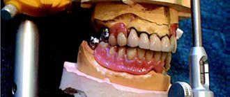
The photograph of the patient’s teeth in the position of maximum tubercular-fissure contact of the antagonist teeth demonstrates the worn surfaces of the denture teeth of both jaws (Fig. 1).
Rice. 1. Clinical view of complete removable dentures of the upper and lower jaws in the position of maximum tubercular-fissure contact of the antagonist teeth
The patient's main complaint was that, despite the presence of complete dentures, they did not feel natural. The mandible showed severe bone atrophy (Fig. 2–5), a rather sharp bone ridge, and a high frenulum attachment in relation to the height of the residual alveolar process.
Rice. 2. Clinical view of the residual anatomy on the right side of the mandible. Note the sharp bony ridge
Rice. 3. Clinical view of the residual anatomy on the left side of the mandible with a high attachment of the frenulum in relation to the height of the residual ridge
Rice. 4. The mandibular model shows excessive atrophy of the residual bone ridge, which has caused the patient's mandibular complete denture to lack stability.
Rice. 5. Panoramic radiograph shows demarcation lines anterior to the mental foramina, marking the anatomical boundaries of implant placement without risk of injury to the inferior alveolar nerve.
In addition, the bone ridge was covered by a thin layer of mucous membrane, characterized by high mobility, with the possibility of displacement of more than 2 mm in the vestibular-lingual direction.
Method
Upon completion of all necessary orthopedic manipulations, a trial removable denture is made for the patient. At the beginning of treatment, an anatomical functional impression is taken; the vertical distance of occlusion (VDO) is assessed; the reproducible position of physiological rest and the minimum interocclusal gap are determined; The centric relation is registered using a special device, which allows obtaining a graphic image of the Gothic arch simultaneously with the deprogramming of the patient’s masticatory muscles to exclude positions of the lower jaw in which wear of the prosthetic teeth occurred due to inadequate VRO.
The functional position of the teeth is determined in accordance with the principles of the neutral zone method, which also allows us to take into account the work of the masticatory muscles during swallowing, speaking and chewing. The prosthodontist attaches the thermoplastic impression material (modeling material) to the acrylic bite block in the same way as a wax bite block. The patient is then asked to imagine drinking warm water and perform several swallowing movements.
This action allows the muscles to shape the thermoplastic impression material and determine the functional labial and lingual positions of the anterior and posterior teeth. Performing this manipulation ensures modeling of the prosthetic wing in the neutral zone and eliminates all unwanted movements. The use of the “neutral zone” method plays a significant role, since it allows the orthopedic dentist to determine the positions of the anterior and chewing teeth of the lower jaw prosthesis.
As a result of using the anatomical and physiological method for determining the height of the bite, the patient was diagnosed with cross occlusion (Fig. 6).
Rice. 6. View of trial removable wax dentures on the main models installed in the articulator. In this clinical case, the occlusal pattern is a crossbite, which was established using an anatomical and physiological method for determining the height of the bite used for the manufacture of the prosthesis, taking into account the neutral zone
The concept of “modeling” a prosthesis for a particular type of treatment involves the production of a cast metal frame for a trial mandibular removable denture, taking into account the labial, lingual and distal boundaries of implant placement (Fig. 7).
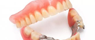
Rice. 7. View of the inner surface of a trial prosthesis of the lower jaw with a metal frame built into a plastic base
Another purpose of using a metal frame is to increase the strength of the prosthesis, which can be weakened after fixation of the attachments required for installation on implants. The use of an ill-fitting old removable denture instead of a new one entails certain difficulties, such as incorrect centric relation, violation of the aesthetic positioning of the denture teeth and inadequate VRO. The orthopedic dentist should eliminate the need to use the patient’s existing removable dentures when using implant-supported prosthetics.
Installation of a complete mandibular denture supported by implants should be carried out in accordance with the most optimal positions of the implants, and not vice versa, when an existing complete denture determines the positions of the implants. The new complete mandibular denture is fabricated with a metal frame to provide additional support for the denture as implants are placed under significant anatomical and functional constraints.
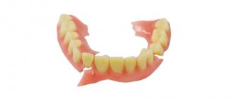
After the manufacture of a complete removable denture of the lower jaw, a significant recess is made on the inside of the base of the prosthesis, necessary for the installation of attachments, while, due to the presence of a metal frame, the risk of reducing the strength of the prosthesis is eliminated (Fig. 8 and 9).
Rice. 8. View of the occlusal and adjusted internal surfaces of the lower jaw removable denture. Thanks to the metal frame of the prosthesis, its strength is maintained, even despite the extensive recess in its base above the implantation site
Rice. 9. View of the prosthesis after taking the impression, the buccal/vestibular and lingual surfaces are modeled with the patient’s masticatory muscles. The metal frame is not visible due to the significant thickness of the base of the plastic prosthesis of the lower jaw
The panoramic radiograph shows the positions of the monolithic implants (diameter 3.25 mm, ERA mini implant [Sterngold Dental]) (Fig. 10).
Rice. 10. Panoramic radiograph showing microimplants immediately after completion of surgery
The implants were installed on the lower jaw in the space between the mental foramina. For the most accurate positioning of implants, complete removable dentures are installed in the patient’s oral cavity, after which the bite is registered in the centric relation position. An occlusal index is made to reproduce the centric relation position. The occlusal index is then placed in the patient's mouth and the patient is asked to close the mandible according to the index. This ensures stable reproduction of the position of central occlusion even if the patient, due to the injected anesthetic, cannot adequately reproduce this position during subsequent dental procedures (Fig. 11).
Rice. 11. Bite registration is performed in the centric relation position using a vinyl polysiloxane-based material (Regisil Rigid Registration Material [DENT-SPLY Caulk]). The resulting index is used for subsequent reproduction and direction of the patient to the position of the central ratio. The photo shows the index before it was trimmed.
In areas with sufficient bone tissue, implants with a diameter of 3.25 mm are installed. The need for additional surgical intervention to increase the volume of bone tissue using osteoplastic material and installation of standard size implants was previously discussed with the patient (Fig. 12).
Rice. 12. Clinical view of the microimplant during its installation in the anterior region of the lower jaw
Laboratory caps are installed in males on the inside of the base of the removable lower jaw denture (Fig. 13). Fixing a metal frame on the inside of the base of a removable lower jaw denture allows the orthopedic dentist to maintain the integrity of the structure and increase its strength.
Rice. 13. View of the inner surface of the complete removable denture of the lower jaw after fixing the attachments; Black caps are installed in the metal males. Pay attention to the location of the metal males relative to the metal frame, which allows you to make the prosthesis more stable
As shown in Fig. 14, the occlusal diameter of the male is 4.4 mm, which provides a gap of 1.0 mm compared to the standard 5.5 mm diameter attachments typically used with the ERA system.
Rice. 14. Photo shows the difference in diameter between the standard metal ERA male and the micro-attachment male
Laboratory caps are removed with a special tool and instead of them, soft retention caps are installed in the metal males of the prosthesis (Fig. 15). The indicator of the minimum holding force is the white color of the caps.
Rice. 15. Black laboratory caps are removed with a special tool and soft retention caps are installed instead.
In the photograph after completion of treatment, a significant improvement in the aesthetic appearance of the patient’s smile with restored buccal corridors is evident (Fig. 16).
Rice. 16. The clinical appearance of the implants three months after surgery, the presence of plaque confirms the lack of proper oral hygiene. Also note the difference in height between implants. The difference in height cannot be corrected because the patient received monolithic implants
The use of monolithic implants requires careful planning to select the required supragingival height during surgery.
The disadvantage of a monolithic implant is the fixed height of its abutment, since this transmucosal/transgingival part cannot be replaced. The positions of the implants are determined in exact accordance with the available amount of residual bone tissue, which eliminates the need to perform additional manipulations to increase the volume of the bone crest.
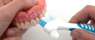
After surgery, the patient is instructed on proper oral hygiene, since one of the reasons for the loss of permanent teeth could also be failure to comply with all the necessary hygiene rules (Fig. 17 and photograph of the patient after treatment). The patient also indicated that he now “didn't even feel like they (the dentures) were there (in the mouth).”
Rice. 17. The patient was re-instructed regarding proper oral hygiene to ensure long-term functioning of the implants. To make caring for the implants easier, the patient was recommended an Oral-B Triumph toothbrush with a FlossAction head (Procter&Gamble)
Discussion and conclusion
The method presented in this article shows how important it is to consider the future orthopedic outcome when planning treatment to meet all the patient's needs. In many cases, the patient's main complaint significantly influences the final outcome of the prosthesis. However, evaluation of an existing complete denture should also be an integral part of a thorough examination of the patient's mouth as you plan to place implants that will provide additional retention to the denture.
The use and modification of prosthetic components, such as a metal frame integrated into the base of a complete mandibular removable denture, provides increased strength to the entire structure, even if it is necessary to create a recess large enough to hold the attachments.
To obtain a functional and aesthetic result from prosthetics, it is important to strictly follow a predictable clinical protocol for the manufacture of the prosthesis.
The result is reflected in the patient’s words: “Food no longer gets under the base of the denture,” “The dentures sit tightly in place and give a feeling of comfort,” “The dentures fit so well to the gums that you forget that they are installed in the oral cavity.”
About the authors:
Joseph Massad is an adjunct member of the Tufts University School of Dentistry and the Department of Prosthodontics at the University of Texas Health Science Center, San Antonio. He is the director of removable prosthodontics at Scottsdale Dental Center. His articles have been published in the Journal of Prosthetic Dentistry, International Journal of Periodontal and Restorative Dentistry, Compendium of Continuing Dental Education, Independent Independent Journal England, The Pankey Gram, Bulletin of the American Association of Dental Examiners, Journal of the Oklahoma Dental Association , Dentistry Today, Dental Economics, etc. Contact: (918) 749-5600 Disclosure: Dr. Massad has been and is an independent paid consultant to many dental companies, such as Harry J. Bosworth, Ivoclar USA and Europe, DENTSPLY Tribute, Chattem Consumer Goods, Laclede Professional Products, Brasseler USA, Simplified Notes Company, Proctor and Gamble, Austenal, Inc, and GC America.
David Bohnenkamp is director of the International Dental Education Program at the University of Texas Health Science Center at San Antonio. He is the author of numerous articles published in peer-reviewed dental journals and is on the editorial board of the Journal of Prosthetic Dentistry. He is currently writing a chapter entitled "Clinical Scenarios for Patients" for the book Osseointegration and Occlusal Rehabilitation and Removable Partial Dentures - A Clinical Guide. Contact Information
Lily Garcia is a professor and chair of the Department of Prosthodontics at the University of Texas Health Science Center at San Antonio. She received her Doctor of Dentistry (DDS) degree from Baylor College of Dentistry and her Master of Dentistry degree from the University of Texas Health Science Center at San Antonio. He is a Diplomate of the American Board of Prosthetics and a Fellow of the American College of Prosthetics. Dr. Garcia has authored numerous articles and reports, edited several dental textbooks, served as a reviewer and editorial board member for several scientific journals, and is co-author of the book Osseointegration and Occlusal Rehabilitation and Removable Partial Dentures - A Clinical Guide). (The book is in press.) Dr. Garcia is in private practice in the field of prosthetics.
Translation – Tatyana Solomennikova