The problem occurs several weeks before the eruption of a baby or permanent tooth. The appearance of a cyst is indicated by a bluish-purple spot (hematoma). This is due to difficulty in tooth eruption.
An eruption cyst is a type of soft tissue benign tumor that appears when a tooth has already begun to erupt through the gum. Scientists and doctors have not agreed on why it occurs. The most likely causes: infection, trauma, improper development of teeth, which causes a lack of space for the “newbie”. In addition, it may be a consequence of caries and improper care.
Eruption cysts occur during the growth of baby teeth or, more often, at 6–9 years of age, when permanent teeth emerge. The fact is that, due to its anatomical structure and location, the chewing group of teeth is the most difficult to erupt.
If you notice a large formation in your baby’s mouth, in the place where a tooth should appear, consult a doctor. Do not resort to self-medication under any circumstances. Infusions, rinses and even strong antibiotics most likely will not help, but ruining a child’s health in this way will cause serious harm.
If you contact a specialist, treatment can follow one of two scenarios. In the most favorable option, the doctor will advise giving the child toys, special teethers, crackers - everything that the baby can chew. This will help break through the mucous membrane.
In difficult situations, surgery may be necessary. Then the surgeon will cut the gum under local anesthesia. This will make teething easier.
It is forbidden to try to cut or tear the mucous membrane on your own; you can cause a serious infection or injure your baby.
Why does a dental cyst form?
The cause of cyst formation is an untreated root canal infection.
The reasons for infection in the root of a tooth can be different:
- As a complication of insufficiently treated or completely neglected
- caries/pulpitis,
- periodontitis,
- periodontal diseases,
- inflammation under the dental crown,
- Error and infection during endodontic treatment (root canal treatment).
- Tooth injury.
- Immune disorders in the body.
- Chronic diseases of the ENT organs: runny nose, sinusitis, tonsillitis.
Children in primary and mixed dentition can also suffer from dental cysts. Even newborns may experience teething with a purulent cyst - the so-called. Bona knots.
Classification of dental cysts
There are not many types of cysts, so understanding them is not difficult. Four main types are distinguished by location:
- wisdom tooth cyst;
— a cyst due to an illiterate (unprofessional) installed dental crown;
- a cyst that arose as a result of the influence of a third-party infectious disease (sinusitis);
- a cyst localized in the area of the front teeth.
At the same time, a classification is used based on previous circumstances preceding the occurrence of the cyst:
- a disease caused by poor-quality removal of one or more teeth;
— periodontal type cyst, provoked by an inflammatory process in the gums;
- a cyst caused by the growth of a wisdom tooth (retromolar or paradental);
- a cyst that occurs in children, first during the period of the appearance of milk (follicular) teeth, and then molars (eruption cyst);
— a cyst that arose as a complication of periodontitis (radicular);
— a situation is also possible in which the tissue forming the tooth degenerates (keratocyst).
What are dental cysts?
What is the difference between a cyst and a granuloma?
A dental cyst is often confused with a granuloma, another dangerous disease in which a round-shaped formation forms in the root area. It is important to distinguish between them, since the diagnosis directly affects the treatment plan and its prognosis. For granuloma, in most cases therapeutic treatment is sufficient. But a cyst requires more radical measures.
Here are the main parameters by which a dental cyst and a dental granuloma differ:
| Difference options | Cyst | Granuloma |
| Size | 0.9 - 3 cm | 0.5 - 0.8 cm |
| Structure | Cavity with fluid or pus | Solid formation covered with connective tissue |
| Clinical picture | The tooth becomes mobile | No mobility observed |
| X-ray result | The image shows a round capsule with clear boundaries | There are no strict outlines of education |
| Condition of periodontal tissues | The mucous membrane is usually not inflamed. In this case, bone loss occurs. | There is severe swelling and redness of the mucous membrane. But bone tissue does not decrease so much that it can be detected clinically. |
Why do you need to remove the cyst?
If a dental cyst is not removed in time, even with an asymptomatic course of the disease, the consequences can be very serious - this is a real “time bomb”.
- The main danger of a cyst is that sooner or later it will destroy the affected tooth, and then “get” to the neighboring teeth.
- Cysts are reborn. Slowly but surely, so in 15-20 years it may already be a malignant formation.
- An infection is raging in the cavity of the cyst, so with a general illness, hypothermia, or a decrease in immunity as a result of simple stress, acute purulent inflammation is possible - flux, abscess, phlegmon.
- The infection can spread to adjacent lymph nodes and cause inflammation - lymphadenitis.
- Gradual thinning of the jaw bone in the affected area, due to purulent melting, turns into osteomyelitis. Even a spontaneous fracture of the jaw is possible.
- The cyst enlarges and can grow into the nasal cavity or maxillary sinus.
- Infection in the cyst can cause sepsis, a blood poisoning.
If treatment is not started on time, the tooth will have to be removed. And this is the minimum “evil”.
What will happen if left untreated?
Ignoring the symptoms of the disease leads to dangerous consequences for health. Thus, the patient may encounter:
- destruction of roots, their strong loosening;
- jaw fracture;
- tumor formation;
- purulent abscess;
- periostat;
- osteomyelitis.
The disease can cause the development of sepsis. This is a very serious condition in which the infection enters the blood and quickly spreads throughout the body.
What are the symptoms of a dental cyst?
For a long time, the cyst may not manifest itself in any way until it becomes large enough in size, so it is very rare that a cyst is diagnosed at an early stage of the disease. This mostly occurs accidentally when the patient is having an x-ray for another reason.
At a later stage, the patient begins to worry about:
- Aching pain that gets worse over time. Such pain is difficult to relieve with regular painkillers.
- Pain when biting.
- Swelling of the gums and/or cheeks.
- An abscess, gumboil, or fistula may form on the gum.
- General malaise, fever.
Summary:
You should not think that if a purulent cyst on the gum spontaneously opens, then you don’t need to go to the dentist and not treat the causative tooth. The source of inflammation at the root tip will still be in place, and it will slowly increase. Over time, this can lead to the formation of a large root cyst at the apex of the tooth root, and even tooth extraction. Therefore, treatment should not be delayed. We hope that our article: Cyst is not a gum treatment - was useful to you.
Sources:
1. Higher prof. the author’s education in therapeutic and surgical dentistry, 2. Based on personal experience as a dentist, 3. National Library of Medicine (USA), 4. “Outpatient surgical dentistry” (Bezrukov V.), 5. “Therapeutic dentistry: Textbook” (Borovsky E.).
How is a dental cyst treated?
The first thing that should be said is that today a dental cyst is successfully treated with a high percentage of preservation of the affected tooth, although in the past the diagnosis of a “cyst” meant 100% removal.
There are 2 main approaches to treatment:
- Therapeutic. Therapeutic treatment of dental cysts is used when
- There is access to the dental canal through the dental crown
- The canals are not sealed or can be opened
This method of treatment is very similar to the treatment of periodontitis, with the only difference being that the doctor additionally removes pus from the cyst cavity and injects special medicinal preparations into the root canal.
This treatment takes at least 1-2 visits, is carried out under sterile conditions, using a rubber dam and a dental microscope, because requires special care from the doctor to prevent relapses.
Treatment of cysts with laser with access through the coronal part of the tooth is another type of therapeutic treatment. Its main difference is that the laser immediately completely disinfects the area of the removed cyst, the treatment is quick and bloodless, the disadvantages include the risk of burns.
There are several methods for surgical removal of cysts, but cystectomy - complete removal of the cyst with all its contents - is the most modern and most effective of them.
Cystectomy is indicated for:
- A sealed root canal that is difficult to open.
- The affected tooth is replaced with a crown
- Pain, swelling of the cheeks/gums.
What is a cyst on the gum?
We have already said above that there is such a disease as apical periodontitis, in which a focus of inflammation forms at the apex of the tooth root. Most often, periodontitis has a sluggish chronic course (often almost asymptomatic), and only against the background of decreased immunity or hypothermia does inflammation worsen. With such an exacerbation, pus begins to actively form at the apex of the tooth root, i.e. A purulent abscess forms in the bone around the apex of the tooth root.
The pressure in the focus of purulent inflammation is increased, so the pus seeks a way out, spreading through the bone tissue. It first penetrates under the periosteum located on the surface of the jaw, and during this period there is only limited, extremely painful swelling on the gum. The next stage is when the pus penetrates through the periosteum under the mucous membrane of the gums. Those. gum cyst – is essentially a purulent abscess formed between the periosteum of the jaw and the mucous membrane of the gums. Because The mucous membrane is elastic, it really looks like a bag filled with pus, soft to the touch.
It should be noted that such a purulent sac can form on the gum not only on the front side of the alveolar process of the jaw (i.e. in front of the teeth), but also, for example, on the side of the palate. Moreover, if this is an ordinary leak of pus under the mucous membrane against the background of apical periodontitis, such a purulent abscess will still look like a small limited swelling, soft to the touch. But occasionally you can encounter other situations when a soft-to-touch swelling deforms a significant area of the gum.
What else can a cyst on the gum look like?
Such deformation of the palate as in the photo above is associated in this case with the formation of a very large root cyst in one of the upper incisors. Such cysts form at the apex of the tooth root in the absence of timely treatment of apical periodontitis. A cyst is a cavity in the bone tissue of the jaw, lined from the inside with a dense fibrous membrane and filled with pus. Cysts grow slowly, and when the growing cyst reaches the oral mucosa, it displaces the gum mucosa.
Variants of the outcome of inflammation - in most cases, purulent melting of the mucous membrane occurs, and pus exits through the formed fistula opening. In this case, after some time, the fistula may close, which will indicate a temporary subsidence of the inflammatory process at the apex of the tooth root, or purulent discharge may leak from the fistula into the oral cavity for a long time. As for the outcome with a root cyst, in this case there cannot be a breakthrough of pus through the mucous membrane, because root cysts have their own fibrous membrane.
How is surgery to remove a cyst (cystectomy) performed?
Removal of the cyst is carried out by a dental surgeon in a surgical office, on an outpatient basis, the operation usually lasts about half an hour, an hour after the manipulation the patient can go home. Improvement in the patient's condition should occur 6-8 hours after the operation. A sick leave certificate is issued for the recovery period - until the stitches are removed.
To relieve all pain, the patient is given local conduction or infiltration anesthesia; we also numb the injection site with an application gel. After surgery, painkillers in tablets are prescribed for the first time.
I Preparatory stage
- The patient is diagnosed by a surgeon, and a consultation with the attending dentist-therapist is required.
- It is advisable, if the operation is not urgent, to provide the patient with professional hygiene and sanitation of the oral cavity before surgery - this reduces the presence of infection in the oral cavity and healing proceeds faster.
II Carrying out the operation
- The patient is given anesthesia.
- The surgical field is treated with an antiseptic.
- The dentist-surgeon separates the mucoperiosteal flap from the bone.
- Access to the affected area is provided, the cyst along with the affected part of the apex of the tooth is removed.
- The cavity is thoroughly cleaned and washed with an antiseptic.
- An antiseptic is injected into the cavity and the wound is sutured.
- During the recovery process, the cavity heals on its own and the bone tissue is restored.
III Postoperative period
- After the operation, you should not eat for 4 hours, then the food should not be hot, liquid and gentle.
- The patient is prescribed antibiotics to fight the infection and painkillers.
- Hygiene can be carried out according to the standard scheme one day after surgery, according to the recommendations of your doctor.
- Antiseptic rinses and healing gels are prescribed.
- Swelling of the gums and face may occur for several days.
- NOT RECOMMENDED:
- Warm the surgical site, because re-development of the infection is possible.
- Take medications without a doctor's prescription.
Diagnostics: basic methods
It is possible to notice a cyst during a routine examination only at the stage of obvious symptoms (swelling of the gums, tubercle, etc.).
Only an X-ray examination can definitively diagnose a cyst and confirm a specialist’s suspicions. As a rule, the picture is taken in the area where the cyst is located.
In some cases, a panoramic photograph of the jaw or an orthopantomogram is additionally required. OPTG is necessary for the maxillofacial surgeon in case of surgical intervention and if the X-ray image does not allow the full picture of the disease to be seen. Such cases often occur when a dental cyst is caused by infections of the nasopharynx and maxillary sinuses.
Help from other specialists
Cysts caused by gum disease require further consultation and treatment with a periodontist. For a cyst caused by ENT diseases, consultation with an otolaryngologist is necessary.
Prevention of dental cysts
Taking standard care of your dental and general health will help prevent the development of cysts.
To prevent dental cysts from bothering you, you must:
- Undergo regular preventive examinations with your dentist every 6 months and a professional hygiene procedure. Do not refuse if the doctor offers you an X-ray examination - he has reasons for this.
- Treat caries and pulpitis in a timely manner.
- To identify hidden caries at an early stage of the disease, for this in our clinic we use digital (not x-ray) diagnostics using the Diagnocam apparatus. We recommend undergoing such diagnostics at least once every six months.
- Brush your teeth well yourself using dental floss, irrigator, and mouthwash. Pay attention to cleaning your tongue.
- Strengthen immunity.
Reasons for the appearance of a purulent sac on the gum -
Let's now take a closer look at what exactly causes purulent inflammation at the root of the tooth, which becomes the cause of the appearance of a purulent abscess on the gum.
- Untreated caries and pulpitis – untreated caries leads to the development of inflammation of the dental pulp (neurovascular bundle located inside the tooth crown and root canals). Dentists call inflammation of the pulp the term pulpitis. In the absence of its timely treatment, apical periodontitis, in turn, occurs, in which a purulent focus of inflammation forms at the apex of the tooth root - let's call it a “purulent periodontal abscess” (Fig. 4).
Pus from such an abscess seeks a way out - as a result, a fistula tract is formed in the bone tissue, through which the pus penetrates first under the periosteum, and then under the mucous membrane (24stoma.ru). As a result, a pus-filled “cyst” appears on the gum, looking like a purulent sac or a purulent lump on the gum (Fig. 5a, b). In this case, treatment of a cyst on the gum will require opening the causative tooth, removing the nerve, treating the source of inflammation at the apex of the tooth root - followed by filling the root canals.
Read more about the treatment of apical periodontitis in the article: → Treatment of inflammation at the apex of the tooth root
- Poorly filled root canals – treatment of diseases such as pulpitis and periodontitis involves filling the root canals in the tooth. But when filling root canals, dentists often make mistakes. For example, according to official statistics alone, root canal filling is performed poorly in at least 60-70% of cases. The main mistake dentists make when filling root canals is that the canals are not filled to the top of the tooth root.
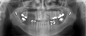
As a result, an infection develops in the unfilled part of the root canals, which causes inflammation to develop at the apex of the tooth root (i.e., apical periodontitis develops again). If a significant amount of pus forms in this focus of inflammation, which can happen during an exacerbation of the inflammatory process against the background of hypothermia, the pus seeks a way out, which can lead to its release under the mucous membrane. Which again will look like a cyst or purulent sac on the gum.→ In Fig. 6-7 you can see poorly filled root canals. Unfilled parts of the canals are marked with white arrows. Black arrows mark the boundaries of the focus of inflammation at the apex of the tooth root (on x-ray, the focus of inflammation looks like intense darkening). → In Fig. 8 you can see the well-filled root canals of the three lower incisors. Black arrows mark the tops of the roots of the teeth, up to which the canals should be filled. We have attached this photo specifically so that you can see how root canals should be filled normally.
How the treatment is carried out - the treatment in this case will consist of unsealing poorly filled root canals, after which the standard treatment of the inflammation at the apex of the tooth root is then carried out (i.e. standard treatment of apical periodontitis). You can read more about this treatment using the link above.
- The appearance of a cyst due to tooth perforation – when filling root canals or when installing pins, dentists often make another mistake - they perforate the wall of the root canal, making a “hole” in the root of the tooth. In Fig. 9-10 you can see how the dentist in both cases brought the pin directly into the bone tissue. As a result, inflammation (periodontal abscess) developed at the site of the perforation, which on an x-ray looks like intense darkening, limited for convenience by black arrows.
Treatment of perforations is a big problem in dentistry, and besides, it cannot be carried out in all cases (for example, teeth such as those in the radiographs above will have to be removed). If small perforations occur, they can be filled with special materials, for example Pro-Ruth material. Such materials are very expensive, but, of course, if you go to the same clinic where this happened to you, you should receive treatment for free, or pay for the cost of dental implantation.
- Gum cyst due to periodontitis – such a cyst in professional language is called “periodontal abscess”. In this case, a purulent abscess under the mucous membrane of the gums is formed in the projection of a deep periodontal pocket, which can form along the tooth root with local or generalized form of periodontitis. Usually, pus from the periodontal pocket is released into the oral cavity along the surface of the root, but if the pocket becomes very deep, the outflow of pus may be disrupted.
If the outflow of pus is disrupted in the projection of the periodontal pocket, a purulent abscess occurs, i.e. the same purulent sac. In the photo below you can see such a periodontal abscess in the projection of the distal root of the 6th lower tooth. On an x-ray of this tooth we see a deep periodontal pocket (to the entire depth of the tooth root). To treat this pathology, it will be necessary to open the purulent abscess and further treatment of periodontitis by a periodontist.Purulent abscess on the gum with local periodontitis -
Treatment
Treatment of dental cysts is carried out through surgery, laser treatment and conservative therapy. The latter has a positive effect only in the initial stages of the disease; overgrown cysts must be removed.
Surgery
To eliminate the pathology, it is not necessary to remove the entire tooth; only the tooth root on which the cyst is located is subject to resection. After removing the affected area, the dentist seals the remaining root, treats the surgical canal through which he removed the bladder with its contents, and stitches it up.
After a few days, the doctor removes the stitches and monitors the wound healing process. It is important to make sure that there are no cyst particles left in the dental canal; to achieve this goal, repeat radiography is performed.
Note! Sometimes it is impossible to remove the root along with the cyst; in these cases, the doctor completely removes the tooth. Indications for complete tooth resection are a difficult-to-reach location of the cyst and a severe course of the disease.
After surgical removal of a cyst, the patient must regularly visit the dentist and follow the recommendations prescribed by the doctor.
Conservative therapy
Tooth cyst - treatment of the disease with conservative methods is possible only in the early stages of its development. In order to eliminate the tumor, the patient is prescribed injections and rinses.
During therapeutic treatment, the dentist opens the dental canal, which leads to a cystic neoplasm, and pumps out exudate from it. The doctor does not fill the canal for ten days; during this period, the patient uses antiseptic solutions and tinctures to rinse the mouth.
Upon completion of the treatment course, the dentist treats the dental canal with medications and then fills the tooth.
Laser removal
Laser treatment is a modern method of treating dental cysts. When performing the method, the doctor opens the dental canal and uses laser irradiation to treat the area where the cystic tumor is located. The laser destroys not only the epithelium of the cyst, but also hundreds of thousands of bacteria that are inside the bladder.
The advantages of laser removal are rapid tissue healing and no risk of secondary infection in the oral cavity and dental canal.
Treatment with antibiotics
In some cases, dental cysts are treated with antibiotics. Taking antibacterial drugs is an auxiliary measure to destroy an expanded infection or the main method of treatment if a dental cyst develops against the background of a primary infectious disease.
Antibacterial drugs can only be prescribed by the attending physician; the following drugs are most often used:
- amoxicillin – has a high antibacterial effect, greatly facilitates the treatment of cysts with other methods;
- Cifroploxacin is a broad-spectrum antibiotic that actively destroys bacteria and relieves inflammation;
- tetracycline - this drug is prescribed more often than others, it actively relieves the inflammatory process, pain syndrome, and facilitates other methods of treating dental cysts.
Sometimes a doctor can prescribe topical antibacterial agents to a patient, but taking such medications is not always advisable - local drugs - antibiotics are quite difficult to distribute evenly over the diseased area.
Note! Antibacterial drugs are potent drugs that also affect beneficial bacteria in the body. You can take such medications only as prescribed by a doctor, without increasing the number of doses or dosage.
Treatment at home
Treatment of dental cysts at home is possible only as an auxiliary therapy. A cyst should not be confused with a granuloma; the latter can resolve on its own, but the cystic formation must be radically removed. Home treatment is not used to remove the cyst, but to eliminate the inflammatory process and destroy harmful bacteria.
The main goal of therapy at home is to provide an antiseptic effect. Propolis tincture, calendula tincture, eucalyptus tincture have an antiseptic effect. Tinctures are used as follows: a small amount of medicine is applied to a cotton swab and applied to the affected area, held for 5-10 minutes.
Medicines with an antiseptic effect can be used before surgery to remove a cyst and after tooth root removal. The antiseptic effect allows these medications to be used in the treatment of caries and other infectious diseases of the oral cavity.
Prevention
It is always easier and faster to prevent any disease than to cure it, so one should not forget about simple preventive rules that will help avoid the development of a dental cyst. The basic rules for preventing dental cysts are based on compliance with the rules of oral care.
How to prevent the formation of pathology:
- do not trigger the course of dental diseases such as caries, periodontitis, pulpitis; if infections occur, consult a doctor immediately;
- Brush your teeth daily and prevent the appearance of plaque, which can later transform into tartar;
- monitor the condition of the teeth and oral cavity after operations and mechanical injuries;
- visit your dentist regularly;
- monitor the condition of filled teeth and dental implants;
Patients who have had their teeth filled or have dental crowns or implants placed are advised to periodically have dental x-rays taken. This will allow timely detection of pathological changes and increase the chances of successful recovery without serious consequences.
Note! All diseases must be treated in a timely manner; inflammatory processes reduce immunity, as a result of which infections move freely from one organ to another; in addition, secondary infections can be added to already developed pathologies. It is important to monitor your health and strengthen local and general immunity.
To strengthen your immune system, strengthen your body, include fresh fruits and vegetables in your diet, play sports and walk outdoors more often. It is more difficult for any infection to get into a hardened body than into a weakened body.
General recommendations after surgery
On the first or second day after tooth extraction, you want to lie down and sleep - this is the body’s natural reaction to the stress experienced. After the release of stress hormones - adrenaline and cortisol - stabilization and rest are needed. Don't deny yourself this.
However, there are recommendations regarding sleep: you should raise the pillow higher and sleep reclining so that your head is elevated. This will prevent unnecessary pressure on the jaw and cause bleeding. To increase the inclination of the bed, place a pillow under the mattress or use two pillows.
Otherwise, maintain a calm regime without physical overload, pressure surges and overheating. That is, for the first time it is better to refuse air travel, hard work, playing sports and lifting weights, visiting the pool, bathhouse or sauna. You should not take a hot bath (a short warm shower would be preferable) and stay in the sun for a long time or sunbathe in a solarium. All this increases blood pressure, which is fraught with new bleeding. It is ideal if you have a couple of days off to recover after your teeth are removed.
The most complete reminder for patients after tooth extraction
So, you left the doctor's office - the surgeon asked you to tightly squeeze a gauze pad with your teeth, which will help stop the bleeding. You need to hold it for about 20-30 minutes, that is, while you fill out the necessary documents at the clinic or go home. Remove the tampon carefully, moving it slightly to the side, without sudden movements, so as not to damage the fresh blood clot.
Very important! A thrombus (usually called a “blood clot”) protects the wound from microbes and prevents pieces of food and saliva from accumulating in the hole during the first week of rehabilitation. This is a natural sterile dressing that starts the process of new tissue formation. The most important rule that will protect you from complications and severe inflammation of the wound is to never remove the clot, do not touch it with your tongue or fingers, do not touch it with a toothbrush and protect it from other influences. Gradually it will resolve on its own.
Do not keep the tampon in place for more than 30 minutes - gauze soaked in blood and saliva will quickly become a breeding ground for infection. In addition, a blood clot is forming in the hole - the presence of a foreign body and additional injury are of no use to it. You need to give the wound the opportunity to heal naturally.
“If you have a complex tooth extraction, especially if you had surgery to remove wisdom teeth, or if you have an underlying condition, such as diabetes, you may need to observe a doctor for a few more hours. For this purpose, we have our own hospital, where you can sit comfortably and relax while we monitor your condition to see if there are any complications. This will give us confidence that the operation will be successful and will reassure you as the patient.”
Vasiliev Alexander Alexandrovich, implant surgeon, work experience more than 9 years make an appointment
Tooth extraction WITHOUT complications! The modern equipment of our clinic allows us to carry out the most accurate and comprehensive diagnostics. Consultation is free!
Sign up online
Swelling after tooth extraction (especially if it was complicated) can be prevented by applying a cold compress - to do this, wrap an ice pack from the freezer in a towel and apply it to the cheek (that is, from the outside) for no more than 20 minutes. After 15 minutes the procedure can be repeated. If you don't have ice on hand, frozen vegetables or meat will do, but be sure to wrap them in a bag and a towel to avoid cold burns. Cold constricts blood vessels, reduces pain and swelling, although it does not completely remove swelling. Compresses are effective immediately after surgery for approximately 3-4 hours.
Do not warm up the injury site under any circumstances! A hot compress will only make the swelling worse by increasing blood flow to the area of inflammation. This will lead to serious complications after surgery.