A dental x-ray is a detailed image of the teeth, bones and soft tissues around them, allowing us to identify various dental and oral problems. X-rays can show cavities, hidden dental structures (such as wisdom teeth), bone loss and other abnormalities that cannot be detected during a visual examination and a routine dental consultation. Dental X-rays are usually performed annually. It is also actively used for follow-up monitoring of teeth after treatment. X-rays may be prescribed more often if your doctor monitors the development of a dental problem directly during treatment.
Types of dental x-rays
Today, there are four main types of dental x-rays according to their intended purpose:
- Bite X-ray – an image of the upper and lower rows of teeth in a closed state. This type of x-ray helps identify malocclusions, subsidence of bones, interdental caries, and the presence of hidden cavities. Also allows visualization of bone loss due to severe gum disease or dental infection.
- A peripheral x-ray of a tooth depicts the entire tooth, its surface and internal parts, that is, it is an image from the crown of the tooth to the part of the bone that supports it. These x-rays are used to identify dental problems below the gum or jaw line, such as affected teeth, abscesses, cysts, tumors, and bone changes associated with certain diseases.
- An occlusal photograph is aimed at identifying the condition of the roots of the teeth and the dental floor. It is actively used for jaw fractures, cleft palate symptoms, abscesses and suspected presence of hidden/not yet erupted teeth (including wisdom teeth). Occlusal x-rays can also be used to detect foreign objects.
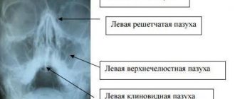
- Panoramic x-ray of teeth. Allows you to obtain a detailed image of the jaws, teeth, nasal cavity and jaw joints. This type of x-ray does not allow visualization of cavities. This type of x-ray is effective in diagnosing tooth fractures, various bone pathologies, cysts and neoplasms. Actively used before orthodontic treatment.
Why does a dentist refer a patient for dental x-rays?
An x-ray gives the doctor the opportunity to most accurately assess the condition of the problem unit or all organs of the oral cavity. And also study the clinical picture in detail. In particular:
- detect foci of inflammation that have not yet manifested themselves;
- identify neoplasms in the bone and soft tissues of the dental system;
- establish the signs and stage of development of periodontitis or periodontal disease;
- establish linear dimensions, volume, structure of bone tissue before implantation;
- more accurately assess the correctness of occlusion (tight closure of the dentition);
- detect anomalies of the dental system organs;
- determine the position of erupting wisdom teeth.
As a rule, radiography is prescribed before the start of orthodontic treatment, when planning implantation and prosthetics, before and after filling the tooth canals. This study is also used to evaluate the results of conservative and surgical treatment.
Free consultation
30-40 minutes
inspection and diagnostics
treatment plan and cost
Make an appointment
What can an x-ray reveal?
Thus, using dental x-rays, the dentist can determine:
- inflammatory processes inside the tooth that are invisible during normal examination;
- presence, size and location of neoplasms (eg, cysts) or foreign bodies;
- quality of dental canal filling;
- the degree of development of caries and the presence of deep, hidden carious areas;
- the presence of an abscess or other purulent-inflammatory processes;
- cracks in teeth;
- abnormalities in bone development;
- the presence of hidden, unerupted teeth (for example, wisdom teeth).
What types of X-ray diagnostics are used in dentistry?
The main types of images obtained during x-ray photography of the dental system:
- Sighting. Provides information about the condition of one or more teeth and adjacent tissues. This helps the specialist assess the condition of dentin, root canals, periodontium, and blood vessels.
- Panoramic (OPTG, orthopantomogram). It is a detailed flat image of the jaws, teeth, joints, and sinuses. This image helps to detect carious areas, defects in fillings, impacted teeth, and cysts. And also draw up a correct plan for implantation, prosthetics, and treatment.
- Occlusal. This is the so-called bite radiography, which is often used when examining children, as well as when it is impossible to obtain clear results using targeted radiography.
In addition, photographs can be film or digital, when the resulting image is transmitted using a special sensor to a computer monitor. In the second case, the study is called radiovisiography.
Features of the appointment of dental x-rays
If you are a new patient, you will likely undergo dental x-rays so that the new dentist can get a clear picture of your dental health. This is especially important if you do not have x-rays from your previous dentist. Children may need dental x-rays more often than adults because their dentists need to monitor the growth and development of their teeth. This is important because it can help the dentist determine whether baby teeth need to be removed to prevent complications, such as adult teeth growing behind baby teeth.
Indications for use
Dental radiography is used for two main purposes: to diagnose and evaluate the quality of treatment performed.
Allows you to detect the following diseases:
- hidden carious lesion;
- pulpitis (inflammation of the neurovascular bundle of the tooth);
- cyst at the roots of the teeth;
- periodontitis (purulent inflammation of the tissue between the root of the tooth and the hole in which it is located);
- gum disease, in which bone tissue atrophies (periodontitis, periodontal disease);
- neoplasm.
Intraoral radiography also helps evaluate the results of procedures such as:
- root canal filling;
- treatment of periodontal diseases.
Risks of Dental X-Rays
Dental X-rays involve radiation, however, the radiation levels are so low that it is considered safe for both children and adults. If your dentist uses digital X-rays instead of film X-rays, the risks from radiation exposure will be even lower.
Also, before the x-ray, the specialist will provide you with a protective apron to put on your chest, abdomen and pelvic area to prevent unnecessary radiation exposure to vital organs. In case of thyroid disease, a special thyroid collar may be used. Children and women of childbearing age may also wear it along with a lead bib.
Pregnancy is a contraindication for radiography. Women who are pregnant or think they may be pregnant should avoid all types of X-rays. Tell your dentist if you think you are pregnant because radiation exposure is not considered safe for the developing fetus.
Can X-ray examinations be performed on pregnant women?
In the first half of the term, the study is done only according to strict indications. In the second half of the term, you can do as much research as you like.
Another interesting fact. People often ask X-ray technicians to wear more protective lead aprons. Let's say right away that this is useless. You receive the same dose of radiation from covered and exposed parts of your body.
And remember that a doctor will never order an x-ray just out of curiosity. X-ray is one of the types of diagnostics of the area of proposed treatment and the anatomical features of the oral cavity.
Come to our clinic and we will make every effort to make your stay comfortable and enjoyable!
Preparation and carrying out the procedure
Dental X-rays do not require special preparation. The specialist will guide you through each step of the x-ray process. He may leave the room briefly during filming. You will be required to stand or sit still for short periods of time. Spacers (film holders), if used, will move and adjust in the mouth to produce correct images.
Once the images are ready—immediately in the case of digital x-rays—your dentist will check them and determine whether or not there are any abnormalities.
Popular methods of radiation diagnostics
Today, the most common and popular method of radiation examination in outpatient practice is intraoral radiography of teeth, or intraoral photograph of the tooth. Sometimes intraoral photographs of teeth are called targeted, which is incorrect. A targeted photograph is a photograph taken outside the standard positioning, and standardized studies are named according to the positioning method.
At a therapeutic appointment during endodontic treatment, at least three intraoral photographs of each tooth under examination must be taken:
- A diagnostic image is necessary to assess the condition of periodontal tissues at the time of examination, make a diagnosis, determine the number and shape of roots, the direction of canals, and choose treatment tactics.
- measuring photograph - a photograph of a tooth at the treatment stage with endodontic instruments inserted into the canals with a fixed stopper length of the working part or verifiers after instrumental treatment of the canals. If the orthogonal projection is performed correctly, provided that the visiograph program is accurately calibrated and there is no projection distortion for incisors and premolars, some measurements can be taken from the diagnostic radiogram. For multi-rooted teeth, it is preferable to measure the length of the canals using endodontic instruments (Fig. 1), an apex locator or from a three-dimensional photograph.
- a control photograph is taken immediately after the end of endodontic treatment in order to determine how well the root canals are filled, as well as after a certain specified time, in order to verify the absence or detect the presence of complications (Fig. 2). When examining multi-rooted teeth and in cases where there is an additional canal, in an image taken with the orthoradial direction of the beam (direct projection), the root canals often overlap each other, which significantly complicates diagnosis and can lead to errors in the treatment process. To obtain a separate image of the root canals, radiography with an oblique (eccentric) direction of the central beam is used (Fig. 1). For each specific case, the mesial or distal inclination (angulation) of the tube in the horizontal plane is selected (for more details, see: Rogatskin D.V., Ginali N.V. The Art of Dental Radiography, 2007).
Ideally, maximum information about the topography of the roots and the condition of periodontal tissues can be obtained by performing polypositional radiography. In this case, for diagnostic purposes, three photographs are taken - one in a straight line, with an orthoradial direction of the beam, and two in an oblique projection - with a distal-eccentric (Fig. 1) and mesial-eccentric direction of the beam (respectively, straight, posterior oblique and anterior oblique projections).
The most important aspects of successful intraoral radiography are standardization and consistent manipulation. Standardization of manipulations means the ability of a specialist conducting a radiation examination to choose the optimal method for each case and take a series of identical images, regardless of the position, condition of the patient and the time separating one examination from another. That is, if a diagnostic or measurement image is considered to be of high quality, each subsequent clarifying and control image must be made with the same spatial and technical settings, and each subsequent image must be identical to the previous one (Fig. 1, 2).
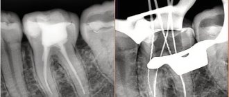
Rice. 1. Diagnostic and measuring images of tooth 36, taken in direct (a) and distal-eccentric projection (b). 36 - chronic apical periodontitis (K04.5) with characteristic changes on the mesial root.
Rice. 2. A control image immediately after treatment of teeth 21, 22 (chronic periapical abscess in a state of suppuration) (a) and a delayed control image 5 months after filling the canal (b), the state of repair at the treatment stage.
Digital radiography
Today there is a new technique for dental radiography using digital technology. The new method eliminates the need to develop X-ray film in a dark room; instead, the X-rays are sent directly to a computer and can be viewed on screen, saved or printed. There are several advantages of using this technology:
- minimal radiation;
- saving time - the image is available on the screen a few seconds after shooting is completed;
- the ability to enlarge images multiple times compared to their actual size on the computer screen;
- the ability to send images by email (for example, to another specialist to get a second opinion, etc.);
- software installed on a computer can help dentists digitally compare current images with previous ones in the process of subtraction radiography. Using this technique, everything that is similar between two images is "subtracted" from them, leaving a clear, detailed image of only the part that is different. This helps dentists easily see subtle changes that might not be immediately noticeable.
Dental radiography is of fundamental importance for correct diagnosis. Thus, today radiography is an integral and extremely important part of professional dental care.
Are dental x-rays safe for human health?
In accordance with SanPiN 2.6.1.1192-03, the maximum permissible annual radiation dose during diagnostic x-ray procedures is 1000 μSv (microsievert) for an adult and 300-400 μSv for a child.
To receive it, within a year you must complete:
- 500 targeted dental examinations using a radiovisiograph;
- 100 procedures of conventional x-rays with image output on film;
- 80 digital or 40 film orthopantomographies (obtaining a panoramic image).
Thus, dental X-rays for almost any purpose do not exceed the permissible doses. Therefore, it does not have a negative effect on health. Even when producing film OPTG, the radiation dose is a maximum of 30 μSv. If it is necessary to take a targeted X-ray of the teeth and use radiovisiography, you can even get several images during one appointment. Without any risk to health, since a single dose in this case is only 2-3 μSv.
Kinds
Contact (pariapical)
It is carried out to obtain an image of the tooth in its true size - from the crown to the root and the hole in which it is located. As a rule, it is used to diagnose pathologies in the area of the tooth root. It also allows you to detect abnormalities in the structure of bone tissue. It is worth noting that this technique is ineffective if it is necessary to examine the condition of periodontal tissues.
When performing contact radiography with a conventional X-ray machine, film sizes of 2x3, 3x4 cm, 5x6 and 6x8 cm are used.
Interproximal
Justified in cases where it is necessary to obtain a clear and undistorted intraoral image to diagnose interdental or cervical caries and determine the degree of bone tissue resorption. Three or four photographs are taken to examine all teeth.
Occlusal (bite)
This technique is used if there is a need to examine several teeth at once (four or more), diagnose dystopic (improperly located in the dentition) and impacted teeth, study the condition of the hard palate, salivary glands, and detect neoplasms and cysts.
Occlusal radiography has proven its effectiveness in examining the dental system in children, as well as in cases where it is not possible to use the contact method due to an increased gag reflex, jaw injuries, and TMJ dysfunction. The main difference is that a film measuring 5x6 or 6x8 cm does not fit into the patient’s oral cavity, but is fixed between the closed teeth.
Long focal length
It differs from contact in that it allows you to get a clear picture of not only periodontal tissues, but also periodontal tissue (this method is actively used in periodontology). This result is achieved by using a device with a more powerful X-ray tube.
Digital radiography (radiovisiography)
Separately, mention should be made of the technology that is based on the use of a radiovisiograph. This computer device allows you to obtain targeted images of teeth with minimal radiation doses - ten times less than conventional film X-rays.
The resulting images are immediately displayed on the computer screen and then transferred to the attending physician's office, saving time.
Images can be stored for an unlimited time, they can be copied to digital media, or sent by e-mail for consultation with other doctors.