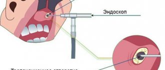
MRI of the sinuses, how a scan is done
The patient is placed on a table that moves inside the tomograph. There is no radiation exposure during this examination. MRI shows sections in different planes, without the need to change the position of the body. The procedure takes about 20 minutes and is very comfortable. There is good air ventilation inside the scanner and soft lighting.
In cases where the use of intravenous contrast is recommended, the study will take a little longer (about 15 minutes additional). After completing the tomography, you can immediately return to your normal lifestyle.
Medical Internet conferences
Relevance.
Inflammatory diseases of the paranasal sinuses are among the most common diseases in this area. Among the many reasons for their occurrence, an important place is given to the topographic and anatomical features of the structure of the sinuses. One of the most variable air cavities of the human skull, according to a number of scientists, is the frontal sinus. This is confirmed by numerous studies conducted at different times in the development of medicine and anatomy, and this fact of variability of the frontal sinus supports the relevance of research in this area. Currently, the study of the size of the frontal sinus and their relationship with the size and structure of the facial skull is becoming increasingly important in justifying surgical interventions, including endoscopic ones. After all, it is individual characteristics that often play a decisive role in the course of the disease, the characteristics of the intervention and the patient’s recovery.
Target.
Identification of patterns of variability in the size and volume characteristics of the frontal sinuses in terms of age and gender.
Tasks.
Calculation of the volume and area of the frontal sinuses using computer craniometry, studying the relationship of the obtained parameters with age and gender.
Materials and methods.
We studied data from high-resolution head computed tomography (CT). For convenience, 83 patients without pathology and surgical interventions in the nasal cavity were divided by gender into 2 groups, each of which was divided into 2 age groups. The technique of computer craniometry was used (RF patent No. 2499558), which consists of obtaining a three-dimensional image of the skull under study, its subsequent introduction into a specially developed computer program “Cranio” (computer program, state registration No. 2015615568) and further determination with its help of the main craniometric parameters parameters. The lower edge of the left orbit and the upper points of the outer edges of the auditory openings (porion) were determined, through which the Frankfurt plane was constructed using the same program. Then, a frontal plane was constructed through the porions and perpendicular to the Frankfurt plane, and a sagittal plane was constructed through the middle of the nasofrontal suture (nasion) and perpendicular to the frontal plane. The necessary craniometric points were marked on the depicted skull, and using the same program the distance from these points to all planes was determined. In this way, the stereotopometric parameters of the facial skull, as well as the volume and surface area of the frontal sinuses, were automatically determined.
Results.
Having processed the data obtained, we found the following correlations. There is a moderately strong positive correlation (0.3) between the Nasion-Hormion size (n-ho) and the volume of the frontal sinus (V). There is a moderately strong positive correlation (0.3) between the Nasion-Dacrion (nd) size and V. There is a moderately strong positive correlation (0.3) between the sizes Nasion - Frontomalar Temporal point (n-fmt) and V. There is a moderately strong positive correlation (0.3) between the size of Nasion - Frontomalar-orbital point (n-fmo) and V. The correlation between other sizes and the volume of the frontal sinus was weaker, indicating a lesser influence of these sizes on the volume of the air sinuses.
Bilateral significantly significant differences in the frontal sinuses in both men and women were not detected in any of the age groups. But significant differences were revealed in the size of the frontal sinuses in men and women depending on age.
The correlation between the volume of the frontal sinuses and their area turned out to be strongly positive (0.9), but not absolute, since these values are influenced by the presence of septations in the sinus, which significantly increase the surface area of the sinus with a slight change in its volume.
Conclusions.
The following results were obtained during the study. In the first age group, the growth of the frontal sinus in men is faster than in women. The enlargement and growth of the sinus is more uniform throughout life in women. Our studies did not show reliable signs of bilateral variability. The septa inside the sinus do not specifically change the volume of the sinus, but they do dramatically increase its surface area. This fact may indicate the complexity of the configuration of the frontal sinus, which in turn is important for predicting the course of diseases and features of surgical tactics.
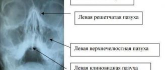
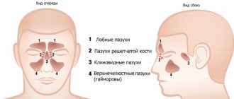
Structure of the sinuses
The paranasal sinuses are air-filled spaces located in the bones of the skull and are an extension of the nasal cavity. Their name corresponds to the name of the bone located nearby.
They are divided into:
- Lower. This is a pair of maxillary (maxillary) sinuses.
- Upper:
- anterior - paired frontal and anterior cells of the ethmoidal labyrinth;
- posterior – unpaired sphenoid sinus and posterior cells of the ethmoidal labyrinth.
Structure of the sinuses
Their main functions:
- Protective. Due to the fact that the inside of the sinuses is covered with a mucous membrane, the air in them is additionally warmed, moistened, and purified. The sinuses protect deeper and more vital structures from external damage.
- Resonator. The sinuses are involved in the formation of individual voice characteristics. Small cavities, such as the sphenoid and ethmoid cavities, create high-pitched sounds, while large cavities (maxillary and frontal) create low sounds. Therefore, during inflammatory processes, the timbre of the voice often changes.
Mycetoma
non-invasive fungal sinusitis of the left maxillary sinus
The hypointense MR signal of a mycetoma may be mistaken for air in the paranasal sinus; Non-invasive fungal sinusitis does not look the same in different sequences.
If they are large, they cause headaches due to the pressure of the cyst shell on the walls of the sinus.
Often combined with allergic rhinitis, hypertrophy of the nasal turbinates and deviated nasal septum
Large cysts located in the lower parts of the maxillary sinus may be asymptomatic, while a small cyst located on the upper wall, in the area of the 2nd branch of the trigeminal nerve, can cause headaches.
Mucocele of the ethmoid labyrinth and frontal sinus on the right
This is a large formation of the paranasal sinus, lined with epithelium and filled with mucus, which is formed as a result of obstruction of the main sinus canal.
The most typical symptom: expansion of the paranasal sinus with smooth, clear contours with thinning and remodeling of the adjacent bone plate.
Indications and contraindications for MRI of the sinuses
Magnetic resonance imaging is necessary in the following cases:
- prolonged headaches, especially pain in the face;
- repeated nosebleeds;
- treatment-resistant nasal congestion;
- impaired sense of smell;
- suspicion of a neoplasm;
- injuries to the facial area;
- congenital anomalies of the nasal cavity and sinuses;
- pathologies identified by X-ray or ultrasound results;
- postoperative observation.
All sinuses communicate with each other, so infectious processes (the most common pathology in this area) can pass from one sinus to another.
Acute sinusitis
Its main signs on MRI are a decrease or increase in signal intensity from the mucous membrane. Changes in the bone walls of the sinuses are possible in the form of thickening or destruction.
Signs of sinusitis and ethmoiditis on MRI
Chronic rhinosinusitis
MRI is of particular importance for inflammation of a fungal nature, which is difficult to diagnose by other methods. It is more common in young people with concomitant bronchial asthma, who are forced to constantly use inhaled corticosteroids, suppressing local immunity. Fungal sinusitis can also result from chronic infection in people with weakened immune systems. This case is characterized by severe difficulty in nasal breathing, thick, difficult to separate secretions in the nasal passages and sinuses, and rapid progression.
On MRI, the inflamed mucous membrane and the exudate contained in the sinus give an intense signal, in the center of which a low-intensity formation is determined, surrounded by a layer of fluid (the so-called “fungus ball”). With other research methods, it can be mistaken for air. MRI is the most informative method of differential diagnosis in this case.
Fungal sinusitis on MRI
Polypous rhinosinusitis
This is a disease of the mucous membrane, which is manifested by the progressive growth of polyps. The local process is often associated with chronic purulent inflammation. Diffuse polyposis, spreading to all paranasal sinuses, is most likely already caused by a decrease in the immunity of the whole organism.
MRI reveals formations of soft tissue density, heterogeneous in structure, sometimes with areas of cartilaginous density. Magnetic resonance imaging in this case helps to differentiate polyps from a tumor process.
Mucocele
This is a benign cyst-like formation of the sinuses. Most often it is a consequence of chronic inflammation and affects the frontal sinus.
On MRI, the sinus appears dilated, but with preserved airiness, and the image intensity is reduced. Magnetic resonance imaging makes it possible to distinguish it from cysts, sinusitis and polyposis. In the case where there is bone damage, it is differentiated from malignant formations. The prognosis is most favorable with early diagnosis and timely treatment.
Tumor diseases
Among benign neoplasms, the greatest clinical significance is:
- papillomas (from respiratory epithelium);
- adenomas (from glandular structures of the mucous membrane);
- vascular tumors;
- osteomas (from bone tissue);
- chondromas (made of cartilage tissue).
Manifestations of nasal and sinus tumors in the early stages are usually minimal and nonspecific. Some types may not produce clinical symptoms for years and are accidentally detected on MRI performed for other indications. Complaints may appear even when the tumor grows beyond the nasal sinuses. May appear:
- deformation of the nose or face;
- double vision;
- lacrimation (with compression of the nasolacrimal duct);
- decreased vision (with compression of the optic nerve);
- meningitis (if it spreads into the cranial cavity).
Often the only symptom may be a headache. This is due to the peculiarities of the innervation of this area. If a benign sinus disease has already been diagnosed, then the appearance of pain may indicate the addition of a secondary infection or malignant degeneration; a mandatory MRI is required.
Treatment of all tumors of the nasal sinuses is surgical and timely accurate diagnosis plays a great role in the prognosis of the disease.
Magnetic resonance imaging is the main diagnostic method for malignant processes in the sinuses and makes it possible to determine the nature, stage, boundaries, choose the right method of surgical treatment and carry out postoperative monitoring.
Hemangioma of the maxillary sinus on MRI (indicated by arrows)
MRI is contraindicated:
- pregnant women in the first trimester;
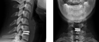
- if there are implants or foreign bodies containing metals with ferromagnetic properties in the patient’s body;
- if the patient has electronic devices installed (pacemaker, insulin pump, cochlear implant or others)
- if the patient suffers from claustrophobia or has an unstable mental state;
- in the presence of vascular clips on the arteries.
Children under 5 years of age and adults with a body weight of more than 130 kg or a waist circumference of more than 150 cm are not examined in our clinic.
Vasomotor sinusitis
It develops in people with disorders of the autonomic nervous system and nasal breathing against a background of negative emotions and stress.
Clinical manifestations
Allergic and vasomotor symptoms are manifested by sneezing, difficulty in nasal breathing, and discharge of large amounts of contents from the nose. Most often this is pansinitis.
Diagnostics
In this case, ACMD-Medox can use both SCT and MRI with equal benefit. With classical radiography, there may be options for under- and over-diagnosis associated with the nature of the visualization of pansinuit in the images.
CT or MRI of the sinuses, which is better?
In rhinology, MRI is most effective for identifying pathologies such as fungal diseases of the mucous membrane, mucoceles, polyps and cysts, and, if necessary, distinguishing tumors from inflammation. Magnetic resonance imaging is informative in case of intracranial and intraorbital spread of a tumor, determines tumor growth beyond the nasal sinuses, while bone tissue and filling materials do not create significant interference with visualization.
CT is more often used for facial injuries, especially in the emergency phase.
Chronic sinusitis
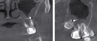
Occurs after untreated or untreated sinusitis; it can also occur in the event of a violation of the outflow of exudate (with a deviated nasal septum) or in the case of contact transmission of infection from the teeth (for example, teeth affected by caries - author's note) to the mucous membrane of the maxillary sinus (odontogenic sinusitis ).
Clinical manifestations
Prolonged runny nose, periodic exacerbations of sinusitis, general malaise, headache, constant sinus discharge, and so on. So outwardly nothing special, but it’s constant.
Diagnostics
It is best to use SCT (especially in cases of odontogenic sinusitis, when it is extremely important to determine the condition of the bone tissue, to exclude the presence of purulent inflammation in the area of the apexes of the roots of the teeth, in general, in those areas where the doctor simply cannot look - author's note). In second place is MRI - most often, it is with the help of magnetic resonance therapy that the diagnosis is made: chronic sinusitis, since it is a “by-product” of an MRI study of the brain, which is one of the most frequently prescribed.